Open Access
Technical advance
Hepatocyte growth factor (HGF) in fecal samples: rapid detection
by surface plasmon resonance
Fariba Nayeri*
1, Daniel Aili
2, Tayeb Nayeri
1, Junyang Xu
1, Sven Almer
3,
Ingemar Lundström
2, Britt Åkerlind
4and Bo Liedberg
2Address: 1Divisions of Infectious Diseases, University Hospital, Linköping, Sweden, 2Department of Physics and Measurement Technology,
University of Linköping, Sweden, 3Department of Gastroenterology & Hepatology, University Hospital, Linköping, Sweden and 4Division of
Clinical Microbiology, University Hospital, Linköping, Sweden
Email: Fariba Nayeri* - [email protected]; Daniel Aili - [email protected]; Tayeb Nayeri - [email protected]; Junyang Xu - [email protected]; Sven Almer - [email protected]; Ingemar Lundström - [email protected]; Britt Åkerlind - [email protected]; Bo Liedberg - [email protected]
* Corresponding author
Abstract
Background: The development of biosensors, based on surface plasmon resonance (SPR) technology, enables monitoring of a variety of biospecific interactions without the need for chemical-, biological- or radiological-labelled reagents.
Method: We utilised SPR to detect hepatocyte growth factor (HGF) in reconstituted faecal samples and studied samples from patients with infectious gastroenteritis (n = 20) and normal controls (n = 10). Mouse anti-human HGF monoclonal antibodies and recombinant human HGF receptor (c-Met)/Fc chimera were immobilised in flow cells of a CM5 biosensor chip.
Results: We found that infectious gastroenteritis produced a higher signal response compared to controls, due to binding of HGF to monoclonal anti-HGF antibody as well as binding of HGF to c-Met receptor (p < 0.01). The SPR signal response correlated with results from ELISA (r = 72%, p > 0.001). The signal response decreased significantly (p < 0.05) when samples were diluted with dextran, because of reduction in both specific as well as unspecific binding of HGF to dextran. The decrease in the specific response might imply that the dextran- binding site for HGF overlaps with the antibody binding epitope, or that dextran binding induces a conformational change of the HGF molecule. Bands corresponding to HGF were found by gel electrophoresis of purified faeces in an affinity chromatography column immobilised by HGF ligands.
Conclusion: Determination of HGF by SPR might be beneficial in diagnosis of acute situations that present with symptoms of gastroenteritis and may, possibly, guide appropriate medical treatments. This is to our knowledge the first report on the use of SPR for detection of HGF in faeces samples.
Background
The SPR technique is suitable for studying biomolecular interactions on, or close to, a surface [1]. SPR enables
rapid detection in real time without any labeling of sam-ples and can be utilised for concentration determination, kinetics studies and epitope mapping. In brief, light
Published: 12 April 2005
BMC Gastroenterology 2005, 5:13 doi:10.1186/1471-230X-5-13
Received: 20 June 2004 Accepted: 12 April 2005
This article is available from: http://www.biomedcentral.com/1471-230X/5/13
© 2005 Nayeri et al; licensee BioMed Central Ltd.
incident on a metal surface at a given angle of incidence can excite a surface-bound electromagnetic wave, a surface plasmon, which propagates along the interface between the metal and the ambient medium. Associated with the surface plasmon is an evanescent field that probes local changes in the refractive index of the ambient medium, induced, for example, by binding of a biomolecule to the surface. The change in refractive index will shift the angle of incidence at which the SPR excitation occurs. This shift in the angle is tracked by monitoring the movement of intensity minima of the reflected light with time using the Kretschmann configuration, and the binding event is pre-sented as a sensorgram [1]. The sensor surface of the SPR apparatus consists of a carboxy-methylated dextran matrix, which is a hydrogel providing a solution-like envi-ronment in which the biospecific interactions occur. The carboxyl groups in the dextran matrix enable covalent coupling of ligands (proteins, receptors, DNA etc) to the surface.
Hepatocyte growth factor (HGF) is a growth factor that is produced by mesenchymal cells during injuries in various organs. It stimulates cell division [2], motility [3], and a normal morphogenic structure [4] in the epithelial cells that have survived an injury (adjacent to the injured area). HGF is translated as a single-chain precursor and activated at the site of injury by proteolytic cleavage, resulting in a double-chained active HGF [5]. Interaction between the active HGF and its specific receptor (c-Met) [6] initiates intracellular signal pathways that result in regeneration and repair of damaged tissue [7]. High amounts of HGF have been determined systemically during injuries caused by infection [8]. At the site of infection, a local production of HGF has been found during bacterial meningitis and pneumonia [9,10].
Using a commercially available ELISA kit, we have shown that the concentration of HGF in faeces increases signifi-cantly during infectious gastroenteritis [11]. The levels decrease to normal after recovery. We have further studied the presence and stability of HGF in faeces samples during infectious processes [12]. Since traditional immunoassays (e.g., ELISA) are time-consuming and laborious, there is a need for quicker and simpler methods. In this study we present a method based on surface plasmon resonance, SPR, which allow us to detect and measure the levels of HGF in faeces in individual samples with the same accu-racy but more rapidly than traditional methods.
Methods
Patients
Nine patients admitted to hospital with signs of acute untreated infectious diarrhoea were included. These patients had participated in a previous study [11]. Faeces samples were taken at admittance and cultured (
Campylo-bacter, Salmonella and Shigella), and laboratory diagnostic tests for Rotavirus (antigen detection) and Calicivirus (elec-tron microscopy) as well as cultures and cytotoxin analy-sis for Clostridium difficile were conducted. Cultures and cytotoxin analysis (toxin A) were positive for Clostridium difficile in 1 case. Cultures revealed growth of Campylo-bacter jejuni in 4 cases. Salmonella art (Salmonella DO) was found in 3 cases. Faeces culture was negative in one patient. This patient had diarrhoea and fever and his wife had gastroenteritis caused by Campylobacter jejuni. A sec-ond group of eleven patients with Clostridium difficile
(positive culture and toxin A) were also analysed. This gave a total of 20 samples of faeces from 20 patients (11 women, 9 men, range 20–85, median 51.5 and mean 52 year). Faeces haemoglobin (Actim Fecal Blood test; Orion Diagnostica) was examined and was positive in 10/13 samples.
Controls
Faeces samples were obtained from 10 healthy vaccina-tion volunteers (six women and four men, 20–60 years) without signs of infection or diarrhoea. Faeces culture and diagnostic tests for Rotavirus and Caliciviruswere negative in all of these cases. These patients had negative Actim Fecal Blood test results (Orion Diagnostica)
Standardizing faeces volume and reconstitution procedure
All faeces samples were stored at -20°C. Prior to handling, the samples were thawed at room temperature and mixed using a Vortex (Vortex-Genie, Scientific Industries Inc., Bohemia, NY, USA). The samples were then placed at -70°C for 15 minutes, followed by room temperature for 2 minutes and dissolved in distilled water at a dilution of 1:6. The suspension was centrifuged at 1000–3000 G for 15 minutes and the supernatant stored at -70°C until analysed.
To avoid the effects of digestive as well as bacterial enzymes, protease inhibitor (1–5%) (Sigma Aldrich) with specific inhibition of serine, cysteine, aspartic proteases and aminopeptidases, containing 4-(2-aminoethyl) ben-zenesulfonyl fluoride (AEBSF), pepstatinA, E-64, bestatin, leupeptin, and aprotinin (but no metal chelators) was added to the thawed samples (room temperature) 30 minutes prior to analysis.
SPR measurements and ligand immobilisation procedures
hydrogel was carried out by conventional carbodiimide chemistry using 200 mM EDC (N-ethyl-N' -(3-diethylami-nopropyl) carbodiimide) and 50 mM NHS (N-hydroxy-succinimide). The activation time was 7 min, followed by a 2–7 min ligand injection. Deactivation of remaining active esters was performed by a 7 min injection of eth-anolamine/hydrochloride at pH 8.5. A flow rate of 5 µl/ min was used during immobilisation. All ligands were diluted in 10 mM acetate buffer pH 4.5, i.e., below the protein isoelectric point, thus enhancing the electrostatic interactions between the dextran matrix and the ligands. The monoclonal anti-HGF (500 µg/ml) was diluted 1:10, the recombinant Met proto-oncogene receptor (100 µg/ ml) 1:5, and the HGF recombinant (5 ug/ml) 1:3. The contact time varied between two and seven minutes resulting in levels of immobilisation between 8000 and 30000 RU (response units). After deactivation, the sur-faces were washed with five subsequent one-minute injec-tions of 5 mM glycine buffer pH 2.0 with 1 M NaCl. One of the flow cells was used as a reference to monitor the response due to buffer and unspecific interactions. This flow cell was treated in the same way as the other during the immobilisation procedure, but omitting the ligand immobilisation step.
Determination of faeces HGF by ELISA
After storage all samples were thawed and centrifuged at 1000 G for 15 minutes prior to analysis. Immunoreactive HGF was determined by ELISA using a commercially available kit (Quantikine HGF Immunoassay, R&D Sys-tems Inc., Minneapolis, USA).
Determination of faeces haemoglobin
Actim Fecal Blood (Orion Diagnostica) is an immuno-chromography technique that is specific for the determi-nation of human haemoglobin using two monoclonal antibodies. The detection limit in faeces is 50 µg haemo-globin/L or 25–50 µg haemoglobin/g. The method detects the intact haemoglobin molecule, but neither haemo-globin that is influenced by the enzymes during gastroin-testinal passage nor animal haemoglobin is detected.
Purification of faecal samples by affinity chromatography, ultrafiltration, and SDS-PAGE
The affinity chromatography columns (Hi-trap Amer-sham Biosciences) were immobilised with monoclonal anti-human HGF and recombinant human HGF receptor (c-MET)/Fc chimera (R&D Systems) respectively. Faeces samples were reconstituted in distilled water. The samples were applied to the 500 µl centrifugal tubes (Amicon Ultra, Millipore, S.A.S. Molsheim, France) and centrifuged at 4000 G for one hour. SDS-PAGE was performed with the faeces samples before and after purification and filtra-tion, using a 4% stacking gel and a 12% running acryla-mide gel [13]. For size estimation prestained protein
standard (Sigma Aldrich) was electrophoresed simultaneously.
Statistics
Non-parametric Friedman and Wilcoxon signed rank tests (absolute values, Statview & SPSS Base 11.0) were used. The Spearman rank correlation coefficient was used for analysis of correlation between parameters. A p-value ≤ 0.05 was regarded as statistically significant.
Results
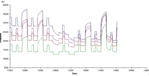
Patients with infectious gastroenteritis had significantly higher signal responses in flow cells immobilised with monoclonal anti-HGF antibody compared to the healthy controls (determined in at least six independent experi-ments)(Friedman p = 0.006, Wilcoxen Signed Rank Tests p = 0.008) (Fig 1 and 2). The signal responses in the flow cells immobilised with c-Met receptor were also signifi-cantly higher in the group with infectious gastroenteritis (Wilcoxen Signed Rank Tests, p = 0.02).
Correlation with ELISA
Determination of HGF in faeces (patients with infectious gastroenteritis) by utilising SPR correlated significantly (n = 20, r = 72%, p < 0.001) with the levels measured by ELISA (range 0.14–8.24, median 0.83 ng/mL) (Fig 3). HGF levels determined by ELISA in healthy controls (n = 10) were low (range 0.01–0.26, median 0.06)
Correlation with age
We found a negative correlation between the SPR signal intensity and age in patients with infectious gastroenteritis (n = 20, r = - 0.55, p = 0.010) (Fig 4). The patients with
Histogram showing signal responses determined by SPR (channel immobilised by mouse anti-human HGF monoclonal antibodies) in infectious gastroenteritis (n = 20, median 1895 ± 429 RU, range 123–6660 RU) and healthy controls (n = 10, median 171 ± 38 RU, range 14–459 RU)
Figure 1
acute gastroenteritis were older than the healthy controls (median 51.5 respective 35.0 years).
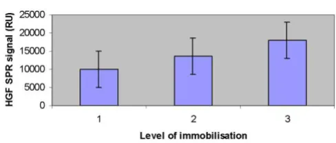
Effects of altering the immobilisation levels
The effect on signal responses due to the amount of mon-oclonal anti-HGF antibody immobilised was determined in different samples (n = 12). Increasing the immobilisa-tion levels by increasing the contact time during immobi-lisation (1, 5, and 10 minutes) caused a significantly higher response of all samples (Friedman p < 0.01, Wil-coxon signed ranks test between 1 and 5 minutes p = 0.058, between 5 and 10 minutes p = 0.003, and between 1 and 10 minutes p = 0.003) (Fig 5). Altering the flow rate (5, 10, and 15 µl/min) did not have a significant effect on the response of the different samples (Friedman p = 0.48) (data not shown). When altering levels of immobilisation of the c-Met receptor (n = 15), no effect in responses in the
case of infection (P = 0.85) was observed. Changing of the flow rate (5, 10, and 15 µl/min) did not influence the sig-nal responses (P = 0.36) (data not shown).
Binding of HGF to dextran
Addition of dextran (0.05%) to the samples resulted in a significant (p < 0.05) decrease in signal responses in flow cells with immobilised anti-HGF but did not affect the HGF binding to c-met (p = 0.11). Diluting recombinant HGF with dextran (0.05%) also resulted in a decrease of binding to the monoclonal anti-HGF antibody (n = 3). The test of significance was not performed.
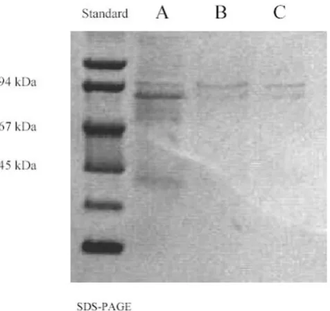
SDS-PAGE
We investigated the presence of HGF in the samples by SDS-PAGE of faeces samples as well as SDS-PAGE of puri-fied faeces samples in an affinity chromatography column
SPR sensogram showing the signal obtained from analysis of faeces from a patient with infectious gastroenteritis
Figure 2
immobilised by either mouse anti-human HGF mono-clonal antibody or recombinant human c-met receptor. Bands corresponding to the HGF were detected with apparent molecular masses of 75–90 kDa (Fig 6).
Protease Inhibitor
Faeces destroyed the proteins immobilised in the flow cells of the CM5 chip (Fig 7). Adding protease inhibitor (1–5%) to the faeces samples within 30 minutes prior to experiments could inhibit this effect completely.
Discussion
In the present work the Surface Plasmon Resonance (SPR) based method has been used for the first time to evaluate faecal samples and monitor the relative amount of HGF in the samples.
HGF has been investigated widely over the past decade. The unique properties of this cytokine, makes it likely to be involved in the recovery process after injuries [3]. Recent studies have strengthened the notion that HGF plays an important role in the regeneration of an injured organ [2]. Several researchers have attempted to treat induced hazardous injuries in animal models by HGF [14,15]. We have studied HGF during infectious diseases and found that HGF was produced in high amounts both systemically and locally during injuries caused by infec-tion [8]. Low amounts of serum HGF in patients with
Correlation between the concentration of HGF in faeces obtained by ELISA and signal responses by SPR (Spearman rank correlation coefficient n = 20, r = 0.72, p < 0.001)
Figure 3
Correlation between the concentration of HGF in faeces obtained by ELISA and signal responses by SPR (Spearman rank correlation coefficient n = 20, r = 0.72, p < 0.001). The shadow points along the regression line are not data points.
Correlation between age of the patients with infectious gas-troenteritis and signal responses by SPR (Spearman rank cor-relation coefficient r = - 0.55, p = 0.01)
Figure 4
Correlation between age of the patients with infectious gas-troenteritis and signal responses by SPR (Spearman rank cor-relation coefficient r = - 0.55, p = 0.01). The shadow points along the regression line are not data points.
Histogram showing the effects of increasing the tion levels by increasing the contact time during immobilisa-tion (1–3 representing 1, 5, and 10 minutes) (Friedman p < 0.01, Wilcoxon signed ranks test between 1 and 5 minutes p = 0.058, between 5 and 10 minutes p = 0.003, and between 1 and 10 minutes p = 0.003)
Figure 5
pneumonia correlated significantly to poor prognosis [16]. Application of HGF locally at the site of an injury such as a chronic ulcer resulted in an accelerated healing process [17].
The gastrointestinal mucosa has a remarkable ability to repair damage, and growth factors play an important role in the regeneration of injured cells in gastrointestinal organs [18]. Nishimura et al. (1998) [19] showed that HGF was the most potent of the cytokines (HGF, TGF-α, TGF-β, and keratinocyte growth factor) in accelerating repair of the damaged monolayer of an epithelial cell line derived from normal rat small intestine. We studied the amounts of HGF in faeces of patients with infectious gas-troenteritis and found that the levels of HGF were signifi-cantly elevated during infection [11]. Although this observation might indicate healing of injury caused by an infection, we could discriminate with high specificity and sensitivity between infectious gastroenteritis and other disorders that present with diarrhoea [11].
In a previous study [12] the concentration and stability of HGF in fecal samples was investigated using ELISA. Although the ELISA method was very reliable, it was labo-rious and gave no information about the form of HGF that was present in the samples. In the present study we
found that there is good correlation between the results obtained from SPR and ELISA measurements of faecal HGF levels. In some cases differences between results obtained by ELISA and SPR are found and this phenome-non is to be further investigated.
Using SPR we were able to monitor the presence of HGF in the samples and the interactions between HGF and dif-ferent ligands with difdif-ferent binding specificity to the HGF molecule. First, a recombinant HGF that showed affinity to the monoclonal anti-HGF antibody as well as c-met receptor was examined (data not shown). The affinity of recombinant HGF disappeared within one week after reconstitution of the lyophilised form (data not shown). On the other hand, the signal responses after analysis of faeces samples in the patients with infectious gastroenteritis were stable after reconstitution. We have tested widely the stability of HGF in faeces samples by ELISA [12] and previously in serum [20]. Unlike our pre-vious observations [11] that showed no significant corre-lation between age of patients and HGF levels determined by ELISA, in this study we found a negative correlation between age and SPR signal in the group with infectious gastroenteritis. This might strengthen the notion that the ability of HGF to interact with its ligand or possibly its activity might change by age. Moreover, in spite of the fact that patients with infectious gastroenteritis were older than healthy controls they had higher SPR signals.
Presence of blood was shown in ten out of thirteen cases with infectious gastroenteritis while it was negative in healthy controls. However we have previously studied the correlation between presence of blood in faeces and HGF levels (determined by ELISA) and did not find any signif-icant correlation [12].
The immoblised layer in the flow cells of the chip were destroyed after analysis of faeces samples (Fig 7). Adding protease inhibitor to the samples before analysis inhibited this effect which might indicate the presence of proteases in faeces. We have previously shown that adding protease inhibitor to faeces does not influence the ability to detect HGF by ELISA [12].
At least two binding sites for HGF on liver cell surfaces have been demonstrated in rat: the heparin-resistant and acid-washable site (HGF receptor, binding site) and the heparin-washable site (low-affinity, cell-surface heparan sulfate proteoglycan binding site) [21]. HGF has also been reported to interact with the extracellular matrix [22]. The low affinity sites may serve as a reservoir for the regulation of endogenous HGF levels and provide a matrix for con-version of promitogen to the two-chain form [21]. The HGF detected during infectious gastroenteritis had bind-ing affinity to dextran (proteoglycan). By addbind-ing
carboxy-SPS-PAGE of faeces
Figure 6
methyl dextran (0.05%) to the samples, the levels of HGF bound to the immobilised monoclonal anti-HGF decreased significantly. Adding the same amount of dex-tran to recombinant HGF gave the same result in signal responses (data not shown). This might indicate a bind-ing affinity of this form of HGF to dextran, which might resemble such affinity to cell-surface heparan sulfate proteoglycan binding sites. These binding sites either overlap with the antibody-binding epitope or induce conformation shift in the HGF upon binding to the dex-tran, resulting in sterical hindrance and thus preventing binding to the antibody.
Conclusion
Infectious gastroenteritis is a common disease in all soci-eties. Determination of HGF by SPR is a reliable method to evaluate the relative amounts of HGF in faeces. This might be beneficial in diagnosis of acute situations that present with symptoms of gastroenteritis and may, possi-bly, guide appropriate medical treatments.
Competing interests
The corresponding author is applying a patent that might be indirectly connected to the manuscript:
-Rapid determination of hepatocyte growth factor (HGF) in the body fluids.
There are no other financial competing interests.
Authors' contributions
1- FN: Participated in design of method, design of study and collecting of samples and drafted the manuscript.
2- DI: Participated in the design of method and in per-forming the statistical analysis.
3- TN: Performed the reconstitution procedure of faeces samples. Participated in design of method. Performed the SPR analysis.
4- JX: Carried out the affinity chromatography and SDS-PAGE studies for HGF in faeces.
SPR sensogram from faeces analysis
Figure 7
Publish with BioMed Central and every scientist can read your work free of charge
"BioMed Central will be the most significant development for disseminating the results of biomedical researc h in our lifetime."
Sir Paul Nurse, Cancer Research UK
Your research papers will be:
available free of charge to the entire biomedical community
peer reviewed and published immediately upon acceptance
cited in PubMed and archived on PubMed Central
yours — you keep the copyright
Submit your manuscript here:
http://www.biomedcentral.com/info/publishing_adv.asp
BioMedcentral 5- SA: Participated in design of study, collecting of
sam-ples and statistical analysis.
6- BÅ: Performed the microbiological assessment of samples.
7- IL: Participated in design and co-ordination of the study
8- BL: Participated in design and co-ordination of the study and evaluation of reliability of the new method as an expert.
All authors read and approved the final manuscript.
Acknowledgements
We are grateful to Lise-Lott Lindvall and Barbro Furuendal for nursing assistance. This study has been supported by a Pharmacia&Upjohn grant and by FORSS (the Research Council in Southeast Sweden).
References
1. Liedberg B, Johansen K: Affinity Biosensing based on Surface Plasmon Resonance Detection, Methods in Biotechnology. In
Affinity sensors: Techniques and ProtocolsVolume 7. Edited by: Rogers KR, Muchandani A. Humana Press Inc., Totowa, NJ .
2. Matsumoto K, Nakamura T: Roles of HGF as a pleiotropic factor in organ regeneration. In Hepatocyte growth factor-scatter factor and the c-Met receptor Edited by: Goldberg ID, Rosen EM. Birkhauser. Basel; 1993:225-50.
3. Jiang WG, Hiscox S, Matsumoto K, Nakamura T: Hepatocyte growth factor/scatter factor, its molecular, cellular and clin-ical implications in cancer. Crit rev Oncol Hematol 1999, 29:209-48. 4. Montesano R, Matsumoto K, Nakamura T, Orci L: Identification of a fibroblast-derived epithelial morphogen as hepatocyte growth factor. Cell 1991, 67:901-908.
5. Mars WM, Zarnegar R, Michalopoulos GK: Activation of hepato-cyte growth factor by the plasminogen activators uPA and tPA. Am J Pathol 1993, 143:949-58.
6. Stuart KA, Riordan SM, Lidder S, Crostella L, Williams R, Skouteris GG: Hepatocyte growth factor-induced intracellular signalling. Int J Exp Pathol 2000, 81:17-30.
7. Faletto DL, Kaplan DR, Halverson DO, Rosen EM, Vande Woude GF: Signal transduction in c-met mediated motogenesis. In Hepa-tocyte growth factor-Scatter factor (HGF-SF) and the c-met receptor Edited by: Goldberg ID, Rosen EM. Basel: Birkhauser; 1993:107-30. 8. Nayeri F, Nilsson I, Brudin L, Fryden A, Söderström C: High
hepa-tocyte growth factor levels in the acute stage of community-acquired infectious diseases. Scand J Infect Dis 2002, 34:127-30. 9. Nayeri F, Nilsson I, Hagberg L, Brudin L, Roberg M, Söderström C,
Forsberg P: Hepatocyte growth factor (HGF) levels in cere-brospinal fluid: a comparison between acute bacterial/non-bacterial meningitis. JID 2000, 181:2092-94.
10. Nayeri F, Millinger E, Nilsson I, Zetterström O, Brudin L, Forsberg P: Exhaled breath condensate and serum levels of hepatocyte growth factor in pneumonia. Respiratory Medicine 2002, 96:115-19.
11. Nayeri F, Almer S, Brudin L, Nilsson I, Åkerlind B, Forsberg P: High hepatocyte growth factor levels in faeces during acute infec-tious gastroenteritis. Scand J Infect Dis 2003, 35:858-62. 12. Nayeri F, Nilsson I, Brudin L, Almer S: Stability of faecal
hepato-cyte growth factor determination. Scand J Clin Lab Invest 2004, 64:589-97.
13. Laemmli UK: Cleavage of structural proteins during the assembly of the head of bacteriophage T4. Nature 1970,
227:680-85.
14. Yanagita K, Matsumoto K, Sekiguchi K, Ishibashi H, Nihol Y, Naka-mura T: Hepatocyte growth factor may act as a pulmotrophic factor on lung regeneration after acute lung injury. J Biol Chem
1993, 268:21212-17.
15. Kondo H, Tani T, Kodama M: Effects of deletion-type human hepatocyte growth factor on murine septic model. J Surg Res
1999, 85:88-95.
16. Nayeri F, Nilsson I, Skude G, Brudin L, Söderström C: Hepatocyte growth factor (HGF) in patients with pneumonia: a compar-ison between survivors and non-survivors. Scand j Infect Dis
1998, 30:405-409.
17. Nayeri F, Strömberg T, Larsson M, Brudin L, Söderström C, Forsberg P: Hepatocyte growth factor might accelerate healing in chronic leg ulcers: a pilot study. J Dermatol Treat 2002, 13:81-86. 18. Jones MK, Tomikawa M, Mohajer B, Tarnawski AS: Gastrointestinal mucosal regeneration: role of growth factors. Front Biosci 1999, 4:D303-9. Review
19. Nishimura S, Takahashi M, Ota S, Hirano M, Hiraishi H: Hepatocyte growth factor accelerates restitution of intestinal epithelial cells. J Gastroenterol 1998, 33:172-78.
20. Nayeri F, Brudin L, Nilsson I, Forsberg P: Sample handling and sta-bility of hepatocyte growth factor in serum during infection.
Cytokine 2002, 19:201-205.
21. Liu K, Kato Y, Narukawa M, Kim DC, Hanano M, Higuchi O, Naka-mura T, Sugiyama Y: Importance of liver in plasma clearance of hepatocyte growth factor in rats. Am J Physiol 1992, 263:G642-G649.
22. Matsumoto A, Yamamoto N: Sequestration of a hepatocyte growth factor in extracellular-matrix in normal adult-rat liver. Biochem Biophys Res Commun 1991, 174:90-5.
Pre-publication history
The pre-publication history for this paper can be accessed here: