R E S E A R C H
Open Access
Molecular characterization and expression
patterns of
Nanog
gene validating its
involvement in the embryonic
development and maintenance of
spermatogonial stem cells of farmed carp,
Labeo rohita
Swagat K. Patra
1, Chakrpani Vemulawada
1, Meenati M. Soren
1, Jitendra K. Sundaray
1, Manoj K. Panda
2and Hirak K. Barman
1*Abstract
Background:The homeobox containing transcription factorNanogplays crucial roles in embryonic development/ proliferation and/or maintenance of spermatogonial stem cells (SSCs) via interacting with transcription factors such asOct4andSox2in mammals. However, knowledge of its exact mechanistic pathways remains unexploited. Very little is known about teleostNanog. Information on theNanoggene of farmed rohu carp (Labeo rohita) is lacking. We cloned and characterized theNanoggene of rohu carp to understand the expression pattern in early developmental stages and also deduced the genomic organization including promoter elements.
Results:RohuNanog(LrNanog) cDNA comprised an open reading frame of 1,161 nucleotides bearing a structural homeodomain; whereas, the genomic structure contained four exons and three introns suggesting that it is homologous to mammalian counterparts. Phylogenetically, it was closely related to freshwater counterparts. Protein sequence (386 AA of 42.65 kDa) comparison revealed its low similarity with other vertebrate counterparts except that of the conserved homeodomain. Tissue distribution analysis revealed the existence ofLrNanogtranscripts only in adult gonads. The heightened abundances in the ovary and proliferating spermatogonia suggested its participations in maternal
inheritance and male germ cell development. The potentiating abundances from fertilized egg onwards peaking at blastula stage vis- à-vis decreasing levels from gastrula stage onwards demonstrated its role in embryonic stem cell development. We also provided evidence of its presence in SSCs by western blotting analysis. Further, the promoter region was characterized, predicting a basal core promoter and other consensus elements.
Conclusion:The molecular characterization ofLrNanogand its documented expression profiling at transcript and protein levels are indicative of its functional linkage with embryonic/spermatogonial stem cell maintenance. This is the first report ofLrNanoggenomic organization including its promoter sequence information with predicted regulatory elements of a large-bodied carp species. This will be useful for elucidating its mechanism expression in future. Nanog could be used as a potential biomarker for proliferating carp SSCs.
Keywords:Embryonic stages, Gene expression, Nanog, Promoter analysis, Rohu (Labeo rohita), Spermatogonial cell
* Correspondence:[email protected];[email protected] 1Fish Genetics and Biotechnology Division, ICAR - Central Institute of Freshwater Aquaculture, Kausalyaganga, Bhubaneswar, Odisha 751002, India Full list of author information is available at the end of the article
Background
Homeobox genes contain a characteristic Helix-Turn-Helix DNA-binding homeodomain encoding a motif of 60 or 63 amino acid (AA) residues [1]. The homeodomain is reported to be highly conserved across species featuring three con-sensusα-helices acting as a binding platform for DNA and an extended non-consensus N-terminal arm provides the basis of functional diversity [2]. Homeodomain-containing proteins are transcription factors (TFs) that regulate diverse developmental programmess by modulating expression patterns of targeted genes in a temporal, spacial and tissue-specific manner [3]. They are involved in cell identity/proliferation and also play a fundamental role in metazoan development [4].
Nanog, belonging to a member of the homeobox family, is believed to be a transcriptional activator. It binds to a 5′-TAAT-3′core DNA motif. In mammals, it is believed to be associated with-finely tuned mechanistic pathways for maintenance of pluripotency and self-renewal of undif-ferentiated embryonic stem cells (ESCs) [5], even in the absence of leukaemia inhibitory factor (LIF) [6,7].Nanog specifically expressed in pluripotent cells of the mouse preimplantation embryo, embryonic germ cells and ESCs of murine [6–8] and human origin [4]. The mammalian TFsNanog,Oct4(known asPou5f1andPou2in teleosts), and Sox2 (Sex determining region Y box 2) constitute a complex reciprocal regulatory network (NOS network) that co-operatively maintains the self-renewal and stem-ness of ESCs [9–11].Nanogshould be a targeted factor for Sox2-Oct4synergism in pluripotent cells. It seems the ex-pression ofNanogis regulated byOct4/Sox2heterodimers, in which Oct4/Sox2 binds to the octamer/sox elements within theNanogproximal promoter region and induces Nanogtranscription [5,12,13]. The exact mechanisms by which Nanog is specifically recruited to its binding sites, the mode of distinction between up-regulated and down-regulated targets; and the wayNanog signals to the RNA polymerase to either initiate or repress transcription are currently unresolved.
Since the last decade, extensive studies on ESCs have revealed/identified several basal TFs that aid the frame-work for retaining the pluripotency and stemness of these cells. As stated above,Nanogis considered one of the major TFs of the core pluripotency transcriptional network in mammals [5]. During mammalian embryonic development, it has been proposed to act as a selector gene during epiblast/primitive endoderm lineage decision [7]. Its overexpression confers LIF-independent self-renewal in mouse ESCs [6]. It was formerly proposed as a transcrip-tion repressor to inhibit the expression of genes important for cell differentiation. The lack of sufficient genotypic sig-natures and phenotypic features have been the bottleneck of Spermatogonial stem cells (SSCs)research advancement, specifically linked to the self-renewal and differentiating
developmental stages in teleosts. Current progressions on purification and cultivation of teleost SSCs [14–17] provided a platform to undertake studies linked to the mechanistic networking pathways of self-renewal or dif-ferentiating features of male germ cell development. Recently, numerous studies were carried out on pheno-typic features and/or genopheno-typic signatures, such asThy1 (thymocyte differentiation antigen 1), Pou2 (POU domain, class 2),Plzf(promyelocytic leukaemia zinc finger), Gfr1α (GDNF family receptor alpha 1) andSsea-1 (stage-specific embryonic antigen 1), preferentially expressed in testes and proliferating undifferentiated spermatogonial cells of medaka (Oryzias latipes) [18], dogfish (Scyliorhinus caniculaL.) [19] and rohu carp (Labeo rohita, a commer-cially important farmed carp) [14,20, 21]. Interestingly, the existence and potentiating activities of Pou2 and Sox2expressions were documented in long-term cultivated rohu spermatogonial cells [21, 22]. In teleosts, Nanog is reported to be a maternally inherited pluripotent gene and its documented gonadal presence during early embryonic development provides clues regarding its participation in regulation of proliferation of the developing embryos [23–26]. In zebrafish (Danio rerio),Nanogregulates the blastomeric division and germ layer patterning and thereby is proposed to be crucial for survival of early embryos [27]. In comparison with mammalian counter-part, functional dissection for teleost Nanog remained insufficient. Fishes, being one of the aquatic diversified species, it is essential to identify and characterize the species-specific Nanog gene, especially for commercially valuable farmed fishes. Even though enriched SSCs of farmed rohu carp (L. rohita) are capable of proliferating in vitro [14], information on theNanoggene is lacking in this carp species. This prompted us to identify and characterize the Nanog gene of rohu carp (hereafter termed asLrNanog) so as to lay a foundation for advanced investigations.
In our present study, we identified and deduced the genomic organization including possible promoter elements of theLrNanog. Its relative expression patterns (mRNA) in the early embryonic developmental stages and various tissues including enriched SSCs (mRNA and LrNanog protein) are also documented.
Methods
Fish and embryos collections
aseptically from healthy adult rohu anaesthetized with MS-222 (100μg/mL tricaine methane sulfonate, Sigma Aldrich, USA) [28]. Fertilized eggs were obtained by induced artificial fertilization and maintained at natural condition (pond water) with aeration. The embryonic developmental stages were observed under a microscope. Eight different embryonic stages (unfertilized eggs, 1 cell, 2 cells, 4 cells, 8 cells, 16 cells, 32 cells, blastula and tail-bud), hatching stage and 24 h of post-hatching stages) were selected. The embryos were microscopically dissected to remove the envelopes. The samples were frozen immedi-ately in liquid nitrogen followed by storage at−80 °C until processed for total RNA isolation.
Cell culture
Rohu spermatogonial cells of undifferentiated nature were purified according to our previously established protocol [14, 22]. Enriched cells were cultivated in vitro for about 1 year in 0.2% (wt/vol) gelatinized flasks containing L15 (Invitrogen, USA) media supplemented with 10 mmol/ L HEPES, 1 mmol/L sodium pyruvate, 1× minimum essential medium nonessential AA solution, 100μmol/L 2-mercaptoethanol, 2 nmol/L sodium selenite, 6 mg/mmol/L D-(+)-glucose, 25 μg/mL insulin, 100 μg/mL transferrin, 0.5% BSA (fraction V), 10% foetal bovine serum, 100μmol/L ascorbic acid (Sigma, USA), 10 ng/mL platelet-derived endothelial cell growth factor (Sigma, USA) and other supplements except Glial cell-derived neurotrophic factor at 28 °C in a humidified carbon dioxide (5%) incuba-tor as described [14,21,22].
Total RNA and genomic DNA isolation and cDNA synthesis
Total RNA was extracted from the sampled tissues and embryos using TRIzol™ RNA extraction reagent (Invi-trogen, UK) following the manufacturer’s guidelines and described elsewhere [14, 21, 22, 29]. Total RNA was also extracted from the enriched rohu SSCs (up to 3 × 10⁶cells) as above. RNA samples were treated with RNase-free DNase I (Invitrogen, UK) to eliminate the possibilities of DNA contaminations, purified, precipi-tated and quantified using the standard protocol. Extracted RNAs were verified by PCR using β-actin house-keeping gene primers. Genomic DNA was iso-lated from the liver of rohu via the phenol-chloroform extraction method [30–32]. The quantity and quality of extracted total RNA and genomic DNA were deter-mined by agarose gel electrophoresis and Nanodrop readings. Total RNA (approximately 1 μg) was reverse transcribed using iScript cDNA Synthesis Kit (Bio-Rad, CA, USA) and Oligo (dT)16 as per the manufacturer’s
guidelines.
Cloning and sequencing ofLrNanogmRNA
cDNA synthesized from testis mRNA was subjected to PCR using the primers (LrNG.F/R) listed in Additional file 1: Table S1, designed from the consensus sequence ofD. rerio, Carassius auratus,O. latipesand other related species avail-able in public databases (http://www.ncbi.nlm.nih.gov/) to obtain the partial sequence of LrNanog. The amplified fragment was isolated and purified using a Gel Extrac-tion Kit (Qiagen, USA) and cloned into a pGEM-T easy vector (Promega, USA) and transformed into chem-ically competent E. coliDH5α cells. Sanger sequencing reactions were performed on the cloned-fragments using an automated ABI 3730 XL analyser. The se-quence was verified as the partial cDNA sese-quence of Nanog using the BLASTn programme (https://blast.ncbi. nlm.nih.gov/Blast.cgi?PROGRAM=blastn&PAGE_TYPE=-BlastSearch&LINK_LOC=blasthome) and aligned using the Clustal Omega ( https://www.ebi.ac.uk/Tools/msa/clus-talo/) program. The 5′- and 3′-ends were amplified using a SMARTer™ RACE cDNA Amplification Kit (Clontech, USA) following the manufacturer’s protocol and as de-scribed [21, 22, 28] using a gene specific primer (GSP) set (including nested GSP) as listed in Additional file1: Table S1. The amplified fragments were processed for bidirectional sequencing as above. The deduced AA se-quence generated by the ExPASy translate tool (http:// expasy.org/tools/dna.html) was verified using the BLASTp program of NCBI.
program (NNPP, http://www.fruitfly.org/seq_tools/pro-moter.html).
Expression profiling ofLrNanogmRNA by quantitative real-time PCR
The differential expression profiling ofLrNanog gene in different tissues including SSCs as well as in different embryonic stages was performed in triplicate for each cDNA sample (three independent experiments) by quanti-tative real-time PCR (qPCR) using SYBR Green Real-time Master Mix II (Roche Diagnostics, Germany) in a Light Cycler® 480 II RT-PCR instrument (Roche Diagnostics, Germany) as per the manufacturer’s instructions and as described elsewhere [21,22,28]. Negative control reactions with respective RNA templates were performed to ensure efficient decontamination of genomic DNA. Because of the specificity and instability of gene expression during early embryonic development, we selected the two most stable reference genes (β-actinandElf1α), based on our previous studies [20, 22], as internal controls so as to obtain more precise results. The LrNanog transcript-specific primers and the house-keeping gene primer sets were listed in Additional file 1: Table S1. The specificity of the primers was confirmed using the melting curve analysis followed by a high-resolution agarose gel electrophoresis to authen-ticate the presence of transcripts of exact sizes, and those were further confirmed by sequencing from both ends.
All the data of triplicate experiments were expressed relative toβ-actin,which was used to normalize any dif-ference in reverse transcriptase efficiency. Threshold cycle (Ct) value (the PCR cycle number at which
fluores-cence was detected above the threshold and decreased linearly with increasing input target quantity) was ob-tained from the qPCR system software (Roche Diagnos-tics, Germany) and used to calculate fold change for the relative gene expression, using the Pfaffl method [33]. The significance of expression of the target gene was analysed using one-way ANOVA test in Microsoft Excel followed by a Student’s pairedt-test. P< 0.05 was consid-ered as statistically significant. All the data were expressed as means ±S.E.
Western blotting
Tissue and spermatogonial cell extracts were prepared by lysing in buffer containing 1% Triton X-100, 140 mmol/L NaCl, 10 mmol/L Tris (pH 8.0) and prote-ase inhibitor cocktail, followed by brief sonication [21,
22]. Western blot was performed using Nanog primary antibody (Cat. No. ab80892; Abcam, UK) following the protocoldescribed elsewhere with minor modifications [22,34–36]. Briefly, the protein samples were suspended in SDS sample buffer (final concentration to 60 mmol/L Tris, pH 6.8, 2% SDS, 100 mmol/L dithiothreitol, and 10% glycerol) and boiled for 5 min. The protein samples
were subjected to 10% SDS-PAGE, electrotransferred onto poly-(vinylidene difluoride) membrane (Millipore, India), and blocked with 5% skim milk in PBS before in-cubation with the primary antibody (1:500 dilution in PBS containing 5% skim milk). Antibody binding was detected using the respective secondary antibody conju-gated with horse-radish peroxidase (1:2,000 dilution in PBS containing 5% skim milk) (Millipore, India) followed by 3,3′-Diaminobenzidine (DAB) staining (Sigma-Aldrich, USA). DAB staining was performed using SIGMA FAST 3,3′-Diaminobenzidine tablets (Sigma-Aldrich, USA) containing 0.7 mg/mL of DAB, 0.17 mg/mL of urea hydro-gen peroxide and 0.06 mol/L Tris buffer in a dark box till bands were visible.
In silico analysis of the rohu Nanog protein and 3D modelling
The deduced AA sequences of known Nanog proteins were retrieved from public databases. The evolutionary relationship of the Nanog gene of different species was analysed using the MEGA package version 6 software [37]. The accession num-bers or Ensembl IDs of the protein sequences used in phylogenetic analyses are mentioned in the correspond-ing figure. The phylogenetic tree was built uscorrespond-ing Pois-son Correction distance based upon the neighbour-joining method with 1,000 bootstrap replicates. The percent of similarity and identity of LrNanog protein with counterparts of other species were calculated using the MatGAT (Matrix Global Alignment Tool) Program [38]. Alignments of deduced AA sequence were achieved with the ClustalW Multiple Alignment program of BioEdit v7 [39]. The domain structure ana-lysis was performed using the SMART program (http:// smart.embl-heidelberg.de/). The secondary structure of LrNanog was predicted by SOPMA and the confidence level was checked in PsiPred (http://bioinf.cs.ucl.ac.uk/ psipred/). Physioco-chemical data were generated from the ProtParam (http://web.expasy.org/protparam/) program of the ExPASy server.
In the absence of a suitable 3D structure in the PDB database, we have generated the tertiary struc-ture of this Nanog protein by using the ab-initio modelling using PHYRE2 (http://www.sbg.bio.ic.ac.uk/
phyre2/). In this server, we submitted a query
se-quence (FASTA) of Nanog protein for obtaining a 3D model and visualized by the PyMOL tool (http://
www.pymol.org/). The structural quality assessment
Analysis) web server (https://prosa.services.came.sbg. ac.at/prosa.php).
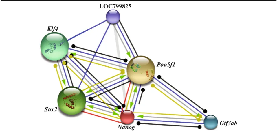
Protein-protein interaction study and identifying post-translation modification sites present on Nanog protein Interacting pathways of the Nanog protein with other pro-teins were depicted using a STRING database (https:// string-db.org/cgi/input.pl). The scores of neighborhood, gene fusion, co-occurrence, co-expression and homology scores with the interacting proteins were considered.
Glycation sites ofεamino groups of lysine residues were predicted using a NetGlycate 1.0 server (http://www.cbs. dtu.dk/services/NetGlycate/). In NetGlycate, a score of N0.5 was considered as glycated. Phosphorylation sites were predicted using a NetPhos2.0 server (http://www. cbs.dtu.dk/services/NetPhos-2.0/). Serine, threonine, and tyrosine residues with a score of≥0.5 were considered as likely phosphorylated AA. Ubiquitylation sites were pre-dicted using a UbPerd (www.ubpred.org). Lysine residues with a score of ≥0.62 were predicted as ubiquitylated. Sumoylation sites were predicted using a SUMOplot (http://www.abgent.com/sumoplot). High probability mo-tifs having a score of 0.5 were adjudged as possible sum oylated [42].
Results
Cloning and characterization ofLrNanogcDNA and deduced protein sequence analysis
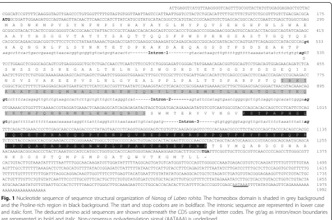
In the absence of sequence information,LrNanogcDNA was partly (consisting of 712 nucleotides) cloned and sequenced from testicular and ovarian cDNA templates, using degenerate primers (Additional file 1: Table S1). The blast analysis showed maximum similarity/identity with the D. rerioand C. auratus counterparts (data not shown). Subsequently, the sequence information for the unknown terminal cDNA regions was generated by imple-menting 5′- and 3´-RACE strategies using gene-specific reverse and forward primers, respectively, designed from the above generated partial 712 bp sequence. The entire cloning strategy has been depicted in Additional file 2: Figure S1. Identifying overlapping RACE sequences, we were able to deduce a full-length cDNA ofLrNanog is of 1992 nucleotides that comprised an open reading frame (ORF) of 1161 nucleotides along with both 5′-untranslated region (UTR) of 175 nucleotides and 3´-UTR of 656 nucleotides. The 3´-UTR contained a putative polyadeny-lation signal (AATAAA) 17 nucleotides upstream of the poly (A) tail (GenBank Accession Number: KX268304) (Fig.1). The ORF initiated with an ATG start codon fulfill-ing the consensus Kozak criterion (A/GNNATGG) of
eukaryotic translation initiation [43], while terminated with a TGA stop codon. The putative translated 386 AA ofLrNanoghas a predicted molecular weight of 42.65 kDa with an estimated isoelectric point of 7.17. In line with earlier evidence, theLrNanogORF contained a character-istic 63 AA long homeobox (HOX) domain positioning between 197 and 259 AA [26] including a proline-rich motif spanning 272-347 AA (Fig.1). These above findings revealed the existence of expressed Nanog gene in the farmed rohu carp,L. rohita.
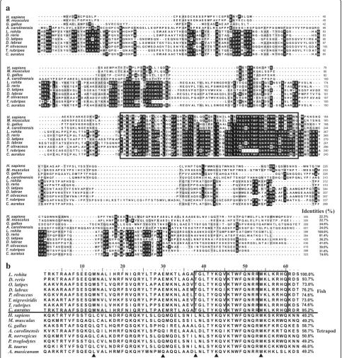
Multiple sequence alignments for the deduced full-length Nanog proteins of available tetrapod and teleosts revealed that LrNanog shared a relatively higher sequence identity withD. rerio(accession no.NP_001091862) andC. auratus(accession no. AEG74407) in the tune of 81.4 and 74.6% respectively, with considerably lower identity with human (accession no. NP_079141) counterpart (Fig.2a). It is noticeable that the N- and C- termini of Nanog proteins were highly variable except that of the conserved homeo-domain (HOX) motif. In addition, the HOX of LrNanog exhibited identities ranging from 46 to 95.2% to the coun-terparts of other vertebrate proteins (Table1). Methionine (M13), glutamic acid (E30), and threonine (T38) residues were noticed to be conserved among fish species; respectively; in place of leucine (L13), glutamine (Q30) and leucine (L38) in tetrapods. In addition, three regions were identified as nearly conserved in teleosts (boxed in Fig.2b). A few AA residues, such as those of methionine (M31), alanine (A35), glycine (G39), threonine (T41), arginine (R53), leucine (L57), histidine (H59) and aspartic acid (D62), were found to be conserved only in fish species. In addition, some AA residues, such as arginine (R5), glutamine (Q12 and Q23), phenylalanine (F20), leucine (L34 and L40) and lysine (K55). were found to be conserved in teleosts and tetrapods.
Evolutionary relationship of Nanog among mammals and teleost
To evaluate the evolutionary relationships, a phylogenetic tree was constructed from the deduced AA sequence of full-length protein using the neighbour-joining method with 1,000 replicates (Fig.3). The teleost and mammalian Nanog proteins are grouped into two distinct clades with above 60% bootstrap support. In the teleost clade, the fresh- and brackish-water species formed two separate clusters. Among the freshwater teleost cluster, LrNanog protein clustered with C. auratus and D. rerio with maximum bootstrap values of 99% and 100%, respect-ively (Fig. 3). The marine teleosts including Tetraodon nigroviridis and Takifugu rubripes, formed a separate cluster, whereas the brackish-water species, including Dicentrarchus labrax, Paralichthys olivaceus and O. latipes, formed a separate tight cluster. Even though O.
latipes is known to be a freshwater species, its habitat also has been reported in salt water [44, 45]. In the mammalian clade, Homo sapiens, Pan troglodytes and Bos taurus showed a tight cluster, whereas two rodents such as Mus musculus and Rattus norvegicus separately clustered. The Nanog of Gallus gallus, Anolis carolinensis and Ambystoma mexicanum clustered in between mam-mals and teleosts, which was in line with the previous findings [26].
The comparative analyses for both deduced protein sequence and homeodomain of LrNanog exhibited high degree (> 70%) of similarity/identity with C. auratus and D. reriocounterparts (Table 1). The LrNanog protein showed merely ~ 50% similarity with the other teleosts, while least with mammalian counterparts. However, the homeodomain was found to be almost conserved among teleosts (> 80% sequence similarity) compared with other vertebrates. The homeodomain showed the highest levels of identity and similarity withC. auratusandD. rerio(≥93%).
Genomic structure including possible promoter of LrNanog
Intron–exon boundaries were derived by aligning cDNA and genomic DNA sequences of LrNanog. It contained four exons and three introns (Fig. 4). All exon–intron boundaries conformed to a 5′-GT/3′-AG splicing rule. Similar to other teleostean and mammalian counterparts, LrNanog gene contained four exons. However, gene sizes in terms of both exons and introns varied from species to species. Its conserved homeodomain region was found to be located between the second and third exons. The above sequence information also detected the TSS (adenosine nucleotide) as marked in Fig.1.
The 2,462 bp upstream sequence, beyond the TSS, was analysed to predict the promoter region using computa-tional tools. We could predict a putative basal core pro-moter containing numerous transcriptional elements, such as, a TATA-box located at−22 to−27 bp, a GC-rich box spanning−44 to−49 bp, and two CAAT-boxes located at
Nanogis expressed prominently in rohu proliferating SSCs and is maternally inherited
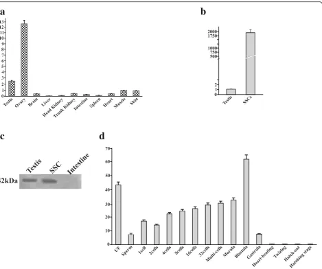
The involvement of Nanog in male germ cell develop-ment in teleosts remained unclear. Hence, we examined the expression profile of LrNanogtranscript in rohu (L. rohita) dividing SSCs including other organs using qPCR. LrNanog mRNA abundance was documented in
the ovary and testis only with negligible detected levels in other somatic tissues including heart, brain, liver, kidney (both head and trunk), intestine, spleen, muscle and skin (Fig. 6a). Furthermore, the signal was relatively stronger (12.5 fold) in ovary than testis. Such an expression pattern was also observed in other species such as Japanese floun-der, goldfish and medaka [23–26]. The LrNanog mRNA
a
b
was most abundantly expressed in proliferating SSCs in tune of 1,978-fold more than testis (Fig.6b). The presence of LrNanog protein was also documented in rohu testis and proliferating SSCs as detected by Western blot analysis with a specific signal of about 42 kDa (Fig.6c), providing an insight into its involvement in stem cell maintenance and development.
The progressive expression pattern ofLrNanogduring early stages of embryonic development was also examined. We found that Nanog mRNA in rohu was detected in metaphase II oocytes (i.e. unfertilized eggs) (Fig.6d) and at all stages preceding the embryonic genomic activation that occurs during blastula. Its elevated level suddenly decreased following the gastrulation stage, possibly because the pluripotent cells started differentiating [46]. These outcomes are in line with the previous data obtained in medaka, goldfish and Japanese flounder [23–26]. The LrNanog transcripts were impressively moderated at the end of the gastrulation stage. From the heart-beating stage onwards, LrNanog could not be documented. Together, our results postulated that the Nanog transcript is most likely inherited maternally and functionally linked to embryonic development, in addition to its participatory roles in SSC maintenance in teleost.
Modelling and validating the structure of the homeobox domain of Nanog protein
The secondary structure predicted by SOPMA docu-mented the absence of beta-sheets in LrNanog protein. It predicted 20.21% alpha helix, 62.18% random coils, 11.66% extended strands and 5.96% beta turns. The con-fidence level of the predicted secondary structure was determined using PsiPred (Additional file3: Figure S2).
The 3D structure of teleost Nanog protein has not yet been determined experimentally. In the absence of crystal structure (3D structures in the PDB database), the in silico analysis is used for the prediction of protein structure to Table 1Similarity (%) and identity (%) of rohu Nanog as well as
its domains with other species
Total amino acid Homeodomain
Similarity Identity Similarity Identity
Danio rerio 86.8 81.4 98.4 93.7
Caracius auratus 77.5 74.6 98.4 95.2
Oryzias latipes 52.6 36.2 85.7 73.0
Dicentrarchus labrax 56.9 41.6 88.9 76.2
Paralichthys olivaceus 54.7 39.0 85.7 74.6
Tetradon nigroviridis 49.0 30.2 84.1 73.0
Takifugu rubripes 52.0 31.6 84.1 74.6
Homo sapiens 33.9 22.3 68.3 49.2
Mus musculus 36.5 22.7 68.3 49.2
Gallus gallus 37.3 24.6 79.4 58.7
Anolis carolinensis 39.2 24.0 79.4 58.7
Rattus norvegicus 36.8 23.0 68.3 50.8
Pan troglodytes 33.9 22.5 68.3 49.2
Bos taurus 35.8 21.7 68.3 46.0
Ambystoma mexicanum 33.2 22.8 60.3 49.2
Fig. 4Comparison of genomic organization ofLrNanogwith otherNanogcounterparts of teleost and mammals. Exons are shown in dark box whereas introns are in straight line. The size of each parts are indicated. The 5′- and 3′- Untranslated regions (UTR) are represented by open box. The 5′- and 3′- region of chicken are not available
a
b
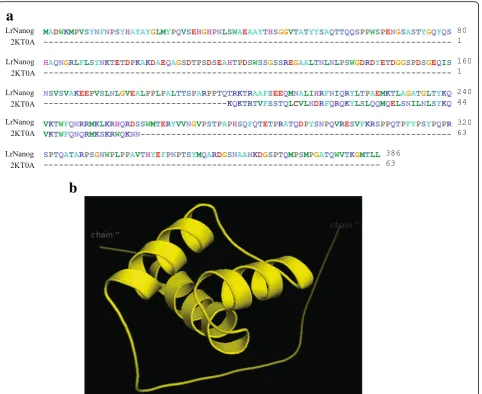
characterize a new protein. We attempted to build a protein structure by abinitio modelling using PHYRE2. The model building was based upon the template 2KT0A (human stem cell TF Nanog homeodomain) that covered a total of 63 residues positioning Thr197 to Lys259, out of a total of 386 AA residues of LrNanog protein (Fig. 7a). After rigorous refinements by means of the electron mi-croscopy technique, a stable structure of the HOX domain could be built (Fig.7b).
The quality of the predicted structured model was assessed/validated by Ramachandran plot in the PROCHECK validation package (Additional file 4: Figure S3). In the plot, the phi/psi angles for 96.7%
residues were with the‘most favoured’regions inclusive of 3.3% residues in the ‘additional allowed’ regions, while none of the residues lay in the‘disallowed conformations’. This indicated that the backbone dihedral angles, phi and psi, in the LrNanog HOX domain model were reasonably accurate. Further, the overall PROCHECK G-factor for the structure was 0.10, implying that the modelled structure is acceptable. The value of the Z-score signified that the 3D model of the HOX domain of LrNanog protein was reliable. Energetic architecture as revealed by ProSA (data not presented) score was negative (−4.3) for the modelled protein. ProSA and Errat analysis implied that the Z-score of our model was very much within the
a
c
d
b
range of scores normally found for proteins of compar-able size. The results, together, ascertained the qualities of a stable and reasonably good model structure.
Protein-protein interactions for Nanog and posttranslational modification on LrNanog protein Nanog is a main member of the NOS triad respon-sible forstem cell pluripotency and maintenance. Protein-protein interacting networks of Nanog along with other factors (Table 2; Fig. 8) involving pluripo-tency was investigated using STRING. This revealed that LrNanog protein interacted with the other pluri-potency marker proteins such as, Pou5f1 (POU do-main, class 5, TF1), Sox2, Klf4 (Kruppel-like factor 4), Gtf3ab (general TF IIIA, b) and LOC799825 (lin-28 homolog A) with scores of 0.996, 0.997, 0.937, 0.909 and 0.908 (Table 2), respectively, that provided a clue
regarding its involvement in pluripotency and mainten-ance of stem cells.
We used different in silico tools to identify/study post-translational modifications of LrNanog protein. NetGlycate predicted that 7 AA residues would undergo glycation. As per NetPhos analysis 30 serine, 13 threonine and 8 tyrosine residues would undergo phosphorylation (Table 3). Simi-larly, UbPred, showing 5 AA positions, had a score of above 0.84, indicating a high possibility of ubiquitination, whereas 1 AA position showed a medium chance of ubi-quitination. Likewise, by using SUMOplot, we were able to predict two positions where there was a moderate level of the chance of sumoylation (Table4).
Discussion
Nanog gene, in association with Oct4 and Sox2 genes, is believed to regulate the stemness properties of
a
b
mammalian stem cells. Presently, its specific underlying mechanisms for pluripotency and maintenance of stem cells are not well understood in teleost fishes.Nanog, as a pluripotent gene, has been characterized in mammals [6] and non-mammalian species [47]. Its orthologs have been characterized in teleostean model organisms, such as zebrafish and medaka [23, 48], including aquarium goldfish [25]. The genomic information for the Nanog
gene of large-bodied farmed rohu carp (L. rohita) was lacking. The current study focused on the identification and characterization ofNanogortholog in rohu carp. We were able to elucidate the Nanog gene structure along with its promoter modules and tissue/cell distributions.
The full-length cDNA ofLrNanogof 1,992 nucleotides comprised of a coding sequence of 1,161 nucleotides translatable to a polypeptide, of 386 AA residues. It Table 2Protein-protein interacting networks as determined by STRING
contained a typical characteristic 63 AA containing HOX domain. The deduced AA sequence of LrNanog showing least homology to other mammalian species, even with other teleost species, demonstrated that Nanog is not highly conserved across mammalian and aquatic species under study. The protein comparison of LrNanog with other species counterparts demonstrated its sequence var-iations amonga wide range of organisms, except that of a relatively conserved HOX-homeodomain. Similar findings were also reported earlier [6, 23]. The HOX domain of LrNanoglacked the conserved tryptophan (W) pentapep-tide repeats in the C-terminus region as documented in human and mouse [4,6,49]. The W-repeats were also not found in lower vertebrates of Japanese flounder, chick and zebrafish [26,47,48].
Phylogenetic analysis revealed that the LrNanog formed a distinct hierarchy along with the other teleost
counterparts depending on habitats (fresh-, brackish-and marine-water), in which the LrNanog was found to be the most closely related to C. auratus and D. rerio counterparts. This is possible becauseL. rohita,C. auratus and D. rerio belong to a common family (Cyprinidae) of freshwater habitat. Tetrapods and teleosts formed differ-ent clusters, indicating a rapid evolution of the Nanog gene that might have begun before the separation of the teleost and tetrapod lineages. These results, together, suggested the W-repeats might have appeared in the higher mammals during evolution. The overall conserved HOX domain, except for W-repeats, possibly evolved from a common ancestor. It is also indicative that its stemness function is associated across species. The overall sequence variations could indicate that its partici-patory regulatory functions in stem cell maintenance may not be identical, but differ/vary from lower to higher vertebrates (species to species). The lack of W-repeats in conserved HOX vis-à-vis three identified conserved regions in teleosts as comparedwith mammalian coun-terparts provided the clue that the binding platform with other important regulatory molecules for teleosteanNanog is less likely to be exactly similar to that of the mammalian counterpart. In such a scenario, two distinct Nanog -mediated networking pathways must be operative between teleosts and mammals.
A total of 9.8 kb long genomic structure, encompassing four exons and three intronic sequences, was generated. Based on genome walking and 5´-RACE data, the transcrip-tion start site (TSS, + 1) with adenosine nucleotide (174 bp upstream from the start codon ATG) was identified. This was in line with verified in silico data. The genomic Table 3Post-translation modified glycation and phosphorylation sites in LrNanog protein
Glycation Phosphorylation
N-Gly Serine Threonine Tyrosine
Pos Score Pos Score Pos Score Pos Score Pos Score Pos Score
5 0.938 29 0.962 124 0.609 259 0.994 58 0.589 16 0.912
99 0.885 37 0.962 126 0.992 260 0.982 110 0.910 43 0.890
167 0.583 62 0. 756 127 0.965 279 0.985 118 0. 534 75 0.621
239 0.926 66 0.993 140 0.913 301 0.965 187 0.687 146 0.826
242 0.773 73 0.958 152 0.979 306 0.984 197 0.937 266 0.953
251 0.896 108 0.847 155 0.987 315 0.655 223 0.935 293 0.946
253 0.910 112 0.990 162 0.799 321 0.882 237 0.624 342 0.681
114 0.938 164 0.788 330 0.940 263 0.740 350 0.669
121 0.987 189 0.840 349 0.658 285 0.979
123 0.924 205 0.958 365 0.985 289 0.886
310 0.856
326 0.910
340 0.955
Table 4Putative ubiquitylation and sumoylation sites in Nanog protein
Ubiquitylation Sumoylation
Residues Score Ubiquitinated Residue Score
93 0.87 High confidence 5 0.54
99 0.85 High confidence 169 0.79
101 0.89 High confidence
167 0.90 High confidence
228 0.63 Low confidence
362 0.94 High confidence
structure ofLrNanogwas found to be conserved in terms of the number of exons and the position of the homeodomain with other counterparts that promoted the probable de-gree of conservative function of the homeodomain. The larger coding exons in teleosts than mammalian counter-parts demonstrated the possible rearrangement of Nanog gene during evolution from fish to tetrapod. The sizes and sequence differences of intronic regions among various species also reflected differential regulatory mech-anisms because introns are known to participate in regu-lating gene expressions.
The in silico sequence analysis of the 5′–flanking gen-omic region led to the identification of consensus TATA-and CAAT-boxes along with GC-box, which is in line with the previous findings in goldfish and Japanese flounder [25, 26], but contrary to the previously docu-mented absence in mouse Nanog [50]. LrNanog has a conventional RNA transcription polymerase II binding site (TATA-box) right upstream (22 bp) of the putative TSS. From these findings, it might be predicted that the 250 bp upstream from the TSS would be the core pro-moter region responsible for theLrNanogtranscriptional activity. In addition, it is likely that theLrNanog gene is driven by a relatively compact regulatory region, because the full-length promoter of rohu (2.4 kb) is about half of the mammalianNanog promoter (~ 5 kb) [50]. Unfortu-nately, we could not detect any mammalian type consen-sus sequence as a binding platform for the Oct4/Sox2 complex [12], which is believed to be the major regula-tory element. However, the identifiedOct4binding motif provided the clue regarding its associated regulatory function with teleostPou2 ortholog. Interestingly, a p53 motif that down-regulates Nanog expression in ESCs during differentiation [51] and Tcf3 binding consensus sequence that limits its self-renewal function [52] were also detected in the LrNanogpromoter. Its own binding site on the promoter region indicated that Nanog not only forms a homodimerization to promote stem cell pluripotency [53] but also could regulate its own expres-sion. Furthermore, some other transcriptional activators (AP-1, SP1, GATA-1 and C/EBP) were also detected in the promoter region that could regulate the expression of Nanog via interacting with the other TFs and en-hancers as suggested earlier [54]. It would be interesting to ascertain the regulatory functions played by each motif in the future. Even though theSox2site could not be predicted, we could detect other SRY (sex-determin-ing region Y)-box factors (Sox5, 6, 7, 8, 9, 21) on the promoter region indicating the potential role of Nanog in the regulation of embryonic and sexual development allied with its mRNA distribution profile. Furthermore, we were able to predict some TFs, including Oct3_4, SIP, PRDM14, FOXP1-ES, LEF1/TCF and ONST that are reported to be involved/associated with stem cell
pluripotency [24, 55–57]. The computational analysis of TFs is mainly based upon the sequence similarity, so fur-ther experiments are necessary to draw any conclusion regarding the functional involvement of these TFs in the regulation of rohuNanoggene expression.
The model structure of LrNanog HOX showed that the alpha helix covered 20.21% of total length and there are no beta-sheets. The value of the Z-score signified the 3D model of the Nanog protein was reliable and precise. The characteristic of good quality model protein was further evident from the Ramachandran plot. This vali-dated that the predicted model protein was well inside the range of typical native structures. Furthermore, the protein-protein interactions, inclusive of pluripotency factors as previously reported in mammals [58], pre-dicted the possible participation of LrNanog in the net-working mechanisms pertaining to pluripotency stem cells. Several post-translational modifications were iden-tified in the LrNanog protein.
Pou2 and Sox2 are also highly expressed in rohu SSCs [22, 61]. Thus, a mammalian-like networking function could not be ruled out. The potentiating presence of LrNanog mRNA in the ovary could be argued as its maternal inheritance, as also reported in other teleos-tean species [23–26, 48]. These observations also sup-ported the computational protein–protein interaction findings. A population of enriched undifferentiated rohu SSCs also produces [14] spermatids in vitro vis-à-vis its abundance in oocytes, and thus the participa-tory role of LrNanog in undifferentiated germ cell development cannot be ruled out. It would be of interest to clarify this particular aspect in future. Because Pou2 (an ortholog of mammalian Oct4) and Sox2 are highly expressed in dividing rohu SSCs [21,
22], it would also be fascinating to carry out the association studies of Nanog, Pou2 and Sox2 in the regulatory mechanisms and pathways involved in proliferation and maintenance of teleost spermato-gonial cells.
Conclusions
In summary, the current study revealed the full-length cDNA sequence, genomic organization, and promoter characterization ofNanoggene of farmed rohu carp,L. rohita. By sequence comparison, phylogenetic analysis and genomic structure, theLrNanog was considered as the mammalian ortholog. The relatively conserved homeodomain present in Nanog of teleosts indicated that it might share some common biological functions with mammalian counterparts, particularly in stem cell maintenance. In support, we have provided evidence that LrNanog is transcriptionally active in proliferating SSCs and also up to the blastula stage of embryonic development. Its restricted abundances in adult gonads also confirmed its participation in stem cell prolifera-tion and/or maintenance. The structural differences including overall conserved HOX domain and phylo-genetic analyses highlighted its possible differential co-ordinating physiological functions between mammalian and teleostean germ cells development. Generation of DNA sequence information and its in silico analyses predicting several potential regulatory TFs in addition to the core promoter elements should provide a road-map for undertaking future experiments linked to its regulated expression. Collectively, the results generated in this study validated the LrNanog as a potential bio-marker for embryonic and SSCs and could be the first step towards elucidating mechanistic pathways in the stemness of SSC maintenance and proliferation in tele-osts. Future studies could be carried out to validate functionally the regulatory mechanisms of Nanog gene in teleost species.
Additional files
Additional file 1:Primers used for various PCR amplifications and mRNA expression analysis of Labeo rohita Nanog. (DOCX 14 kb)
Additional file 2:Figure S1.Schematic representation of different strategies opted to characterize the structural organization of rohuNanog
gene. Various fragments ofLrNanogwas obtained as illustrated by the closed and open boxes for the sense and antisense primers, respectively. The start and stop codons are represented as ATG and TGA, respectively. The genomic DNA is depicted with exons as rectangular boxes and the introns as solid lines. The exon-intron boundaries are determined by the alignment of cNDA sequence and genomic DNA sequence. RT-PCR, re-verse transcription–polymerase chain reaction; UTRs, untranslated regions; RACE, rapid amplification of cDNA ends. (EPS 761 kb)
Additional file 3:Figure S2.Secondary structure of LrNanog protein predicted by SOPMA, confirming the absence of beta-sheets inLrNanog
gene. (EPS 1350 kb)
Additional file 4:Figure S3.Ramachandran plot of LrNanog protein showing residues in most favored (white), additionally allowed (yellow), generously allowed (pale yellow), and disallowed region, generated by PROCHECK (SAVES, server). (EPS 2536 kb)
Abbreviations
EGC:Embryonic germ cell; EM: Electrone microscopy; ESC: Embryonic stem cell; HOX: Homeobox; LIF: Leukemia inhibitory factor; ORF: Open reading frame; Plzf: Promyelocytic leukemia zinc finger; qPCR: Quantitative real time PCR; RACE: Rapid amplification of cDNA ends; SSC: Spermatogonial stem cell; TF: Transcription factor; UTR: Untranslated region
Acknowledgments
We would like to thank Dr. K. D. Mohapatra, ICAR-Central Institute of Freshwater Aquaculture, Bhubaneswar, Odisha for providing embyos ofLabeo rohita.
Funding
Funding for this study was provided by grant from Indian Council of Agricultural Research (ICAR) and Department of Biotechnology (DBT), Government of India. SKP was a recipient of Senior Research Fellow of Council of Scientific and Industrial Research (CSIR), Govt. of India. The funding sources had no role in the study design.
Availability of data and materials
All data generated or analysed during this study are included in this published article [and its supplementary information files].
Authors’contributions
HKB conceived and designed the experiments. SKP, VC and MMS performed the experiments and collected the samples. SKP and VC analyzed the data, wrote the main manuscript, and prepared the figures. JKS and MKP corrected the grammatical errors and general support. All authors reviewed and approved the final manuscript.
Ethics approval
Not applicable.
Competing interests
The authors declare that they have no competing interests.
Author details
1Fish Genetics and Biotechnology Division, ICAR - Central Institute of Freshwater Aquaculture, Kausalyaganga, Bhubaneswar, Odisha 751002, India. 2Center of Biotechnology, Siksha‘O’Anusandhan University, Bhubaneswar, India.
Received: 18 November 2017 Accepted: 25 April 2018
References
2. Shang Z, Isaac VE, Li H, Patel L, Catron KM, Curran T, et al. Design of a
“minimAl”homeodomain: the N-terminal arm modulates DNA binding affinity and stabilizes homeodomain structure. Proc Natl Acad Sci U S A. 1994;91(18):8373–7.
3. Gehring WJ, Affolter M, Burglin T. Homeodomain proteins. Annu Rev Biochem. 1994;63:487–526.
4. Hart AH, Hartley L, Ibrahim M, Robb L. Identification, cloning and expression analysis of the pluripotency promoting Nanog genes in mouse and human. Dev Dyn. 2004;230(1):187–98.
5. Do JT, Scholer HR. Regulatory circuits underlying pluripotency and reprogramming. Trends Pharmacol Sci. 2009;30(6):296–302. 6. Chambers I, Colby D, Robertson M, Nichols J, Lee S, Tweedie S, et al.
Functional expression cloning of Nanog, a pluripotency sustaining factor in embryonic stem cells. Cell. 2003;113(5):643–55.
7. Mitsui K, Tokuzawa Y, Itoh H, Segawa K, Murakami M, Takahashi K, et al. The homeoprotein Nanog is required for maintenance of pluripotency in mouse epiblast and ES cells. Cell. 2003;113(5):631–42.
8. Wang SH, Tsai MS, Chiang MF, Li H. A novel NK-type homeobox gene, ENK (early embryo specific NK), preferentially expressed in embryonic stem cells. Gene Expr Patterns. 2003;3(1):99–103.
9. Niwa H. How is pluripotency determined and maintained? Development. 2007;134(4):635–46.
10. Silva J, Smith A. Capturing pluripotency. Cell. 2008;132(4):532–6. 11. Young RA. Control of the embryonic stem cell state. Cell. 2011;144(6):940–54. 12. Kuroda T, Tada M, Kubota H, Kimura H, Hatano SY, Suemori H, et al.
Octamer and sox elements are required for transcriptional cis regulation of Nanog gene expression. Mol Cell Biol. 2005;25(6):2475–85.
13. Kashyap V, Rezende NC, Scotland KB, Shaffer SM, Persson JL, Gudas LJ, et al. Regulation of stem cell pluripotency and differentiation involves a mutual regulatory circuit of the NANOG, OCT4, and SOX2 pluripotency transcription factors with polycomb repressive complexes and stem cell microRNAs. Stem Cells Dev. 2009;18(7):1093–108.
14. Panda RP, Barman HK, Mohapatra C. Isolation of enriched carp
spermatogonial stem cells fromLabeo rohitatestis for in vitro propagation. Theriogenology. 2011;76(2):241–51.
15. Santos Nassif Lacerda SM, Costa GM, da Silva Mde A, Campos-Junior PH, Segatelli TM, Peixoto MT, et al. Phenotypic characterization and in vitro propagation and transplantation of the Nile tilapia (Oreochromis niloticus) spermatogonial stem cells. Gen Comp Endocrinol. 2013;192:95–106. 16. Li Z, Li M, Hong N, Yi M, Hong Y. Formation and cultivation of medaka
primordial germ cells. Cell Tissue Res. 2014;357(1):71–81.
17. Liu R, Li M, Li Z, Hong N, Xu H, Hong Y. Medaka Oct4 is essential for pluripotency in blastula formation and ES cell derivation. Stem Cell Rev Rep. 2014;11:11–23.
18. Hong N, Li Z, Hong Y. Fish stem cell cultures. Int J Biol Sci. 2011;7(4):392–402. 19. Bosseboeuf A, Gautier A, Auvray P, Mazan S, Sourdaine P. Characterization of spermatogonial markers in the mature testis of the dogfish (Scyliorhinus caniculaL.). Reproduction. 2014;147(1):125–39.
20. Mohapatra C, Barman HK, Panda RP, Kumar S, Das V, Mohanta R, et al. Cloning of cDNA and prediction of peptide structure of Plzf expressed in the spermatogonial cells ofLabeo rohita. Mar Genomics. 2010;3(3–4): 157–63.
21. Mohapatra C, Barman HK. Identification of promoter within the first intron of Plzf gene expressed in carp spermatogonial stem cells. Mol Biol Rep. 2014;41(10):6433–40.
22. Patra SK, Chakrapani V, Panda RP, Mohapatra C, Jayasankar P, Barman HK. First evidence of molecular characterization of rohu carp Sox2 gene being expressed in proliferating spermatogonial cells. Theriogenology. 2015;84(2): 268–76.
23. Camp E, Sanchez-Sanchez AV, Garcia-Espana A, Desalle R, Odqvist L, Enrique O'Connor J, et al. Nanog regulates proliferation during early fish
development. Stem Cells. 2009;27(9):2081–91.
24. Gabut M, Samavarchi-Tehrani P, Wang X, Slobodeniuc V, O’Hanlon D, Sung HK, et al. An alternative splicing switch regulates embryonic stem cell pluripotency and reprogramming. Cell. 2011;147(1):132–46.
25. Marandel L, Labbe C, Bobe J, Le Bail PY. Nanog 5′-upstream sequence, DNA methylation, and expression in gametes and early embryo reveal striking differences between teleosts and mammals. Gene. 2012;492(1):130–7. 26. Gao J, Wang J, Jiang J, Fan L, Wang W, Liu J, et al. Identification and
characterization of a nanog homolog in Japanese flounder (Paralichthys olivaceus). Gene. 2013;531(2):411–21.
27. Tian J, Chng SC, Ong J, Reversade B. Cloning and functional study of Nanog in zebrafish. Dev Biol. 2011;356:234.
28. Panda RP, Chakrapani V, Patra SK, Saha JN, Jayasankar P, Kar B, et al. First evidence of comparative responses of toll-like receptor 22 (TLR22) to relatively resistant and susceptible Indian farmed carps toArgulus siamensis
infection. Dev Comp Immunol. 2014;47(1):25–35.
29. Rasal KD, Chakrapani V, Patra SK, Mohapatra SD, Nayak S, Jena S, et al. Identification of Deleterious Mutations in Myostatin Gene of Rohu Carp (Labeo rohita) Using Modeling and Molecular Dynamic Simulation Approaches. 2016. BioMed Research International. 2016;2016:10. 30. Barman HK, Das V, Mohanta R, Mohapatra C, Panda RP, Jayasankar P.
Expression analysis of beta-actin promoter of rohu (Labeo rohita) by direct injection into muscle. Curr Sci. 2010;99:1030–2.
31. Mohanta R, Jayasankar P, Mohapatra KD, Saha J, Barman HK. Molecular cloning, characterization and functional assessment of the myosin light polypeptide chain 2 (mylz2) promoter of farmed carp,Labeo rohita. Transgenic Res. 2014;23:601–7.
32. Rasal KD, Chakrapani V, Patra SK, Jena S, Mohapatra SD, Nayak S, et al. Identification and prediction of consequences of non-synonymous SNP in glyceraldehyde 3-phosphate dehydrogenase (GAPDH) gene of zebrafish,
Danio rerio. Turk J Biol. 2016;40:43–54.
33. Pfaffl MW. A new mathematical model for relative quantification in real-time RT-PCR. Nucleic Acids Res. 2001;29(9):e45.
34. Barman HK, Takami Y, Ono T, Nishijima H, Sanematsu F, Shibahara K, et al. Histone acetyltransferase 1 is dispensable for replication-coupled chromatin assembly but contributes to recover DNA damages created following replication blockage in vertebrate cells. Biochem Biophys Res Commun. 2006;345(4):1547–57.
35. Sanematsu F, Takami Y, Barman HK, Fukagawa T, Ono T, Shibahara K, et al. Asf1 is required for viability and chromatin assembly during DNA replication in vertebrate cells. J Biol Chem. 2006;281(19):13817–27.
36. Barman HK, Takami Y, Nishijima H, Shibahara K, Sanematsu F, Nakayama T. Histone acetyltransferase-1 regulates integrity of cytosolic histone H3-H4 containing complex. Biochem Biophys Res Commun. 2008;373(4): 624–30.
37. Tamura K, Stecher G, Peterson D, Filipski A, Kumar S. MEGA6: molecular evolutionary genetics analysis version 6.0. Mol Biol Evol. 2013;30(12):2725–9. 38. Campanella JJ, Bitincka L, Smalley J. MatGAT: an application that generates
similarity/identity matrices using protein or DNA sequences. BMC bioinformatics. 2003;4:29.
39. Hall TA. BioEdit: a user friendly biological sequence alignment ed-itor and analysis program for windows 95/98/NT. Nucl Acids Symp Ser. 1999;41:95–8. 40. Rasal KD, Chakrapani V, Patra SK, Jena S, Mohapatra SD, Nayak S, et al.
Identification and prediction of consequences of non-synonymous SNP in glyceraldehyde 3-phosphate dehydrogenase (GAPDH) gene of zebrafish,
Danio rerio. Turk J Biol. 2015;40:43–54.
41. Rasal KD, Chakrapani V, Patra SK, Mohapatra SD, Nayak S, Jena S, et al. Identification of Deleterious Mutations in Myostatin Gene of Rohu Carp (Labeo rohita) Using Modeling and Molecular Dynamic Simulation Approaches. BioMed research international. 2016;2016:10. 42. Kelly JN, Barr SD. In silico analysis of functional single nucleotide
polymorphisms in the human TRIM22 gene. PLoS One. 2014;9(7):e101436. 43. Kozak M. An analysis of vertebrate mRNA sequences: intimations of
translational control. J Cell Biol. 1991;115(4):887–903.
44. Parenti LR. A phylogenetic analysis and taxonomic revision of ricefishes, Oryzias and relatives (Beloniformes, Adrianichthyidae). Zool J Linnean Soc. 2008;154(3):494–610.
45. Rainer F, Daniel P.Oryzias latipes. http://www.fishbase.se/summary/Oryzias-latipes.html. April 2014.
46. Kane DA, Kimmel CB. The zebrafish midblastula transition. Development. 1993;119(2):447–56.
47. Canon S, Herranz C, Manzanares M. Germ cell restricted expression of chick Nanog. Dev Dyn. 2006;235(10):2889–94.
48. Schuff M, Siegel D, Philipp M, Bundschu K, Heymann N, Donow C, et al. Characterization ofDanio rerioNanog and functional comparison to Xenopus vents. Stem Cells Dev. 2012;21(8):1225–38.
49. Medvedev SP, Elisaphenko EA, Shevchenko AI, Mazurok NA, Zakian SM. Nanog gene: genomic organization and expression in the vole Microtus rossiaemeridionalis. Dokl Biochem Biophys. 2009;425:102–5.
51. Lin T, Chao C, Saito S, Mazur SJ, Murphy ME, Appella E, et al. p53 induces differentiation of mouse embryonic stem cells by suppressing Nanog expression. Nat Cell Biol. 2005;7(2):165–71.
52. Pereira L, Yi F, Merrill BJ. Repression of Nanog gene transcription by Tcf3 limits embryonic stem cell self-renewal. Mol Cell Biol. 2006;26(20):7479–91. 53. Wang J, Levasseur DN, Orkin SH. Requirement of Nanog dimerization for
stem cell self-renewal and pluripotency. Proc Natl Acad Sci U S A. 2008; 105(17):6326–31.
54. Wu DY, Yao Z. Functional analysis of two Sp1/Sp3 binding sites in murine Nanog gene promoter. Cell Res. 2006;16(3):319–22.
55. Tsuneyoshi N, Sumi T, Onda H, Nojima H, Nakatsuji N, Suemori H. PRDM14 suppresses expression of differentiation marker genes in human embryonic stem cells. Biochem Biophys Res Commun. 2008;367(4):899–905. 56. Xu RH, Sampsell-Barron TL, Gu F, Root S, Peck RM, Pan G, et al. NANOG is a
direct target of TGFbeta/activin-mediated SMAD signaling in human ESCs. Cell Stem Cell. 2008;3(2):196–206.
57. Kim CG, Chung IY, Lim Y, Lee YH, Shin SY. A Tcf/Lef element within the enhancer region of the human NANOG gene plays a role in promoter activation. Biochem Biophys Res Commun. 2011;410(3):637–42. 58. Saunders A, Faiola F, Wang J. Concise review: pursuing self-renewal and
pluripotency with the stem cell factor Nanog. Stem Cells. 2013;31(7):1227–36. 59. Komatsu K, Fujimori T. Multiple phases in regulation of Nanog expression
during pre-implantation development. Develop Growth Differ. 2015;57(9): 648–56.
60. Gagnon JA, Obbad K, Schier AF. Zebrafish nanog is primarily required in extraembryonic tissue. Development. 2017.https://doi.org/10.1242/dev. 147793.