R E S E A R C H A R T I C L E
Open Access
Differences in sex distribution, anatomic
location and MR imaging appearance of
pediatric compared to adult chordomas
Ronnie Sebro
1, Thomas DeLaney
2,3, Francis Hornicek
3,4, Joseph Schwab
3,4, Edwin Choy
3,5, G. Petur Nielsen
3,6and Daniel I. Rosenthal
3,7*Abstract
Background:Chordomas are rare malignancies that primarily affect adults, but also rarely affect pediatric patients. We compared the imaging appearance, demographic and anatomic distributions of adult and pediatric chordomas in a large cohort.
Methods:We performed a retrospective review of medical records of 220 subjects with histologically confirmed chordomas of the axial skeleton and pre-treatment magnetic resonance imaging studies. Age, sex, type of chordoma (conventional, chondroid or dedifferentiated), the anatomic location of the chordoma, as well as whether the lesion was primarily extra-osseous were recorded. Pediatric subjects were less than 21 years at the time of diagnosis. Binomial two-sample tests of proportions and Fisher’s exact tests were used to compare proportions between the pediatric and adult subjects.
Results:Fifty six pediatric subjects (58.9 % female) and 164 adult subjects (42.1 % female) were identified. The proportion of female subjects with chordomas was significantly higher in the pediatric cohort compared to the
adult cohort (P= 0.04). Most chordomas occur in Caucasians, however African-Americans were more represented
in the pediatric cohort than in the adult cohort (P= 0.01). 69.6 % (39/56) of the pediatric chordomas involved the clivus/skull base and cervical spine compared to 29.3 % (48/164) of the adult chordomas (P= 1.99 × 10⁻⁷). Only 1.8 % (1/56) of the pediatric chordomas was in the sacrococcygeal region compared to 36.0 % (59/164) of the adult chordomas (P= 2.55 × 10⁻⁸). In cases where pre-treatment imaging was available, 93.8 % (16/17) of pediatric
chordomas were predominantly extra-osseous compared to 76.7 % (46/60) of adult chordomas (P= 0.17).
Conclusions:Pediatric chordomas more often affect females and occur most frequently at the craniocervical junction with decrease in incidence distally in the spine, whereas adult chordomas most frequently involve the craniocervical and sacrococcygeal regions.
Keywords:Chordoma, Pediatric, Adult, Chondroid, Dedifferentiated, Conventional, Spine, MRI
Background
Chordomas are rare, malignant tumors that bear a histological resemblance to cells of notochordal origin [1–3]. Chordomas have an incidence of approximately 1 in 1 million with approximately 300 new cases in the United States each year [4]. Chordomas predominantly
arise in the clivus/skull base and/or spine [5, 6] with rare reports of extra-axial chordomas [7, 8]. Although chordomas predominantly affect middle-aged or elderly adults, they are also seen in the pediatric population (age < 21 years) [4, 9].
Pediatric chordomas are not well characterized in the literature due to the relative paucity of cases [4]. Anec-dotal evidence suggests that pediatric chordomas tend to be predominantly extra-osseous with some involvement of the surface of the bone rather than an osseous tumor
* Correspondence:[email protected]
3
The Harris Center for Chordoma Care, Massachusetts General Hospital, 55 Fruit Street, Yawkey 3B, Boston, MA 02114, USA
7Department of Radiology, Massachusetts General Hospital, Yawkey 6E, 55
Fruit Street, Boston, MA 02114, USA
Full list of author information is available at the end of the article
© 2016 The Author(s).Open AccessThis article is distributed under the terms of the Creative Commons Attribution 4.0 International License (http://creativecommons.org/licenses/by/4.0/), which permits unrestricted use, distribution, and reproduction in any medium, provided you give appropriate credit to the original author(s) and the source, provide a link to the Creative Commons license, and indicate if changes were made. The Creative Commons Public Domain Dedication waiver (http://creativecommons.org/publicdomain/zero/1.0/) applies to the data made available in this article, unless otherwise stated. Sebroet al. BMC Medical Imaging (2016) 16:53
with a small soft tissue component and therefore greater than 50 % of the lesion tends to be extra-osseous.
We utilize a large cohort of subjects evaluated and/or treated for chordomas at the corresponding author’s in-stitution to assess whether the sex and anatomic distri-bution of chordomas in pediatric subjects mirrors that seen in adult subjects, and evaluate the imaging appear-ance of chordomas to assess whether chordomas in pediatric subjects are more likely to be predominantly extra-osseous than chordomas in adult subjects.
Methods
Subjects
The study was approved by the Institutional Review Board (IRB) at the corresponding author’s institution and need for signed informed consent was waived.
This was a retrospective cohort study. Subjects had to have a pathologically proven diagnosis of chordoma for inclusion into the study. Histories were verified by searching a text database of all medical records and by review of all available imaging at our institution. Data was collected on subjects treated and/or evaluated at our institution between 01/01/1996 to 06/30/2014.
Data was collected on each subject’s age at diagnosis (based on age at the time of the initial pathology report), sex, self-reported race/ethnicity (Asian, African-American, Hispanic or Caucasian), type of chordoma (conventional, chondroid or dedifferentiated), anatomic location of the initial lesion (clivus/skull base, cervical spine, thoracic spine, lumbar spine, sacrococcygeal spine), and whether greater than 50 % of the pre-treated lesion was intra-osseous. Lesions that involved the clivus and skull base (above McGregor’s line) were considered clivus/skull base tumors. Lesions that involved the skull base and any component of the cervical spine (that is, lesions that extended inferior to McGregor’s line) were considered as cervical spine/skull base tumors.
Imaging
Tumors were imaged using contrast-enhanced magnetic resonance imaging (MRI) performed on both 1.5 and 3 T systems. Measurements were obtained using the T2 imaging sequences. To assess whether a tumor was pri-marily intra-osseous or extra-osseous, the maximal intra-osseous component was identified in the axial plane and at that level, the maximal intra- and extra-osseous components were measured along the same plane. A lesion was considered primarily extra-osseous if there was more extra-osseous (>50 %) tumor at the level of the maximal intra-osseous tumor. All images were reviewed by a fellowship trained musculoskeletal radiologist.
Statistical analyses
Binomial two sample tests for proportions were used to compare proportions between the pediatric and adult subjects. χ2and Fisher’s exact tests were used to assess whether the anatomic location of the chordomas varied between pediatric subjects (subjects that were younger than 21 years of age) and adult subjects (subjects that were greater than or equal to 21 years of age at the time of diagnosis) and to test whether chordomas in pediatric subjects were more likely to be predominantly extra-osseous than chordomas in adult subjects.
P-values <0.05 were considered statistically significant. Statistical analyses were performed using R v2.9 software (https://www.r-project.org/).
Results
We identified a total of 220 subjects (46.4 % female) with pathologically proven chordomas. Summary statis-tics were calculated for demographic variables and are shown in Table 1.
There were significantly more female subjects in the pediatric cohort (58.9 %) compared to the adult cohort (42.1 %) (P= 0.04). The majority of subjects in the
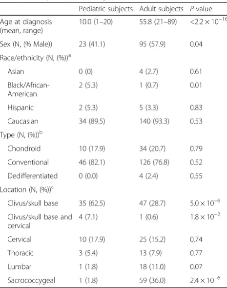
Table 1Study sample demographics
Pediatric subjects Adult subjects P-value Age at diagnosis
(mean, range)
10.0 (1–20) 55.8 (21–89) <2.2 × 10⁻¹⁶
Sex (N, (% Male)) 23 (41.1) 95 (57.9) 0.04 Race/ethnicity (N, (%))a
Asian 0 (0) 4 (2.7) 0.61
Black/African-American
2 (5.3) 1 (0.7) 0.01 Hispanic 2 (5.3) 5 (3.3) 0.83 Caucasian 34 (89.5) 140 (93.3) 0.53 Type (N, (%))b
Chondroid 10 (17.9) 34 (20.7) 0.79 Conventional 46 (82.1) 126 (76.8) 0.52 Dedifferentiated 0 (0.0) 4 (2.4) 0.55 Location (N, (%))c
Clivus/skull base 35 (62.5) 47 (28.7) 5.0 × 10⁻⁶ Clivus/skull base and
cervical
4 (7.1) 1 (0.6) 1.8 × 10⁻² Cervical 10 (17.9) 25 (15.2) 0.74 Thoracic 3 (5.4) 13 (7.9) 0.77 Lumbar 1 (1.8) 18 (11.0) 0.07 Sacrococcygeal 1 (1.8) 59 (36.0) 2.4 × 10⁻⁶
a
14 adult and 18 pediatric subjects declined to report race/ethnicity b
60 adult and 32 pediatric subjects’pathology reports listed chordoma as final diagnosis. These were categorized as conventional chordomas based on discussion with the pathologist
c
combined cohorts were Caucasian (92.6 %), with rela-tively few Asian (2.1 %), African-American (1.6 %) or Hispanic (3.7 %) subjects. The ethnic/racial distribution of chordoma subjects in the pediatric cohort was signifi-cantly different from that in the adult cohort, with a higher proportion of pediatric subjects being Hispanic or African-American and a lower proportion of pediatric subjects being Asian or Caucasian compared to the adult subjects. We found that the proportion of African-Americans was statistically significantly higher in the pediatric cohort compared to the adult cohort (P= 0.01). The majority of the chordomas (78.2 %) were of the con-ventional subtype, which were approximately four times more numerous than the chondroid subtype in both co-horts. Dedifferentiated chordomas were not seen in the pediatric cohort, and only accounted for 2.4 % of the chordomas in the adult cohort. Approximately 20.7 % (34/164) and 76.8 % (126/164) of the adult chordomas, and 18.5 % (10/54) and 81.5 % (44/54) of the pediatric chordomas were of the chondroid and conventional subtypes respectively but there was no significant difference in frequency between adult and pediatric chordomas (P= 0.57).
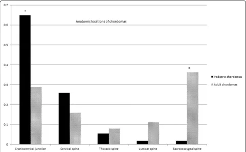
Analysis of the anatomic location of the primary chor-doma lesions showed that in the pediatric cohort, the majority (72.2 %) of lesions were at the craniocervical junction (clivus and/or skull base and cervical spine)
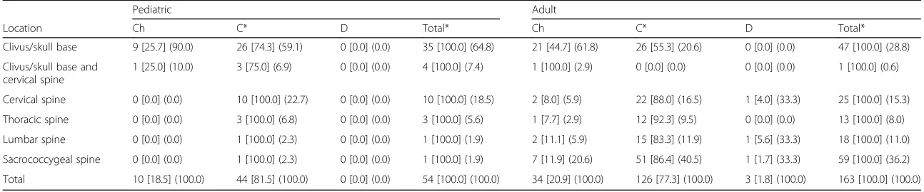
compared to only 29.6 % of the lesions in the adult co-hort (P= 5.1 × 10⁻⁸) (Fig. 1). There was no significant difference in the anatomic location of the chondroid chordomas in the adult cohort compared to the pediatric cohort (P= 0.29), however there was a highly significant difference in the anatomic location of the conventional chordomas in the adult cohort compared to the pediatric cohort. There was a higher proportion of chordomas at the craniocervical junction in the pediatric cohort com-pared to the adult cohort (P= 1.24 × 10⁻⁷) and a higher proportion of chordomas as the sacrococcygeal region in the adult cohort compared to the pediatric cohort (P= 2.45 × 10⁻⁷) (Table 2).
35 % (76/220) of the subjects had pre-treatment im-aging (prior to any radiation, surgery or chemotherapy) available for determining whether the chordoma was pri-marily intra-osseous or extra-osseous. Table 3 shows that most chordomas tend to have a substantial extra-osseous component, with 93.3 % of pediatric chordomas having a predominatly extra-osseous component com-pared to 76.7 % of adult chordomas. This difference was not statistically significant (P= 0.17).
The proportion of predominantly extra-osseous chor-domas was not statistically different by sex (P> 0.05) or subtype of chordoma (P> 0.05) in the combined cohort. However, sacrococcygeal chordomas and chordomas at the craniocervical junction were more likely to be
Fig. 1Anatomic location of chordomas. *P-value < 0.05
Table 2Comparison between anatomic location and type of chordomaa
Pediatric Adult
Location Ch C* D Total* Ch C* D Total*
Clivus/skull base 9 [25.7] (90.0) 26 [74.3] (59.1) 0 [0.0] (0.0) 35 [100.0] (64.8) 21 [44.7] (61.8) 26 [55.3] (20.6) 0 [0.0] (0.0) 47 [100.0] (28.8)
Clivus/skull base and cervical spine
1 [25.0] (10.0) 3 [75.0] (6.9) 0 [0.0] (0.0) 4 [100.0] (7.4) 1 [100.0] (2.9) 0 [0.0] (0.0) 0 [0.0] (0.0) 1 [100.0] (0.6)
Cervical spine 0 [0.0] (0.0) 10 [100.0] (22.7) 0 [0.0] (0.0) 10 [100.0] (18.5) 2 [8.0] (5.9) 22 [88.0] (16.5) 1 [4.0] (33.3) 25 [100.0] (15.3)
Thoracic spine 0 [0.0] (0.0) 3 [100.0] (6.8) 0 [0.0] (0.0) 3 [100.0] (5.6) 1 [7.7] (2.9) 12 [92.3] (9.5) 0 [0.0] (0.0) 13 [100.0] (8.0)
Lumbar spine 0 [0.0] (0.0) 1 [100.0] (2.3) 0 [0.0] (0.0) 1 [100.0] (1.9) 2 [11.1] (5.9) 15 [83.3] (11.9) 1 [5.6] (33.3) 18 [100.0] (11.0)
Sacrococcygeal spine 0 [0.0] (0.0) 1 [100.0] (2.3) 0 [0.0] (0.0) 1 [100.0] (1.9) 7 [11.9] (20.6) 51 [86.4] (40.5) 1 [1.7] (33.3) 59 [100.0] (36.2)
Total 10 [18.5] (100.0) 44 [81.5] (100.0) 0 [0.0] (0.0) 54 [100.0] (100.0) 34 [20.9] (100.0) 126 [77.3] (100.0) 3 [1.8] (100.0) 163 [100.0] (100.0)
Chchondroid Cconventional Ddedifferentiated a
1 adult with a dedifferentiated chordoma and 2 pediatric subjects did not have imaging confirmation of the anatomic location of their lesions Numbers in square brackets [] represent the percentage of each chordoma histological subtype for each anatomic location i.e., row percentages
Numbers in round brackets () represent the percentage of chordomas in a given anatomic location for each histological subtype of chordoma, i.e., column percentages *P-value <0.05 comparing pediatric to adult chordomas
BMC
Medical
Imaging
(2016) 16:53
Page
4
of
predominantly extra-osseous compared to lesions in the thoracolumbar spine (P< 0.05).
Discussion
Our work suggests that there are differences between pediatric and adult subjects with chordoma. Unlike adult subjects, pediatric subjects with chordomas are statisti-cally more likely to be female. We also noted that there were slightly higher proportions of African-American/ Black and Hispanic and slightly lower proportions of Caucasian and Asian subjects with pediatric chordomas compared to subjects with adult chordomas. It is unclear whether this change in ethnic/racial distribution is re-lated to the referral pattern.
Our study shows that the distribution of the anatomic location of chordomas in pediatric subjects is different from that in adult subjects, with pediatric chordomas being primarily cranially located around the craniocervi-cal junction decreasing in frequency towards to caudal spine whereas adult chordomas primarily occur at the most cranial and most caudal aspects of the spine. We do not know what accounts for the differences in chor-doma anatomic location between the pediatric popula-tion and the adult populapopula-tion. Finally, we showed that the majority of the chordomas at the craniocervical junction and sacrococcygeal regions are predominantly extra-osseous compared to those in the thoracolumbar spine.
Our findings are in concert with previously published reports. Prior reports noted that there was a female pre-dominance amongst pediatric subjects with chordomas [4, 10], and a male predominance amongst adult subjects [11] which is similar to our study. A report investigating the Surveillance, Epidemiology, and End Results Pro-gram (SEER) database of chordomas over approximately 30 years (1973–2003) showed that chordomas predom-inantly affect Caucasians with few African-American and
Native American cases [11]. This is similar to our own results; however we also find that the self-reported race/ ethnicity distribution changes in the pediatric population with chordomas.
It is sometimes stated that one of the diagnostic features of chordoma is that it involves the midline [12, 13]. It has been postulated that this is in some way related to residual notochord cells within the nu-cleus pulposus [12, 13]. The predilection for arising within fused segments (clivus, C2, sacrum) tends to support this idea, suggesting that notochordal remnant cells are somehow protected by the encasing bone as noted in our results. We noted that chordo-mas arising from the craniocervical junction and sacrococcygeal spine can be entirely or almost entirely extra-osseous or surface-based, and that this was less likely in lesions arising from the thoracic or lumbar spine.
It is unclear as to why the chordomas at the cranial and caudal aspects of the axial skeleton have substantial soft tissue components. It can be argued that sacrococ-cygeal tumors can reach a large size before becoming symptomatic given the potential large pre-sacral/pre-coccygeal space, and therefore large soft tissue masses remain clinically occult. However, there is substantially less potential pre-vertebral space in the craniocervical spine to accommodate a soft tissue mass and there are several adjacent neurovascular structures which should mean craniocervical chordomas should present with less of an extra-osseous component. Further research is re-quired to understand whether this difference is clinically significant and whether thoracolumbar chordomas share similar genotyping and genomic profiles as well as re-sponse to therapy as craniocervical and sacrococcygeal chordomas.
Exophytic chordomas (chordomas with a predominant soft tissue component) at the cervical and craniocervical Table 3Comparison between pediatric and adult pre-treatment chordomas based on whether the lesion was predominantly intra-osseous or extra-osseous
Location Pediatric subjects Adult subjects Predominantly
intra-osseous
Predominantly extra-osseous*
Total* Predominantly intra-osseous
Predominantly extra-osseous*
Total* Clivus/skull base* 0 [0.0] (0.0) 11 [100.0] (73.3) 11 [100.0] (68.8) 3 [50.0] (21.4) 3 [50.0] (67.4) 6 [100.0] (10.0) Clivus/skull base and cervical spine 0 [0.0] (0.0) 2 [100.0] (13.3) 2 [100.0] (12.5) 0 [0.0] (0.0) 1 [100.0] (2.2) 1 [100.0] (1.7) Cervical spine 1 [100.0] (100.0) 0 [0.0] (0.0) 1 [100.0] (6.3) 0 [0.0] (0.0) 9 [100.0] (19.6) 9 [100.0] (15.0) Thoracic spine* 0 [0.0] (0.0) 2 [100.0] (13.3) 2 [100.0] (12.5) 5 [100.0] (35.7) 0 [0.0] (0.0) 5 [100.0] (8.3) Lumbar spine 0 [0.0] (0.0) 0 [0.0] (0.0) 0 [0.0] (0.0) 5 [71.4] (35.7) 2 [28.6] (4.3) 7 [100.0] (11.7) Sacrococcygeal spine 0 [0.0] (0.0) 0 [0.0] (0.0) 0 [0.0] (0.0) 1 [3.1] (7.1) 31 [96.9] (67.4) 32 [100.0] (53.3) Total 1 [6.3] (100.0) 15 [93.4] (100.0) 16 [100.0] (100.0) 14 [23.3] (100.0) 46 [76.7] (100.0) 60 [100.0] (100.0)
Numbers in square brackets [] represent the percentage of chordomas with a particular MR imaging appearance for each anatomic location i.e., row percentages Numbers in round brackets () represent the percentage of lesions in a given anatomic location for chordomas with a particular MR imaging appearance, i.e., column percentages
*P-value < 0.05 comparing pediatric to adult chordomas
junction have been shown to be associated with in-creased risk of recurrence [14]. In addition, chordomas associated with the upper cervical spine and craniocervi-cal junction have been shown to be independently asso-ciated with worse prognosis after adjusting for preoperative Frankel score, intralesional surgery, greater extent of invasion and revision surgery [15]. Our results show that pediatric chordomas are more likely to be in the upper cervical spine and at the craniocervical junc-tion, and although these factors are associated with worse prognosis in the adult population, overall survival is longer and overall mortality is lower in pediatric pa-tients [16], again highlighting a difference between pediatric and adult chordomas.
There are a few limitations to the analyses. The study is retrospective in nature and therefore subject to ascer-tainment bias. Our institution is a tertiary care center, and the majority of subjects were referred from other in-stitutions for surgery, radiation therapy and/or second opinions, and the referral pattern may introduce a selec-tion bias. We suspect that the self-reported ethnicity of the patients may be explained by selection/referral bias, and it is unclear whether this would influence sex and anatomic distribution. The race/ethnicity was not known for a large proportion of the pediatric subjects. Another limitation is that we did not distinguish between tumors that arise in bone and have a large soft tissue mass, from tumors that have an extra-osseous origin with some os-seous involvement, because this differentiation can be very difficult even for musculoskeletal radiologists and sometimes contentious. However, we feel that our as-sessment presents the data in a format that can be reproduced by other researchers. Finally, because chor-domas are rare, the small sample size limits the statis-tical power to detect other subtle differences that may exist between pediatric and adult subjects with chordomas.
Conclusion
Chordomas in pediatric subjects are more likely to occur in females, the ethnic/racial distribution and anatomic distribution of chordomas differs between pediatric and adult subjects, with the majority of chordomas in pediatric patients occurring at the craniocervical junc-tion, decreasing in incidence distally in the spine, whereas in adult subjects chordomas were more likely to occur in the sacrococcygeal region and craniocervical junctions, with very rare involvement of the thoracic and lumbar spine.
Abbreviation
MRI, magnetic resonance imaging
Acknowledgements
This work was funded by a Ralph Schlaeger Research Fellowship Award.
Funding
Dr. Sebro was funded by a Ralph Schlaeger Research Fellowship Award. The funding body had no role in the design of the study and collection, analysis, and interpretation of data or in writing the manuscript.
Availability of data and materials
Data to replicate findings are presented in the Tables of the main paper. Due to patient privacy protection, any additional materials of the study are only available upon individual request directed to the corresponding author.
Authors’contributions
RS acquired the data, reviewed MRI studies and performed statistical analyses. RS and DR drafted the manuscript. All authors have read and approved of the final manuscript.
Competing interests
RS was funded by a Ralph Schlaeger Research Fellowship Award. JS has been a consultant for Stryker, Biom’up, and Synthes, plus a speaker for Synthes. TD functions and an Editor/author for UpToDate and prepares on-line medical text. TD also acts as a consultant for the Amgen T-VEC Sarcoma Advisory Board. The other authors have no disclosures.
Ethics approval and consent to participate
The study was approved by the Institutional Review Board (IRB) at the corresponding author’s institution and need for signed informed consent was waived.
Author details
1Musculoskeletal Radiology, Department of Radiology, Perelman School of
Medicine, University of Pennsylvania, 3737 Market Street, Philadelphia, PA 19104, USA.2Department of Radiation Oncology, Massachusetts General
Hospital and Harvard Medical School, 55 Fruit Street, Boston, MA 02114, USA.
3The Harris Center for Chordoma Care, Massachusetts General Hospital, 55
Fruit Street, Yawkey 3B, Boston, MA 02114, USA.4Department of Orthopedic Surgery, Massachusetts General Hospital and Harvard Medical School, 55 Fruit Street, Boston, MA 02114, USA.5Department of Hematology/Oncology, Massachusetts General Hospital and Harvard Medical School, 55 Fruit Street, Boston, MA 02114, USA.6Department of Pathology, Massachusetts General Hospital and Harvard Medical School, 55 Fruit Street, Boston, MA 02114, USA.
7
Department of Radiology, Massachusetts General Hospital, Yawkey 6E, 55 Fruit Street, Boston, MA 02114, USA.
Received: 10 April 2016 Accepted: 1 August 2016
References
1. Barresi V, Ieni A, Branca G, Tuccari G. Brachyury: a diagnostic marker for the differential diagnosis of chordoma and hemangioblastoma versus neoplastic histological mimickers. Dis Markers. 2014;2014:514753. 2. Jo VY, Hornick JL, Qian X. Utility of brachyury in distinction of chordoma
from cytomorphologic mimics in fine-needle aspiration and core needle biopsy. Diagn Cytopathol. 2014.
3. Aydin AL, Sasani M, Oktenoglu T, Solaroglu I, Ozer AF. A case of chordoma invading multiple neuroaxial bones: report of ten years follow up. Turk Neurosurg. 2013;23(4):551–6.
4. McMaster ML, Goldstein AM, Bromley CM, Ishibe N, Parry DM. Chordoma: incidence and survival patterns in the United States, 1973–1995. Cancer Causes Control. 2001;12(1):1–11.
5. Williams BJ, Raper DM, Godbout E, Bourne TD, Prevedello DM, Kassam AB, Park DM. Diagnosis and treatment of chordoma. J Natl Compr Canc Netw. 2013;11(6):726–31.
6. Walcott BP, Nahed BV, Mohyeldin A, Coumans JV, Kahle KT, Ferreira MJ. Chordoma: current concepts, management, and future directions. Lancet Oncol. 2012;13(2):e69–76.
7. Lantos JE, Agaram NP, Healey JH, Hwang S. Recurrent skeletal extra-axial chordoma confirmed with brachyury: imaging features and review of the literature. Skeletal Radiol. 2013;42(10):1451–9.
8. van Akkooi AC, van Geel AN, Bessems JH, den Bakker MA. Extra-axial chordoma. J Bone Joint Surg (Br). 2006;88(9):1232–4.
10. Hoch BL, Nielsen GP, Liebsch NJ, Rosenberg AE. Base of skull chordomas in children and adolescents: a clinicopathologic study of 73 cases. Am J Surg Pathol. 2006;30(7):811–8.
11. Mukherjee D, Chaichana KL, Parker SL, Gokaslan ZL, McGirt MJ. Association of surgical resection and survival in patients with malignant primary osseous spinal neoplasms from the Surveillance, Epidemiology, and End Results (SEER) database. Eur Spine J. 2013;22(6):1375–82.
12. Hirosawa RM, Santos AB, França MM, Fabris VE, Castro AV, Zanini MA, Nunes VS. Intrasellar chondroid chordoma: a case report. ISRN Endocrinol. 2011;2011:259392.
13. Sun X, Hornicek F, Schwab JH. Chordoma: an update on the
pathophysiology and molecular mechanisms. Curr Rev Musculoskelet Med. 2015;8(4):344–52.
14. Wang K, Wang L, Wu Z, Tian K, Ren C, Jia G, Zhang L, Zhang J. Bone invasiveness is associated with prognosis in clivus chordomas. J Clin Neurosci. 2016;27:147–52.
15. Zou MX, Huang W, Wang XB, Li J, Lv GH, Deng YW. Prognostic factors in spinal chordoma: a systematic review. Clin Neurol Neurosurg. 2015;139:110–8.
16. Lau CS, Mahendraraj K, Ward A, Chamberlain RS. Pediatric chordomas: a population-based clinical outcome study involving 86 patients from the Surveillance, Epidemiology, and End Result (SEER) database (1973–2011). Pediatr Neurosurg. 2016;51(3):127–36.
• We accept pre-submission inquiries
• Our selector tool helps you to find the most relevant journal
• We provide round the clock customer support
• Convenient online submission
• Thorough peer review
• Inclusion in PubMed and all major indexing services
• Maximum visibility for your research
Submit your manuscript at www.biomedcentral.com/submit