classification system based on a literature review
Myroslava Kumka, MD, PhD*
Jason Bonar, BScKin, DC
Fascia is virtually inseparable from all structures in the body and acts to create continuity amongst tissues to enhance function and support. In the past fascia has been difficult to study leading to ambiguities in nomenclature, which have only recently been addressed. Through review of the available literature, advances in fascia research were compiled, and issues related to terminology, descriptions, and clinical relevance of fascia were addressed. Our multimodal search strategy was conducted in Medline and PubMed databases, with other targeted searches in Google Scholar and by hand, utilizing reference lists and conference proceedings. In an effort to organize nomenclature for fascial structures provided by the Federative International Committee on Anatomical Terminology (FICAT), we developed a functional classification system which includes four categories of fascia: i) linking, ii) fascicular, iii) compression, and iv) separating fasciae. Each category was developed from descriptions in the literature on gross anatomy, histology, and biomechanics; the category names reflect the function of the fascia.
An up-to-date definition of fascia is provided, as well as descriptions of its function and clinical features. Our
*Canadian Memorial Chiropractic College, Department of Anatomy Correspondence should be addressed to:
Myroslava Kumka, 6100 Leslie Street, Toronto ON M2H 3J1, Canada Tel: 416-482-240 ext: 175
Email: [email protected] ©JCCA 2012
Le fascia est pratiquement inséparable de toutes les structures du corps, et il sert à créer une continuité entre les tissus afin d’en améliorer la fonction et le soutien. Il a déjà été difficile d’étudier le fascia, ce qui a donné lieu à des ambiguïtés dans la nomenclature, qui n’ont été abordées que récemment. Grâce à un examen de la documentation disponible, les avancées dans la recherche sur le fascia ont été compilées, et les problèmes relevant de la terminologie, des
descriptions et de la pertinence clinique du fascia ont été traités. Nous avons adopté une stratégie de recherche multimodale pour nos recherches dans les bases de données Medline et PubMed, avec des recherches ciblées dans Google Scholar et manuelles, au moyen de listes de références et de comptes rendus de congrès.
classification demonstrates the use of internationally accepted terminology in an ontology which can improve understanding of major terms in each category of fascia.
(JCCA 2012;56(3):179-191)
k e y w o r d s : fascia, connective tissue, classification,
anatomy, histology, terminology, innervations, manual therapy
acceptée à l’échelle internationale dans le cadre d’une ontologie qui peut améliorer la compréhension des termes importants dans chaque catégorie de fascia.
(JCCA 2012;56(3):179-191)
m o t s c l é s : fascia, tissu conjonctif, classification,
anatomie, histologie, terminologie, innervations, thérapeutique manuelle
Introduction
Fascia is an uninterrupted viscoelastic tissue which forms a functional 3-dimensional collagen matrix.1-3 It
sur-rounds and penetrates all structures of the body extending from head to toe, thus making it difficult to isolate and develop its nomenclature.1 The Federative International
Committee on Anatomical Terminology (FICAT), in the 1998 edition of Terminologia Anatomica, points out significant flaws in the nomenclature system for fascia, largely stemming from the anglocentric nature of the terms used, and lack of formal international applicability.4
The usage of the terms superficial fascia and deep fascia is considered incorrect by the FICAT because histological terminology referring to layers of the connective tissue varies too much internationally to be generalized by these two terms.4 Common names of certain fascia are also
considered to be inaccurate, e.g., Scarpa’s, Camper’s, and
Colles’. It is suggested that they be replaced with cutaneous tissue of abdomen membranous layer, sub-cutaneous tissue of abdomen fatty layer and membranous layer of perineum, respectively.4 Terminologia
Anatom-ica gives a long list of terms related to their definition of fascia, and attempts to offer a system for grouping vari-ous fasciae based on embryological origins and modes of development. However, clear details on the groupings and justification for this strategy are not given, and it re-mains difficult to organize and properly use the multiple fascial terms. Furthermore, the application of these terms for communication in research, education, and clinical practice remains difficult and impractical. In light of the contribution made by the FICAT on fascial terminology, it is important to utilize their work on terminology and suggest how it may be further arranged and used in
prac-tice. Indeed, a number of experts have called for further development on the description and nomenclature of all fasciae and have made significant contributions of their own.5-7
Recent advances in research within the fields of bio-mechanics, gross anatomy, and histology, provide the international research community with an opportunity to improve the terminology associated with fascia, thereby improving intra- and inter-professional communications.5, 8-10 However, discrepancies still exist concerning the
of-ficial definition, terminology, classification and clinical significance of fascia.5-6
For example, the FICAT broadly defines fascia as sheaths, sheets or other dissectible connective tissue ag-gregations. The First International Fascia Research Con-gress (2007) formulated a comprehensive definition of fa-scia as the soft tissue component of the connective tissue system, emphasizing its uninterrupted, three-dimensional web-like extensions and highlighting its functional at-tributes.1,4 The Congress went on to include joint and
organ capsules, muscular septa, ligaments, retinacula, aponeuroses, tendons, myofascia, neurofascia and other fibrous collagenous tissues as forms of fascia, inseparable from surrounding connective tissues.1 The broad nature
of these two definitions is controversial, and not all of the tissues have been widely accepted into common usage. Thus, a consistent terminology to classify and categor-ize fascia has not been formally established and accepted internationally.5-6 Gray’s textbook approach to naming
identifying regional fascia, but it is not used consistently in practice internationally, nor does it help to describe the functional role of the regional fascia.
A more recent ontology highlights the importance of two functional forms of fascia, connecting and
discon-necting, for which fiber orientation and descriptions of
fa-scia’s role in proprioception are key justifications for each category.7 In support of this principle, there is an
exten-sive body of work demonstrating that significant amounts of force are transmitted amongst multiple antagonist and synergistic muscle groups across joint capsules through various sections of extra- and intramuscular fascia.7-8,13-15
Although the recent ontologies grounded in fascial function represent an advance in classification, the com-plexities of fascia make it necessary to expand on these two categories (connecting and disconnecting) to further sort and describe all groups of fascia according to their attributes reported recently in the literature.7 These
addi-tional attributes include: collagen type ratio, extracellular matrix proteins, nerve fiber types, myofascial force trans-mitting potential, details on fiber orientation, and influ-ence on the circulatory system.8,16-21
Lack of consistent terminology has a negative effect on communication within health professions, and im-pedes collaborations in research.22 Since fascia represents
a topic of growing interest worldwide, it should be a pri-ority to reduce ambiguity in fundamental terms so that this field of research can properly advance. For example, the lack of common definitions and nomenclature hinders communication between those involved in the process of diagnosis and treatment of pelvic pathologies in every-day practice.23 One study revealed this by comparing the
unofficial/common terms being used in the field and the terms provided by the Terminologia Anatomica, demon-strating how this text can be used to improve consistency in the nomenclature. While we do accept the validity of a topographical approach in naming fascia, it does not take into account the fact that fascia doesn’t have a beginning and end like muscle, nor does a topographical approach account for fascia’s microscopic features, or diversity of its functional characteristics.5-7,12
Purpose
The aim of the present study is to develop a classifica-tion system for fascial structures, based primarily on their functional properties, but also incorporating
morphologic-al characteristics identified in the literature, and utilizing internationally accepted terminology from the FICAT.4,24
Methods:
A review of the literature on fascia was conducted with electronic searches of EBSCO databases, PubMed, and Google Scholar, along with hand searches of proceedings from the past two International Fascia Research Con-gresses (2007, 2009). Search strategies in EBSCO and PubMed consisted of individual and combined MeSH term searches for “Fascia” and “Connective Tissue” using specific limiters for our topic to emphasize anat-omy. These search terms were also paired separately with various keywords – “terminology”, “extracellular ma-trix”, “collagen”, and “function”. Results were limited to English, full text peer reviewed journal articles, confer-ence proceedings, and textbooks. Inclusion was limited to articles with abstracts related to morphology, termin-ology, and/or clinical aspects of fascia. Information was then used to help organize fascia terminology, utilizing terms from two texts: Terminologia Anatomica and Ter-minologia Histologica.4,24
Results
In section A below, we present the results of our literature review, discussing the anatomical, histological, and bio-mechanical features of fascia, and its innervation. Then in section B, we outline our resulting proposal for a new classification system (Table 1) incorporating these fea-tures of fascia. It is important to first understand the key characteristics and the divergent classifications of fascia at gross anatomical, histological, and biomechanical per-spectives before outlining the details of these four cat-egories.
A. Literature Review
I. Overview
The range of research advances revealed in the literature includes observations on imaging, advanced dissection and staining techniques, as well as modeling of tissue de-formation, and in vitro cellular processes.10,12,25-30 Areas
research, we see that fascia is not a passive structure, but a functional organ of stability and motion, virtually insepar-able from all surrounding tissue.
II. Gross Anatomy
Gross anatomical studies of fascia demonstrate an ar-ray of characteristics based on location, density, fiber direction, and fascia’s relationship to surrounding struc-tures.19,26-27 Based on location, the FICAT describes the
following fasciae: i) in relation to the body regions: fascia of head and neck, fascia of trunk, and fascia of limbs, ii) in relation to the surrounding structures: subcutaneous fascia, fascia of muscles, visceral fascia, parietal fascia, and fascia extraserosalis which represents any other fa-scia which lies inside the parietal fafa-scia and outside the visceral fascia.4
Fascia’s key characteristic, continuity, helps explain concepts such as myofascial force transmission.3 Hunjing
describes biomechanical features of extramuscular and intramuscular force transmission in rat specimens where surrounding connective tissues are seen to influence force potential and connect muscle groups.13-15 Dissectional
ob-servations of the intramuscular connective tissue (IMCT) show this fascia to influence length of sarcomeres to im-prove force production.15 Other morphological
descrip-tions of fascia characterize it as being optimally designed to take up tensional forces in the musculoskeletal system. In the past, classical dissection techniques have essen-tially ignored fascia , simply removing it to get to muscle and deeper structures.5 Subsequently, it was revealed
through careful dissection that fascia commonly occurs as an undulating layered system of different connective tissue types.19,28-29 Three-dimensional models of the
cru-ral and thoracolumbar fasciae demonstrate that the “deep fasciae” are formed of three sub-layers of connective tis-sue with different densities and orientations.19,29 It was
discovered that in each sub-layer the collagen fibers are parallel to each other, whereas the orientation between the fibers of adjacent layers changes, forming an angle of approximately 70-80 degrees with each other.29 This
al-lows denser fascial sheets to slide freely over underlying layers, without significant friction, and enhances fascia’s ability to take up strain in virtually all directions.29
It is difficult to gain an appreciation for the true appear-ance of fascia, aside from basic structure, in embalmed cadavers. Direct observation of fascia’s appearance and
behavior in a living, hydrated body, has been conducted with recent fluoroscopic imaging under the skin of the dorsal forearm, shedding new light on how this sliding collagenous system works.32-33 These observations
dem-onstrate that fascia incorporates a water dense vacuolar system able to slide independent of the rate of contraction in muscle around it and able to conduct structures like ca-pillaries throughout sections of myofascia.32-33 The
visco-elastic nature of fascia can only be observed in hydrated tissue, and in embalmed tissue we are only observing an artifact of the living tissue. A better appreciation for the true gross appearance of fascia can be gained through fresh body dissections, and in vivo via direct imaging techniques.34
Pathological changes in fascia have been observed with special imaging techniques, such as ultrasonic elas-tography that displays deformation and the elasticity of soft tissues.34 This technology allows a non-invasive
esti-mation of tissues stiffness based on the fact that soft tis-sue has greater tistis-sue displacement than hard tistis-sue when externally compressed.10 By further quantifying the
prop-erties of the soft tissues, as with recent 3-D mathematical models of fascial deformation, insight will be gained into the effects of manual treatments beneath the skin and how the body responds to various forces.30,35-36
III. Histology
Fascia has specific cells, ground substance, and fiber types that make it a form of connective tissue proper.16,37-38
A better understanding of fascia at the cellular level gives insight into its functional properties.39 Clear changes to
the extracellular matrix (ECM) in the form of adhesive sites between microscopic filaments have been studied in “scarred” fascia.40-41 Collagen types have also been shown
to vary with mechanical forces and strains.39 We
hypoth-esize that the functional properties of fascia are reliant on the composition of the ECM, specific cells, and filaments, including but not limited to the ratio of collagen types. Collagen, a triple helix glycoprotein, is the key struc-tural fiber that gives connective tissue its ability to resist tension.17,37-38 There are twenty five distinct collagen types
recognized in the Ross histology textbook and atlas, and twenty eight collagen types recognized in the latest re-view by Gordon.16,37 Although, type I collagen is the main
in-cluding, but not limited to, types I, III, IV, V, VI, XI, XII, XIV, XXI.16-17,37-38 Collagen provides resistance to tension
and stretch, which commonly occur in fascial tissues, such as ligaments, tendons, sheaths, muscular fascia and deeper fascial sub-layers.37
Collagen type III, also known as reticular fiber, is in-volved in forming the scaffolding for the cells of the loose connective tissues related to the endoneurium, vascular walls, and smooth muscle.1-2,17A collagen fibril needs the
support of not only fibrillar collagen types, but also a mix of non-fibrillar forms known as fibril-associated collagens with interrupted triple helices (FACITs).16 The functions of
FACITs include: i) anchoring to the basement membrane, ii) regulating the diameter of fibrils, iii) forming lattice networks, and iv) acting as transmembrane structures.16
These fibrils are important to the integrity and function of fascia within the ECM. Elastic fibers within the ground substance give fascia its characteristic stretch.29,37-38,42
A combination of multiple types of collagen within the extracellular matrix forms a unique structure, like a blue-print that reflects the function and compliance of fascia in various regions.16-17 Without a characteristic fiber
arrange-ment and composition for each fascial region, it is likely that fascia would not withstand stresses or have the same function.
The cells within fascia include fibrocytes (fibroblasts, myofibroblasts), adipocytes, and various migrating white blood cells.27,41-42 Fibroblasts are highly adaptable to their
environment, and show a capacity to remodel in response to the direction of various mechanical stimuli, produ-cing biochemical responses.29,41,43-45 If function changes,
as with increased mechanical stress, or prolonged im-mobilization, deoxyribonucleic acid (DNA) transcription of pro-collagen in the fibroblasts will change types (e.g., collagen type I into collagen type III), or undifferentiated cell types may adapt towards a more functionally appro-priate lineage (e.g., chondrocyte).42,45-48
Benjamin et al, studied the morphological changes ob-servable in various tendons and ligaments in response to biomechanical stresses.46 It was established that the
tis-sue structure and the molecular composition of ECM are directly correlated with the local mechanical forces.46,47
Under significant states of compression, tissue once populated with fibroblasts, becomes invested predomin-ately with chondrocytes and forms specialized connect-ive tissue, cartilage, with further solid mineral
depos-ition.46,47 These adaptations have been demonstrated in
the supraspinatus tendon, transverse acetabular ligament, transverse ligament of atlas, as well as various other liga-ments and tendons throughout the body.47,48
Myofibroblasts within fascia demonstrate contractile properties and contain actin-myosin filaments typically seen in smooth muscle.49-51 The significance of these
con-tractile properties remains unclear, however in-vitro ob-servations of autonomous contraction of myofibroblasts harvested from porcine and rat fascia when stimulated with various pharmacological agents (i.e., mepyramine, angiotensine, glyceryltrinitrate) have been repeated in different labratories.52-53 An estimation of tension created
by contraction of myofibroblasts when extrapolated to a large fascial sheet (i.e., thoracolumbar fascia) may pro-duce tension within the musculoskeletal system between 30-40N.51 The significance of this contractile property
re-mains hypothetical and reproduction of these contractile forces in-vivo in response to efferent neural stimulus is yet to be done.
Increased concentration of myofibroblasts in patho-logical fascia has been observed, suspected to create tis-sue contractures in clinical conditions like palmar fascial fibromatosis (Dupuytren’s disease), plantar fascial fibro-matosis (Ledderhose’s disease), and adhesive capsulitis (frozen shoulder).49,54-56 Fascia is also susceptible to the
actions of typical cells of inflammation influencing com-munication, growth, and function.37,38
Fiber orientation in fascia is important to its overall structure and function, and can be viewed with the un-aided eye, polarized light, or various microscopic tech-niques.18,27,57 It is a consistent observation that the fibers
are oriented parallel to predicted force vectors, and are likely to resist tension.18,27,57 Based on certain common
characteristics, including the fiber arrangement, connect-ive tissue proper is classified by the Terminologia Histo-logica as loose connective tissue and dense connective tissue.24 The dense connective tissue is sub-categorized
as: i) unidirectional parallel ordered dense connective tis-sue, ii) multidirectional parallel ordered dense connective tissue, iii) woven connective tissue, iv) irregular fuso-cellular connective tissue.24
IV. Mechanotransduction
compression. These stimuli exert their effects within the cells through filaments of the ECM, and so mechanical stimulation leads to a cascade of events which eventu-ally influences the activity in the nucleus.48,58,59
Mechano-transduction is produced as cells convert a diversity of mechanical stimuli, transmitted throughout the ECM, into chemical activity to regulate morphology and function of tissues.60-61 The cellular responses include the release of
interleukins, adhesion kinases and other biochemicals.61-62
Local injury in a tissue can have widespread conse-quences via mechanotransduction’s role in stimulating quiescent cells to form active fibroblasts.62 This
mechan-ism of cellular activation via mechanical force has been hypothesized to play a role in embryological develop-ment as part of the induction process of mesenchymal cells throughout the mesoderm.63 Clinically, the effects of
mechanotransduction have been observed with the inter-vention of acupuncture.64 There is evidence to suggest
that the insertion of acupuncture needles into the fascia stimulates the activity of fibroblasts, presumably through physical strain exerted on the transmembranous micro-filaments in the ECM.65-66 Needles will also cause
dis-placement of that tissue, and twisting of the needle once it is inserted can cause further displacement of the fascia as measured with ultrasonic elastomyography.35 Simple
manual pressure has also been demonstrated, through deformation, to cause alterations in the viscoelasticity of tissue.30,36 These cellular and filamentous responses may
provide a theoretical framework for the therapeutic mech-anisms of soft tissue therapies.36,67-68 An understanding of
mechanotransduction reveals that for healing and injury it is not only the gross observable reaction of tissues that is of interest, but also the biomechanical response of the ECM within fascia.
V. Innervation
Electron microscopy and special staining procedures demonstrate that fascia is populated by sensory neural fib-ers, suggesting that fascia contributes to proprioception and nociception, and may be responsive to manual pres-sure, temperature, and vibration.18,26,69,70 Some receptors
found within fascia may be responsive to, and influence some autonomic responses, such as lowering blood pres-sure.71-72
On a structural basis, two classes of sensory receptors are recognized: free nerve endings as terminal branches of
the axons, and encapsulated endings with distinctive ar-rangements of non-neuronal cells that completely enclose the terminal parts of the axons.73 Some of these
recep-tors function as both a mechanoreceptor and a nociceptor (types III and IV receptors).73 Pain that arises in muscles,
tendons, ligaments, and bones is detected by these recep-tors. There have been conflicting results in the research, but most recent evidence has revealed small diameter free nerve endings in the thoracolumbar fascia of rats and hu-mans.69 One investigation has revealed the presence of
these fibers on electronmicroscopy18, meanwhile another
group has demonstrated calcitonin gene-related peptide (CGRP) and substance P (SP) within the same fibers, sug-gesting afferent function, including nociception.69 Further
work must be done on human specimens as currently the best evidence is heavily reliant on animal models.
Langevin et al, developed a pathophysiological model of low back pain based on connective tissue nociception, after demonstrating on ultrasound the structural altera-tions of the low back connective tissues.34,70 Fascial
con-nections within different motor units, and different func-tional synergists, can provide an alternative explanation for referred pain distributions, which often do not fol-low either nerve pathways or the morphology of a single muscle.74
Many encapsulated endings found in fascia are mech-anoreceptors that respond to mechanical pressure or deformation, and include Golgi receptors, Pacinian cor-puscles, and Ruffini’s corpuscles.18,26,75-76 Different
tech-niques of tissue manipulation may stimulate the above re-ceptors: the high-velocity thrust manipulations and vibra-tory techniques likely stimulate Pacinian receptors, while slow, deep soft tissue techniques likely target Ruffini’s bodies.75-76 Knowing what receptors are more
signifi-cantly concentrated in a particular target tissue can help a manual practitioner choose the method of stimulus and technique (e.g., deep pressure, light stroke, stretch, ten-sion, or vibration).75-76 Understanding that certain types of
fascia are more densely populated with certain particular receptors can aid in the overall understanding of the body and creating more effective approaches to manual treat-ments.
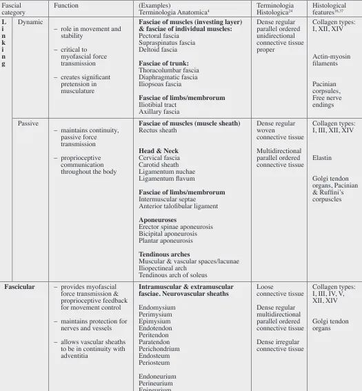
B. Classification System
system which includes four categories of fascia: i)
link-ing, ii) fascicular, iii) compression, and iv) separating
fasciae.
All fascia-related terminology provided in the Ter-minologia Anatomica can be subsumed within these four categories (Table 1).4 This system is not meant to be a
reductionist approach to the fascial system, but a mode of exploring and better understanding the complex inter-action of functions that exist within the system. Each re-gion of the body contains multiple categories, suggesting that every region of the body has a complex mixture of different fascial types. To illustrate this concept, the thigh is an example of a body region which contains all four fascial categories: Illiotibial band (Linking), perimysium of the quadriceps femoris muscle (Fascicular), fascia lata (Compression), and subcutaneous tissue (Separating).
I. Linking Fascia
The linking category is predominantly dense regular par-allel ordered unidirectional connective tissue proper with a significant amount of collagen type I.16,24 This includes
fasciae of muscles, fasciae of regions (head & neck, trunk, limbs), aponeuroses, tendinous arches and neurovascular sheaths.4
This category is subdivided into dynamic and passive divisions. The dynamic division includes major fascial groups more significantly related to movement and joint stability, and characterized by higher concentrations of contractile and proprioceptive fibers. The dynamic div-ision is composed of fasciae of muscles (investing layer, fascia of individual muscle), and fasciae of the trunk.4 The
innervation of dynamic linking fascia functionally differ-entiates it from other categories, permitting it to contrib-ute to nociception and proprioception. For example, the thoracolumbar fascia (TLF) contributes to spinal stabil-ity and makes firm connections between the trunk and limbs.77 It is also densely innervated by free nerve
end-ings and Paciniform corpuscles which respond to rapid pressure and vibration.2,18,73,78
The passive division is acted on by other extramuscu-lar tissues to maintain continuity throughout the body or form tunnels and sheaths.7 The passive division
incorpor-ates fasciae of muscles (muscle sheaths), fasciae of the head and neck, fasciae of limbs, aponeuroses, tendinous arches, and retinaculae.4 This group can act as muscular
insertion points, such as the epicranial aponeurosis, and
as joint linkages and tendinous arches ultimately provid-ing proprioceptive information when tension is exerted.7
The passive linking fasciae can only transmit force when they are stretched and loaded, while dynamic fasciae can theoretically contract more autonomously like smooth muscle, thereby affecting tension in the musculoskeletal system, but not significant enough to be the primary mov-er of limbs.51
II. Fascicular Fascia
Fascicular fascia forms adaptable tunnels which bundle
vessels as well as fascicles within muscle, tendon, bone and nerves. Fascicular fascia plays an important role in organization, transport, strength and locomotion.39 This
category is organized as a mixture of both loose and dense regular multidirectional connective tissues.24 Types I and
III collagen are the major components of these tissues with lesser amounts of Types V, VI, XII, and XIV.16,39,79
Fascicular fascia of the muscle comprises three
dis-tinct layers of IMCT: epimysium surrounding whole muscles, perimysium separating fascicles or bundles of muscle fibers within the muscle, and endomysium cov-ering the individual muscle fibers.39 Forming the muscle
architecture, this network of collagen fibers can be seen as an extensive matrix of tunnels that connects and dis-sipates force within muscle, provides intramuscular path-ways and mechanical support for large and small nerves, blood vessels and lymphatics.32,39,79 The fascicular fascia
of the muscle converges into a dense regular connect-ive tissue link at the myotendinous junction to become
fascicular fascia of the tendon, comprising endotendon,
peritendon and epitendon.5,7,79 At this junction, fascicular
fascia is richly innervated by Golgi tendon organs which are stimulated by muscle contraction.37,38 Tension in the
tendon results in a reflex decrease in tonus in contiguous striated muscle fibers.70
IMCT is essential for myofascial force transmission (as outlined in Results section A I), enhancing the forces produced by muscles.8 Fascicular fasciae allow forces to
be transferred from within muscle to synergistic muscles, and also, via the extramuscular pathway, through the
link-ing fascia, to antagonistic muscles.8,13-14 The fascicular
fascia forms the connective tissue envelope for nerve fascicles and whole peripheral nerves: perineurium and epineurium, respectively.37,38 The perineurium serves as a
Table 1 Fascial categories: function, terms, and histological features Fascial
category Function (Examples)Terminologia Anatomica4 Terminologia Histologica24 Histological features16,37
L i n k i n g
Dynamic
– role in movement and stability
– critical to myofascial force transmission – creates significant
pretension in musculature
Fasciae of muscles (investing layer) & fasciae of individual muscles:
Pectoral fascia Supraspinatus fascia Deltoid fascia
Fasciae of trunk:
Thoracolumbar fascia Diaphragmatic fascia Iliopsoas fascia
Fasciae of limbs/membrorum
Iliotibial tract Axillary fascia
Dense regular parallel ordered unidirectional connective tissue proper
Collagen types: I, XII, XIV
Actin-myosin filaments
Pacinian corpsules, Free nerve endings Passive
– maintains continuity, passive force transmission – proprioceptive
communication throughout the body
Fasciae of muscles (muscle sheath)
Rectus sheath
Head & Neck
Cervical fascia Carotid sheath Ligamentum nuchae Ligamentum flavum
Fasciae of limbs/membrorum
Intermuscular septae Anterior talofibular ligament
Aponeuroses
Erector spinae aponeurosis Bicipital aponeurosis Plantar aponeurosis
Tendinous arches
Muscular & vascular spaces/lacunae Iliopectineal arch
Tendinous arch of soleus
Dense regular woven
connective tissue Multidirectional parallel ordered connective tissue
Collagen types: I, III, XII, XIV
Elastin
Golgi tendon organs, Pacinian & Ruffini’s corpuscles
Fascicular – provides myofascial force transmission & proprioceptive feedback for movement control – maintains protection for
nerves and vessels – allows vascular sheaths
to be in continuity with adventitia
Intramuscular & extramuscular fasciae. Neurovascular sheaths
Endomysium Perimysium Epimysium Endotendon Peritendon Paratendon Perichondrium Endosteum Periosteum Endoneurium Perineurium Epineurium
Loose
connective tissue Dense regular multidirectional parallel ordered connective tissue Dense irregular connective tissue
Collagen types: I, III, IV, V, XII, XIV
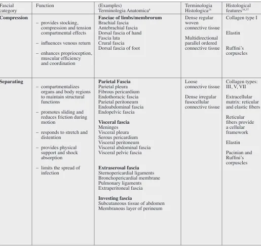
Table 1 (Continued) Fascial
category Function (Examples)Terminologia Anatomica4 Terminologia Histologica24 Histological features16,37
Compression
– provides stocking, compression and tension compartmental effects – influences venous return – enhances proprioception,
muscular efficiency and coordination
Fasciae of limbs/membrorum
Brachial fascia Antebrachial fascia Dorsal fascia of hand Fascia lata
Crural fascia Dorsal fascia of foot
Dense regular woven
connective tissue Multidirectional parallel ordered connective tissue
Collagen type I
Elastin
Ruffini’s corpuscles
Separating
– compartmentalizes organs and body regions to maintain structural functions
– promotes sliding and reduces friction during motion
– responds to stretch and distention
– provides physical support and shock absorption
– limits the spread of infection
Parietal Fascia
Parietal pleura Fibrous pericardium Endothoracic fascia Parietal peritoneum Endoabdominal fascia Endopelvic fascia
Visceral fascia
Meninges Visceral pleura Serous pericardium Visceral peritoneum Visceral abdominal fascia Visceral pelvic fascia
Extraserosal fascia
Sternopericardial ligaments Bronchopericardial membrane Pulmonary ligaments
Extraperitoneal fascia
Investing fascia
Subcutaneous tissue of abdomen Membranous layer of perineum
Loose
connective tissue Dense irregular fusocellular connective tissue
the formation of a blood-nerve barrier.80 The blood
ves-sels that supply the nerves travel in the epineurium.80
These two layers of the fascicular fascia are innervated by the nervi nervorum, which can evoke nociception through the release of CGRP and may create neurogenic inflam-mation.81 An inflammation of the nervi nervorum causes
the inflammatory reaction of the nerve’s fascial envelopes to induce the mechanical sensitivity, which can manifest as local, radicular, or neuropathic pain.75,82,83
III. Compression Fascia
Compression fascia is a mixture of dense regular woven
and multidirectional parallel ordered connective tissue layers that ensheath whole limbs to create a stocking ef-fect.24,84 This fascial category plays an important role in
locomotion and venous return due to its influence on com-partmental pressure, muscle contraction and force distri-bution.20,29,84 For example, the crural fascia is composed
of two or three layers of parallel ordered collagenous fiber bundles, each layer being separated by a thin layer of loose connective tissue.19,29 The spatial orientation of
the collagen fibers changes from layer to layer within the
compression fascia.29 The presence of loose connective
tissue interposed between adjacent layers permits local sliding, allowing the single layers to respond more effect-ively.29
Examples of this type of fascia are observed in the limbs and are observed as fascia lata, crural fascia, bra-chial fascia, and antebrabra-chial fascia. While there are pro-prioceptors embedded in this fascia, its role as a sensory organ is less significant than that of the linking, or
fascicu-lar categories.
IV. Separating Fascia
Separating fascia is generally loose connective tissue
and dense irregular fusocellular connective tissue.24 The
reticular Type III collagen fibers and elastic fibers are the major components of the ECM of separating fascia, with small amounts of collagen Types V, VII.16-17 While
the reticular fibers provide a supporting framework for the cellular constituents, the elastic fibers form a three-dimensional network to allow separating fascia to re-spond to stretch and disten tion.28,37 Separating fascia
div-ides the body in visible sheets and layers of varying fibers allowing it to take up forces and friction in all directions. While its major function is to allow more efficient sliding
of tissues over one another, it may still form adhesions from faulty movement patterns or injury.54
FICAT’s terms for separating fascia include: parietal fascia, visceral fascia, extraserosal fascia, investing/sub-cutaneous fascia, formerly known as fascia superficialis.4
This category also includes synovial sheaths and fasciae of limbs.4 Parietal fascia lies outside the parietal layer of
serosa such as pericardium, pleura and peritoneum, and lines the wall of a body cavity.4 Visceral fascia lies
im-mediately outside the visceral layer of the serosa and sur-rounds the viscera.4,85 Extraserosal fascia lies within the
space between the visceral and parietal fasciae.4
This fascia class is a complex connective tissue matrix, ensheathing everything from body cavities to individual organs. It separates, supports, and compartmen talizes or-gans and regions in order to maintain proper structural and functional relationships throughout the body. This group of fascia has a unique appearance and texture upon observation, ranging from transparent woven sheets to a fuzzy cotton-like consistency.28
The innervation of separating fascia serves primarily to sense distension and compression of tissues. More de-tailed histological analyses are necessary to reveal with certainty the fascial innervations of these deep layers. However, concentrations of Pacinian corpuscles (de-tecting deep pressure) and Ruffini’s corpuscles, which responding slowly to sustained pressure and tangential forces, are thought to be present in much of separating fascia, for example, in subcutaneous tissue.28,70,86 Deep
sustained pressure may be necessary for manual practi-tioners to affect this fascial tissue.
Conclusion
Through this article, we have reviewed advances in fa-scia research and addressed issues related to terminology and classification of fascia. The literature supports defin-ing fascia as an innervated, continuous, functional organ of stability and motion that is formed by 3-dimensional collagen matrices. In an effort to organize the nomencla-ture of fascia, we devised a functional classification sys-tem which includes four categories of fascia: i) linking, ii)
fascicular, iii) compression, and iv) separating fasciae.
cat-egory. Such a classification system based on functional properties of fasciae may have morerelevance to the clin-ical experiences of manual therapists.
All fascial related terminology provided in the Ter-minologia Anatomica4 can be subsumed within these four
fascial categories (Table 1).
Each region of the body contains multiple categories. This suggests that the complex interaction of different fascial types improves the musculoskeletal system’s ef-ficiency. It is our hope that this classification system will add clarity, improve diagnostic precision and contribute to manual therapists’ understanding of fascia as a target of pathology and treatment.
References
1. LeMoon K. Terminology used in Fascia Research. J Bodyw Mov Ther. 2008; 12(3):204–212.
2. Yahia L, Pigeon P, DesRosiers E. Viscoelastic properties of the human lumbodorsal fascia. J Biomed Eng. 1993; 15(5):425–429.
3. Stecco A, Macchi V, Stecco C, et al. Anatomical study of myofascial continuity in the anterior region of the upper limb. J Bodyw Mov Ther. 2009; 13(1):53–62.
4. Terminologia Anatomica: international anatomical terminology. Federative Committee of Anatomical Terminology (FCAT). Stuttgart, New York: Thieme, 1998:1–292.
5. Langevin H, Huijing P. Communicating about fascia: history, pitfalls and recommendations. Int J Ther Massage Bodywork. 2009; 2(4):3–8.
6. Mirkin S. What is fascia? Unveiling an obscure anatomical construct. J Bodyw Mov Ther. 2008; 12(4):391–392. 7. van der Wal J. The architecture of the connective tissue
in the musculoskeletal system – an often overlooked functional parameter as to proprioception in the locomotor apparatus. Int J Ther Massage Bodywork. 2009; 2(4):9–23.
8. Huijing PA. Epimuscular myofascial force transmission: historical review and implications for new research. International society of biomechanics Muybridge award lecture, Taipei. J Biomechanics. 2009; 42(1):9–21. 9. Stecco A, Masiero S, Macchi V, et al. The pectoral fascia:
anatomical and histological study. J Bodyw Mov Ther. 2009; 13(3):255–261.
10. Ophir J, Cespedes I, Ponnekanti H, et al. Elastography: a quantitative method for imaging the elasticity of biological tissues. Ultrasound Imaging. 1991; 13:111–134.
11. Standring S, editor. Gray’s Anatomy: The Anatomical Basis of Clinical Practice. 40th. ed. Churchill–Livingstone, Elsevier, 2008:1–1576.
12. Langevin H, Rizzo D, Fox J, et al. Dynamic morphometric
characterization of local connective tissue network structure in humans using ultrasound. BMC Systems Biology. 2007; 1:25.
13. Huijing PA, van de Langenberg R, Meesters J, Baan G. Extramuscular myofascial force transmission also occurs between synergistic muscles and antagonistic muscles. J Electromyogr Kinesiol. 2007; 17(6):680–689.
14. Huijing PA. Epimuscular myofascial force transmission between antagonistic and synergistic muscles can explain movement limitation in spastic paresis. J Electromyogr Kinesiol. 2007; 17(6):708–724.
15. Huijing P, Baan G. Extramuscular myofascial force transmission within the rat anterior tibial compartment: proximo-distal differences in muscle force. Acta Physiol Scand. 2001; 173:297–311
16. Gordon M, Hahn R. Collagens. Cell Tissue Res. 2010; 339(1):247–257.
17. Gelse K, Poschl E, Aigner T. Collagens—structure, function, and biosynthesis. Adv Drug Deliver Rev. 2003; 55:1531–1546.
18. Yahia L, Rhalmi S, Newman N, Isler M. Sensory innervation of human thoracolumbar fascia. An immunohistochemical study. Acra Orthop Scad. 1992; 63(2):195–197.
19. Benetazzo L, Bizzego A, De Caro R, et al. 3D
reconstruction of the crural and thoracolumbar fasciae. Surg Radiol Anat. 2011. Published Online 4 January. Doi:10.1007/s00276–010–0757–7.
20. Caggiati A. Fascial relations and structure of the tributaries of the saphenous veins. Surg Radiol Anat. 2000; 22:191– 196.
21. Hocking D, Titus P, Sumagin R, Sarelius I. Extracellular matrix fibronectin mechanically couples skeletal muscle contraction with local vasodilation. Circ Res. 2008; 102(3):372–379.
22. Wendell-Smith C. Fascia: An illustrative problem in international terminology. Surg Radiol Anat. 1997; 19(5):273–277.
23. Ercoli A, Delmas V, Fanfani F, et al. Terminologia
Anatomica versus unofficial descriptions and nomenclature of the fasciae and ligaments of the female pelvis: a
dissection-based comparative study. Am J Obstet Gynecol. 2005; 193(4):1565–1573.
24. Terminologia Histologica. International terms for human cytology and histology/ Federative International Committee on Anatomical Terminology (FICAT). Baltimore: Wolters Kluwer/Lippincott Williams & Wilkins, 2008:1–207.
25. De Zordo T, Lill SR, Fink C, et al. Real-time
sonoelastography of lateral epicondylitis: comparison of findings between patients and healthy volunteers. AJR. 2009; 193:180–185.
of the upper limb. Second part: study of innervation. Morphologie. 2007; 91(292):38–43.
27. Stecco C, Porzionato A, Lancerotto L, et al. Histological study of the deep fascia of the limbs. J Bodyw Mov Ther. 2008; 12(3):225–230.
28. Hedley G. Demonstration of the integrity of human superficial fascia as an autonomous organ. J Bodyw Mov Ther. 2008; 12(3):258.
29. Stecco C, Pavan P, Porzionato A, et al. Mechanics of crural fascia: from anatomy to constitutive modeling. Surg Radiol Anat. 2009; 31(7):523–529.
30. Chaudhry H, Schleip R, Ji Z, Bukiet B, Maney M, Findley T. Three-dimensional mathematical model for deformation of human fasciae in manual therapy. J Am Osteopath Assoc. 2008; 108(8):379–390.
31. Huijing PA, Hollander P, Findley T, Schelip R, eds. Proceedings of the 2nd International Fascia Research
Congress. Fascia Research II: Basic Science and
Implications for Conventional and Complementary Health Care. Munich: Elsevier, 2009:1–11.
32. Guimberteau J, Delage J, McGrouther D, Wong J. The microvacuolar system: how connective tissue sliding works. J Hand Surg Eur. 2010; 35(8):614–622.
33. Guimberteau J, Sentucq-Rigall J, Panconi, B. Introduction to the knowledge of subcutaneous sliding system in humans. Ann Chir Plast Esth. 2005; 50(1):19–34. 34. Langevin H, Stevens-Tuttle D, Fox J, et al. Ultrasound
evidence of altered lumbar connective tissue structure in human subjects with chronic low back pain. BMC Musculoskelet Disord. 2009; 10(1):151.
35. Langevin H, Konofagou E, Badger G, et al. Tissue displacements during acupuncture using ultrasound elastography techniques. Ultrasound Med Biol. 2004; 30(9):1173–1183.
36. Chaudhry H, Huang C, Schleip R, et al. Viscoelastic behavior of human fasciae under extension in manual therapy. J Bodyw Mov Ther. 2007; 11(2):159–167. 37. Ross MH, Pawlina P. Histology: a text and atlas: with
correlated cell and molecular biology.6th ed. Baltimore: Wolters Kluwer/Lippincott Williams & Wilkins, 2011:158–217.
38. Gartner L, Hiatt J. Color Textbook of Histology.3rd. ed. Edinburgh: Saunders & Elsevier, 2007:1–592.
39. Purslow P. The structure and functional significance of variations in the connective tissue within muscle. Comp Biochem Phys A. 2002; 133(4):947–966.
40. Koz´ma K, Olczyk K, G1owacki A, Bobinski R. An accumulation of proteoglycans in scarred fascia. Mol Cell Biochem. 2000; 203(1–2):103–112.
41. Chirasatitsin S, Engler AJ. Detecting cell-adhesive sites in extracellular matrix using force spectroscopy mapping. J Phys Condens Matter. 2010; 22(19). Doi:10.1088/0953– 8984/22/19/194102.
42. Jarvinen TA, Jozsa L, Kannus P, et al. Organization
and distribution of intramuscular connective tissue in normal and immobilized skeletal muscles. An
immunohistochemical, polarization and scanning electron microscopic study. J Muscle Res Cell Motil. 2002; 23(3):245–254.
43. Eagan T, Meltzer K, Standley P. Importance of strain direction in regulating human fibroblast proliferation and cytokine secretion: a useful in vitro model for soft tissue injury and manual medicine treatments. JMPT. 2007; 30(8):584–592.
44. Meltzer K, Thanh V, Cao B. In vitro modeling of repetitive motion injury and myofascial release. J Bodyw Mov Ther. 2010; 14:162.
45. Mammoto A, Ingber D. Cytoskeletal control of growth and cell fate switching. Curr Opin Cell Biol. 2009; 21(6):864– 870.
46. Benjamin M, Ralphs JR. Fibrocartilage in tendons and ligaments—an adaptation to compressive load. J Anat. 1998; 193(4):481–494.
47. Milz S, Benjamin M, Putz R. Molecular parameters indicating adaptation to mechanical stress in fibrous connective tissue. Adv Anat Embryol Cell Biol. 2005; 178:1–71.
48. Bank R, TeKoppele J, Oostingh G, Hazleman B, Riley G. Lysylhydroxylation and non-reducible crosslinking of human supraspinatus tendon collagen: changes with age and in chronic rotator cuff tendinitis. Ann Rheum Dis. 1999; 58:35–41.
49. Klinge U, Si ZY, Zheng H, Schumpelick V, et al. Collagen I/III and matrix metallopro-teinases (MMP) 1 and 13 in the fascia of patients with incisional hernias. J Invest Surg. 2001; 14(1):47–54.
50. Schleip R, Rankl S, Zorn A, et al. Myofibroblast Density in Fasciae. In: Huijing PA, Hollander, P, Findley, T, Schelip, R, eds. Proceedings of the 2nd International Fascia
Research Congress. Fascia Research II: Basic Science and Implications for Conventional and Complementary Health. Munich: Elsevier; 2009:1–219.
51. Schleip R, Klingler W, Lehmann-Horn F. Active fascial contractility: Fascia may be able to actively contract in a smooth muscle-like manner and thereby influence musculoskeletal dynamics. Med Hypotheses. 2005; 65:273–277.
52. Masood N, Naylor IL. The in vitro reactivity of fascia from the rat and guinea-pig to calcium ions and mepyramine. Br J Pharmacol. 1994;112:416P. 53. Masood N, Naylor IL. Effect of adenosine on rat
superficial and deep fascia and the effect of heparin on the contractile responses. Br J Pharmacol. 1994;113:112. 54. Hedley G. Notes on visceral adhesions as fascial
pathology. J Bodyw Mov Ther. 2010; 14(3):255–61. 55. Gabbiani G, Majno G. Dupuytren’s contracture: fibroblast
56. Bunker T. Time for a new name for frozen shoulder— contracture of the shoulder. Shoulder & Elbow. 2009; 1(1):4–9.
57. Gerlach, UJ, Lierse, W. Functional construction of the superficial and deep fascia system of the lower limb. Acta Anat. 1990; 139(1):11–25.
58. Ingber DE. Tensegrity I. Cell structure and hierarchical systems biology. J Cell Sci. 2003; 116:1157–1173. 59. Engler A, Sen S, Sweeney H,, Discher D. Matrix elasticity
directs stem cell lineage specification. Cell. 2006; 126(4):677–689.
60. Katsumi A, Orr AV, Tzima E, Schwartz MA. Integrins in mechanotransduction. J Biol Chem. 2004; 279(13):12001– 12004.
61. Ingber DE. Mechanosensation through integrins: Cells act locally but think globally. Proc Natl Acad Sci USA. 2003; 100(4):1472–1474.
62. Langevin H. Connective tissue: A body-wide signaling network? Medical Hypothesis. 2006; 66(6):1074–1077. 63. Lelean P.The migratory fascia hypothesis. J Bodyw Mov
Ther. 2009; 13(4):304–310.
64. Langevin H, Churchill D, Cipolla M. Mechanical signaling through connective tissue: a mechanism for the therapeutic effect of acupuncture. FASEB J. 2001; 15(12):2275–2282. 65. Langevin H, Bouffard N, Badger G, Churchill D,
Howe A. Subcutaneous tissue fibroblast cytoskeletal remodeling induced by acupuncture: Evidence for a mechanotrasduction –based mechanism. J Cell Phys. 2006; 207:767–774.
66. Julias M, Edgar L, Buettner H, Shreiber D. An in vitro assay of collagen fiber alignment by acupuncture needle rotation. BioMedical Engineering OnLine. 2008; 7(19). 67. Barnes M. The Basic Science on myofascial release:
morphological change in connective tissue. J Bodyw Mov Ther. 1997; 1(4):231–238.
68. Simmonds N, Miller P, Gemmell H. A theoretical framework for the role of fascia in manual therapy. J Bodyw Mov Ther. 2010; doi:10.1016/j.jbmt.2010.08.001. 69. Tesarz J, Hoheisel U, Wiedenhöfer B, Mense S. Sensory
innervation of the thoracolumbar fascia in rats and humans. Neuroscience. 2011;194:302–308.
70. Langevin H, Sherman K. Pathophysiological model for chronic low back pain integrating connective tissue and nervous system mechanisms. Med Hypotheses. 2007; 68(1):74–80.
71. Coote J, Perez-Gonzalez J. The response of some sympathetic neurons to volleys in various afferent nerves. J Physiol. 1970; 208(2):261–278.
72. Mitchell J, Schmidt R. Cardiovascular reflex control by afferent fibers from skeletal muscle receptors. In: Shepherd
J, Abboud F, eds. Handbook of Physiology section 2, The Cardiovascular System. III. Bethesda: American Physiological Society, 1983:623–658.
73. Kiernan JA. Barr’s the human nervous system: an anatomical viewpoint.9th.ed. Baltimore: Wolters Kluwer/
Lippincott Williams & Wilkins, 2009:35–48.
74. Han D. The other mechanism of muscular referred pain: The “connective tissue” theory. Med Hypotheses. 2009; 73(3):292–295.
75. Schleip R. Fascial plasticity—a new neurobiological explanation: Part 1: J Bodyw Mov Ther. 2003; 7(1):11–19. 76. Schleip R. Fascial plasticity—a new neurobiological
explanation: Part 2. J Bodyw Mov Ther. 2003; 7(2):104– 116.
77. Vleeming A, Pool-Goudzwaard AL, Stoeckart R, et al. The posterior layer of the thoracolumbar fascia, its function in load transfer from spine to legs. Spine. 1995; 20(7):753– 758.
78. Barker P, Guggenheimer K, Grkovic I, et al. Effects of tensioning the lumbar fasciae on segmental stiffness during flexion and extension. Spine. 2006; 31(4):397–405.
79. Loukas M, Shoja M, Thurston T, Jones V, et al. Anatomy and biomechanics of the vertebral aponeurosis part of the posterior layer of the thoracolumbar fascia. Surg Radiol Anat. 2008; 30(2):125–129.
80. Passerieux E, Rossignol R, Chopard A, et al. Structural organization of the perimysium in bovine skeletal muscle: Junctional plates and associated intracellular subdomains. J Struct Biol. 2006; 154(2):206–216.
81. Bove G. Epi-perineurial anatomy, innervation, and axonal nociceptive mechanisms. J Bodyw Mov Ther. 2008; 12 (3):185–190.
82. Sauer SK, Bove M, Averbeck W. Rat peripheral nerve components release calcitonin gene-related peptide and prostaglandin e2 in response to noxious stimuli: evidence that nervi nervorum are nociceptors. Neuroscience. 1999; 92(1):319–325.
83. Bove GM, Light AR. The nervi nervorum: missing link for neuropathic pain? Pain Forum. 1997; 6:181–190.
84. Fourie W. Fascia lata: Merely a thigh stocking, or a coordinator of complex thigh muscular activity? J Bodyw Mov Ther. 2008; 12(3):265.
85. Johnson G, Zhang M, Barnett R. A Comparison between Epoxy Resin Slices and Histology Sections in the Study of Spinal Connective Tissue Structure. J Int Soc Plastination. 2000; 15(1):10–13.