This is an Accepted Manuscript of an article published by Taylor & Francis in British Poultry Science on 29 January 2015, available online at
http://www.tandfonline.com/doi/full/10.1080/00071668.2014.1000822.
The full details of the published version of the article are as follows:
TITLE: Effect of species-specific sound stimulation on the development and hatching of broiler chicks
AUTHORS: Tong, Q; McGonnell, I M; Romanini, C E B; Bergoug, H; Roulston, N; Exadaktylos, V; Berckmans, D; Bahr, C; Guinebretiere, M; Eterradossi, N; Garain, P; Demmers, T G M
JOURNAL TITLE: British Poultry Science VOLUME/EDITION: 56/2
PUBLISHER: Taylor & Francis
Effect of species-specific sound stimulation on the development and hatching of broiler
chicks
Qin Tong1, Imelda M. McGonnell 1, Carlos E.B. Romanini2,Hakim Bergoug3, Nancy
Roulston4, Vasileios Exadaktylos2, Daniel Berckmans2, Claudia Bahr2, Maryse Guinebretière
3, Nicolas Eterradossi5, Pascal Garain4, Theo Demmers 1*
1 The Royal Veterinary College, Hawkshead Lane, North Mymms, Hatfield, AL9 7TA
Hertfordshire, United Kingdom
2 Division M3-BIORES: Measure, Model & Manage Bioresponses, KU Leuven, Kasteelpark
Arenberg 30 - box 2456, B-3001 Leuven, Belgium
3 Anses, Ploufragan-Plouzané Laboratory, Avian and Rabbit Epidemiology and Welfare Unit,
BP 53 Ploufragan, 22440 France.
4 Research and Development, Petersime N.V., Centrumstraat 125, B-9870 Zulte (Olsene),
Belgium
5 Anses, Ploufragan-Plouzané Laboratory, Avian and Rabbit
Virology-Immunology-Parasitology Unit,BP 53 Ploufragan, 22440 France.
Abstract
1. Previous research has reported that chicken embryos develop a functionary auditory
system during incubation and that prenatal sound may play an important role in embryo
development and alter the hatch time. In this study the effects of prenatal auditory
stimulation on hatch process, hatch performance, the development of embryo and blood
parameters were investigated.
2. Four batches of Ross 308 broiler breeder eggs were incubated either in control or
sound-stimulated groups. Sound-sound-stimulated embryos were exposed to a discontinuous sound
of species-specific calls by means of a speaker at 72dB for 16 hours a day: maternal
calls from day 10 to day 19 of incubation time and embryo/chick calls from day 19 until
hatching. The species-specific sound was excluded in the control group.
3. The onset of hatch (IP) was delayed (P=0.05) in the sound-stimulated group compared
to controls. This was also supported by comparison of the exact hatching time of
individual focal chicks within the two groups. However, the sound-stimulated embryos
had a lower hatchability than the control group, mainly due to significant increased
numbers of late deaths (P<0.01). The embryos exhibited a similar growth pattern
between the sound-stimulated group and the control group. Although sound exposure
decreased body weight at day 16, no consistent effect of sound on body weight at
incubation stage was observed. Species-specific sound stimulation also had no impact
Introduction
In nature, a clutch of eggs incubated under the mother hen hatches within a short ‘hatch
window’ (HW), which is defined as the time between the early-hatching and late-hatching
chicks. During artificial incubation, maternally-derived components of incubation control and
sound communication are excluded. In the industrial hatchery setting, the HW can be as long
as 48 h due to differences in genetics and handling (i.e. storage conditions of the eggs before
incubation) between batches of eggs. As the spread of hatch increases and thus the HW, the
time of first access to feed and water also increases. This delay in access to feed for day-old
chicks ultimately impairs post hatch growth (Decuypere et al., 2001, Gonzales et al., 2003,
Willemsen et al., 2010).
Several studies report that the auditory development in birds is precocious during incubation
(Friauf and Lohmann, 1999, Konishi, 1973, Rubel and Fritzsch, 2002). In domestic chickens,
the ontogeny of hearing is thought to begin as early as day 10 of incubation (Alladi et al.,
2002). Chicken embryos have been reported to respond to external sound below 90 dB from
late day 16 (Jones et al., 2006). Early studies indicated that the specific interactions between
hen and embryo take place the day before hatching, by means of vocal communication
(Tuculescu and Griswold, 1983, Gottlieb, 1965). Perception of vocalised communication by
the embryo may result in physiological and/or behavioural changes. The determination of
physiological parameters in relation to vocalised communication, for instance blood values
and hormones, can lead to a deeper understanding of how the embryo responds to
vocalisation and the significance of this with respect to the well-being of the animal
(Manteuffel et al., 2004).
In some avian species, parents exert considerable control (vocalisation, movement and
(Reed and Clark, 2011). Research has shown that hen’s vocalisations delay internal pipping
(IP) when the chick penetrates the air cell membrane of the egg with its beak until most
embryos reach the hatching phase well-developed and begin hatching in sync (Greenlees,
1993). In addition to maternal vocalisation, embryo vocalisation plays a role in the
synchronisation of hatching (Veterany et al., 1999; Vergne and Mathevon, 2008). Avian
embryos produce the first sounds at IP and this true vocalisation via the syrinx gradually
develops into a species specific sound (Rumpf and Tzschentke, 2010). In addition, embryos
begin to regularly produce clicking sounds at external pipping (EP) due to the egg tooth
tapping against the eggshell. Clicks are accompanied by the development of breathing and
respiration movements and are not a real vocalisation (Tong et al., 2013). Earlier studies
(Vince, 1966; White, 1984) have demonstrated that accelerated hatch during artificial
incubation is only in response to clicking sound produced by the embryos and not maternal
calls.
The aim of this study was to achieve a delayed and narrowed hatch window through
manipulation of maternal and embryo sounds during incubation. We wanted to reveal the
underline mechanism via embryonic parameters and hatch related hormone. Hatch window,
hatching time of individual chicks, hatchability, body and organ weights, blood values and
plasma corticosterone (CORT) concentrations were compared between the sound-stimulated
MATERIAL AND METHODS
Incubation and sound protocols
Four experiments were conducted and each experiment consisted of two incubators. In total,
4 batches of fertilised Ross 308 eggs (n = 600 each batch) were obtained from a local supplier
(Henry Stewart & Co. Ltd, Lincolnshire, UK). Eggs were incubated in the small custom-built
“BioStreamer” incubators (Petersime NV, Zulte, Belgium) under standard incubation
conditions with an eggshell temperature of 37.8°C and a relative humidity around 60%.
A background sound of 70 dB, which emanates from the motor and fan, was present in all
groups and could not be eliminated. In the control incubator, there was no additional
species-specific sound stimulation. In the sound-stimulated incubator, embryos were exposed to
pre-recorded files based on natural incubation sounds which were pre-recorded from 9 Ross broody
hens at 53 weeks of age and their incubated eggs (Greenlees, 1993). Any effect of the
individual incubator was negated by swapping the incubator used between control group and
sound group in the four experimental repeats. The sound stimulations were given in two
phases. The maternal calls, which are of low-frequency range (500-1000 Hz), were delivered
from day 10 to internal pipping (IP) followed by embryo/chick calls, which are of high
frequency (2000 – 4500 Hz), from IP until hatch. The one hour maternal sound file was a
composition of several call types (cluck sound, beak-clapping and alarm sounds) with 65%
silence. The one hour embryo/chick sound file consisted of distress and pleasure calls with
11% silence (Collias, 1987, Wood-Gush, 1971). The auditory stimulation was given at 72dB
and over a continuous period of 16 hours per day through a built-in speaker connected to the
Monitoring of hatching process
Animal experiments were performed with ethical approval from the Royal Veterinary
College Animal Ethics Committee.
The onset of hatch (IP) and the end of hatch were detected and recorded by the incubator
controller (Petersime BIO-IRISTM) which indicates the start and the end of hatching process.
The HW of entire batch is defined as the duration between IP and Hatch for each incubation.
In total, 40 focal eggs of each group in four experiments were randomly selected and
individually labeled. The focal eggs were placed at fixed location on the tray and after
transfer they were placed separately in a specially constructed area (8 x 8 x 8 cm metallic
mesh grid) of the top basket. The hatching time of individual focal eggs was determined
using an analogue colour video camera (VDC 413, Inter M, Korea) which was attached to the
ceiling of the incubator. Additional light (intensity 80 lux) was provided from day 18 of
incubation time to ensure a clear view of the baskets. The video image was recorded every 5
minutes for 5 seconds at a frame rate of 1 frame per second (fps) using Milestone surveillance
software (NW Systems Group Limited, Scotland). The labeling of hatching time was based
on seeing the chick just emerge from the egg and recorded as the incubation time (hours).
Twenty out of 40 focal eggs in each group were successfully hatched and hatching times of
these focal chicks were determined.
Hatch performance
All eggs were candled at day 18 and those with evidence of a living embryo were transferred
from the turning trays to hatching baskets. Both machines were stopped after 512h (21 days
and 8 hours) of incubation. Hatchability (the percentage of fertile eggs that hatch), early death
(ED) from day 0 to day 7, middle death (MD) from day 8 to day 15 and late death (LD) from
hatched chicks were scored for quality using a standard method (Tona et al., 2003). This
method assesses chick quality based on several physical conditions (activity, feather, eye, leg,
comb, navel area and remaining yolk) and chicks with full score (100%) were considered as
first class chicks.
Embryo and blood parameters
Samples of five eggs or chicks selected randomly from each group were collected at eight
incubation stages: day 10, day 12, day 14, day 16, day 18, day 19 (IP), day 20 (external
pipping; EP) and day 21. Embryos or chicks were killed and their organs (heart, liver and
stomach) were dissected and weighed. Arterialised blood of embryos at d 18, IP, EP and
chicks at d 21 was collected from allantoic veins or the left ventricle, respectively. Blood was
collected into heparin-coated syringes and 200 μl whole blood was immediately analysed
using epoc Portable Blood Gas Electrolyte and Critical Care Analyser (Woodley Equipment
Company Ltd, UK) for the blood values including pH, partial pressure of carbon dioxide
(pCO2; mmHg), partial pressure of oxygen (pO2; mmHg), bicarbonate (HCO3-; mmol/l),
total carbon dioxide (TCO2; mmol/l), base excess (BE; mmol/l), sodium (Na; mmol/l),
potassium (K; mmol/l), ionised calcium (iCa; mmol/l), glucose (Glu; mmol/l), lactate (lac;
mmol/l), haematocrit (Hct; %) and haemoglobin (Hb; g/dl). The remaining blood was
centrifuged at 3000 rpm for 10 min. The plasma was decanted into 1.5 ml tubes and frozen at
−20°C for CORT analysis. Plasma CORT was measured using a commercially available
double antibody RIA-kit (IDS Ltd, Boldon, England) (Tona et al., 2007)
Statistical analysis
Data were analysed using SPSS (PASW statistics 20) and expressed as mean ± SE of the
mean (SEM). Hatch window, hatchability, mortality, chick score and chick weight at the end
of incubation were obtained from 4 experiment repeats. Hatching time, embryo and blood
mixed model, taking into account sound treatment, incubator and incubation stage as fixed
effects, and batch as a random effect, was used to determine the statistical significance
between the control and the sound-stimulated groups. When the means of the linear mixed
model were statistically different, the means were compared using the least significant
difference (LSD) test. Significance was based on P ≤ 0.05.
Results
Effect on hatch performance
No effect of incubator was observed on hatch performance. The IP and HW detected in
groups are presented as the hours of incubation time in the Table. IP occurred in the
sound-stimulated group approximately 4 h later than that of the control group (P = 0.05). However,
there was no statistical difference in HW between the control group and the sound-stimulated
group (P = 0.5).
Table. Mean (±SE) time of onset of internal pipping (IP) and length of hatch window (HW)
in additional natural sound exposed embryos compared to controls
Table 1.
Group IPa HW (h)
Control 465.3±1.5 27.0±2.0
Sound-stimulated 469.5±1.0 25.5±1.3
P-value 0.05 0.5
a hours of incubation time
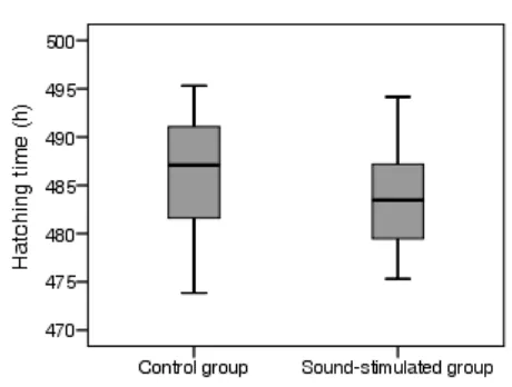
The individual hatching time of 20 focal chicks of each group is shown in Figure 1. The
sound-stimulated focal chicks started hatching later than the control focal chicks. However,
the average hatching time of the sound-stimulated focal chicks (483.9 ± 1.3 h) was not
Figure 1. Boxplot of hatching time of focal chicks in the control group and the sound
stimulated group (n=20 chicks of each group).
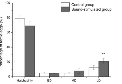
The mean values of hatchability, early death, middle death and late death of the control group
and the sound-stimulated groups of four repeats are shown in Figure 2. No significant
differences in hatchability were detected between groups and incubators. However,
significantly higher late death was found in the sound-stimulated group than the control
group (P < 0.01). No differences in chick quality and chick weight were found between the
Figure 2. Mean of hatchability and mortality in the control group and the sound-stimulated
group over 4 experiments (n = 1200 eggs at setting of each group, fertility was about 85%–
95%). ED: early death, d 0–7; MD: middle death, d 8–15; LD: late death, d 16–21; **values
were significantly different between the control and sound-stimulated groups at P < 0.01.
Effect on embryonic development
The average egg weight at setting in the control group and the sound-stimulated group was
59.7 ± 0.5 g and 59.3 ± 0.5 g, respectively. Because results of the relative body and organ
weights were very similar to those of absolute body and organ weights, only the findings of
absolute organ weights were compared between different treatments. Except for incubation
stage (P < 0.01), sound treatment and incubator had no significant effect on body weight and
organ weights. In both groups, embryonic body weight increased steadily up to d 19 before
Effects on blood values and corticosterone levels
No significant differences in blood values were found between the different sound treatments
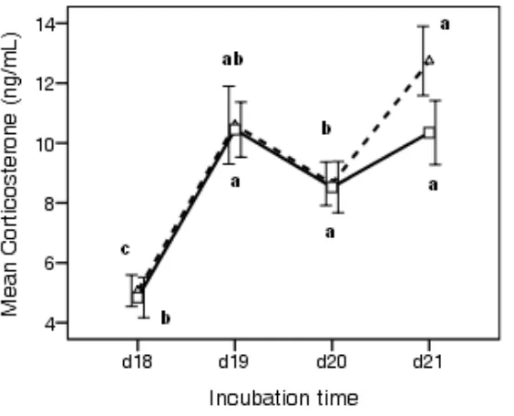
and incubators (data not shown). Figure 3 shows the general profile of the plasma CORT
levels during hatch. There were no significant differences in CORT between control and
sound-stimulated groups at the specific time points measured (d 18, d 19, d 20 and d 21).
However, within the control group and within the sound group, there were significant
differences between the individual time points, and the pattern of these differences was not
identical between the groups. In both groups, plasma CORT levels increased significantly (P
< 0.01) from d 18 (5 ng/ml) and reached a peak at IP (10.5 ng/ml) before dropping to 8.6
ng/ml when chicks emerged from the eggshell at d 20. However, CORT showed slight
differences in newly hatched chicks on d 21, being 12.7 ng/ml in control chicks and 10.3
ng/ml in the sound-stimulated chicks; however, this was not significantly different.
Figure 3. Effect of prenatal auditory stimulation on plasma corticosterone (CORT) levels
during hatch. Each value represents the mean ± SEM (n = 11 to 15). Mean values sharing a
Discussion
Balaban et al. (2012) found that chick embryos show selective sensitivity to maternal
vocalisations before the forebrain becomes active and maternal vocalisation causes the entire
brain to become active as an integrated system earlier than expected.
There has been little documentation on how incubating hen sound can affect the hatching of
domestic chickens. Greenlees (1993) reported that acoustic enrichment during incubation
may be responsible for the initial delay in hatch, but the functional incubating hen calls were
unclear. The delayed pipping observed in the sound-stimulated embryos in this study could
be due to the exposure of maternal calls before pipping. Hen vocalisations may serve as an
evolutionary parenting mechanism to prevent some eggs from needing additional incubation
time while hatched chicks were ready to explore. In this study, the sound was switched to
embryo/chick vocalisation calls around 469.5 h of incubation. The intent was to synchronise
the hatching process when the whole group started pipping. From this stage, embryos were
expected to be well developed and able to adjust pipping behaviour due to vocalisation cues.
Whereas a shorter HW was achieved via manipulation of real vocalisation, there was no
synchronising effect of the embryonic/chick calls. The individual hatching time of focal
chicks was not accelerated in the present study, although there are some other embryo sound
communications that have been reported to affect hatching synchronisation. Veterany et al.
(2005) demonstrated that the onset and time intervals of synthetic pipping sounds have
different influences on chicken hatching but without affecting hatchability. It was concluded
that the stimulation by a synthetic sound beginning after 433 h of incubation or with a time
interval of 176 min resulted in an earlier pipping and shorter hatch window. Furthermore, to
achieve acceleration of hatch, the rate and amplitude of artificial click stimulation are also
pointed out that the synchronisation effect due to chick clicking sounds depends on physical
contact between siblings in the egg.
A dramatic increase in the plasma CORT concentrations from day 18 to IP was observed in
both control group and sound-stimulation group. This confirms the findings of Kalliecharan
et al. (1974, 1976) and Scott et al. (1981) that CORT concentrations reach a peak at IP. The
CORT profile during hatching did not show any significant difference between treatments,
indicating that this sound exposure did not influence plasma corticosterone levels. After
hatching, CORT levels increased in both control and sound-stimulated groups. In the control
group, the CORT levels of chicks at take-off increased significantly from the external pipping
embryos. Furthermore, the plasma CORT levels in control chicks was also higher than
sound-stimulated chicks but were not statistically significant which was likely due to limited sample
size. The higher CORT levels of control chicks might be due to the early hatch and longer
holding period in the incubator.
In this study, prenatal auditory stimulation did not physically improve chick quality and
embryonic growth in terms of body weight and organ weight during incubation. However,
significantly higher late death was observed in the sound-stimulated embryos.
The causes of increased mortality in sound-stimulated group were unclear. An increased
mortality occurred in duck eggs which were incubated under the artificial sound stimulation
(Veterany et al., 1999). Stress, caused by prenatal sound, could be a factor influencing
mortality. However, there is no evidence that stress was related to this increased rate of
embryo death. The timing of presentation of prenatal auditory stimulation and type of sound
are important. If the sounds were not in a proper sequence, rate, frequency and duration
compared to the natural timing and patterning, it might have a negative impact and cause
prenatal stress, which has the potential to impair embryo development and animal well-being.
what degree and via which mechanisms, prenatal auditory stimulation affects hatchability and
hatching behaviour.
Acknowledgements
We are grateful to Dr Yumei Chang for statistical advice and the support of Biological
Services Unit at the Royal Veterinary College.
References
ALLADI, P. A., WADHWA, S. & SINGH, N. (2002). Effect of prenatal auditory enrichment
on developmental expression of synaptophysin and syntaxin 1 in chick brainstem
auditory nuclei. Neuroscience,114: 577-90.
BALABAN, E., DESCO, M. & VAQUERO, J. J. (2012). Waking-like Brain Function in
Embryos. Current Biology,22: 852-861.
COLLIAS, N. E. (1987). The Vocal Repertoire of the Red Junglefowl - a Spectrographic
Classification and the Code of Communication. Condor,89: 510-524.
DECUYPERE, E., TONA, K., BRUGGEMAN, V. & BAMELIS, E. (2001). The day-old
chick: a crucial hinge between breeders and broilers. Worlds Poultry Science Journal,
57: 127-138.
FRIAUF, E. & LOHMANN, C. (1999). Development of auditory brainstem circuitry.
Activity-dependent and activity-independent processes. Cell Tissue Research,297:
187-95.
GONZALES, E., KONDO, N., SALDANHA, E. S., LODDY, M. M., CAREGHI, C. &
DECUYPERE, E. (2003). Performance and physiological parameters of broiler
GOTTLIEB, G. (1965). Prenatal auditory sensitivity in chickens and ducks. Science,147:
1596-1598.
GREENLEES, B. (1993). Effects of enriching the acoustic environment during incubation on
hatching and the post-hatch chick responses.Master of Science, The University of
Guelph.
JONES, T. A., JONES, S. M. & PAGGETT, K. C. (2006). Emergence of hearing in the
chicken embryo. Journal of Neurophysiology,96: 128-41.
KALLIECHARAN, R. & HALL, B.K. (1974) A developmental study of the levels of
progesterone, corticosterone, cortisol, and cortisone circulating in plasma of chick
embryos. General and Comparative Endocrinology, 24: 364–372.
KALLIECHARAN, R. & HALL, B.K. (1976) A developmental study of progesterone,
corticosterone, cortisol, and cortisone in the adrenal glands of the embryonic chick.
General and Comparative Endocrinology, 30: 404–409.
KONISHI, M. (1973). Development of Auditory Neuronal Responses in Avian Embryos.
Proceedings of the National Academy of Sciences of the United States of America,70:
1795-1798.
MANTEUFFEL, G., PUPPE, B. & SCHON, P. C. (2004). Vocalization of farm animals as a
measure of welfare. Applied Animal Behaviour Science,88: 163-182.
OCKLEFORD, E. M. & VINCE, M. A. (1985). Acceleration of Hatching in Fowl and Quail -
Relationship between Artificial and Natural Stimulus Amplitude. British Poultry
Science,26: 57-63.
REED, W. L. & CLARK, M. E. (2011). Beyond Maternal Effects in Birds: Responses of the
Embryo to the Environment. Integrative and Comparative Biology,51: 73-80.
RUBEL, E. W. & FRITZSCH, B. (2002). Auditory system development: primary auditory
RUMPF, M. & TZSCHENTKE, B. (2010) Perinatal acoustic communication in birds: why
do birds vocalize in the egg? The Open Ornithology Journal, 3: 141–149.
SCHWAGMEYER, P. L., MOCK, D. W., LAMEY, T. C., LAMEY, C. S. & BEECHER, M.
D. (1991). Effects of Sibling Contact on Hatch Timing in an Asynchronously
Hatching Bird. Animal Behaviour,41: 887-894.
SCOTT, T.R., JOHNSON, W.A., SATTERLEE, D.G. & GILDERSLEEVE, R.P. (1981)
Circulating levels of corticosterone in the serum of developing chick embryos and
newly hatched chicks. Poultry Science, 60: 1314–1320.
TONA, K., BAMELIS, F., DE KETELAERE, B., BRUGGEMAN, V., MORAES, V. M.,
BUYSE, J., ONAGBESAN, O. & DECUYPERE, E. (2003). Effects of egg storage
time on spread of hatch, chick quality, and chick juvenile growth. Poult Sci,82:
736-41.
TONA, K., ONAGBESAN, O., BRUGGEMAN, V., DE SMIT, L., FIGUEIREDO, D. &
DECUYPERE, E. (2007). Non-ventilation during early incubation in combination
with dexamethasone administration during late incubation: 1. Effects on physiological
hormone levels, incubation duration and hatching events. Domestic Animal
Endocrinology,33: 32-46.
TONG, Q., ROMANINI, C.E., EXADAKTYLOS, V., BAHR, C., BERCKMANS, D.,
BERGOUG, H., ETERRADOSSI, N., ROULSTON, N., VERHELST, R.,
MCGONNELL, I.M. & DEMMERS, T. (2013) Embryonic development and the
physiological factors that coordinate hatching in domestic chickens. Poultry Science,
92: 620–628.
TUCULESCU, R. A. & GRISWOLD, J. G. (1983). Prehatching Interactions in Domestic
VERGNE, A. L. & MATHEVON, N. (2008). Crocodile egg sounds signal hatching time.
Current Biology,18: R513-R514.
VETERÁNY, L., HLUCHÝ, S., JEDLIČKA, J. & ČERVEŇANOVÁ, E. (2005). Effect of
the use of synthetic sound during incubation in chicken. Journal of Agricultural
Sciences,50: 131.
VETERANY, L., HLUCHY, S. & WEIS, J. (1999). The influence of sound stimulation
during hatching on the mortality of ducks. Acta Physiologica Hungarica,86: 105-10.
VINCE, M., READER, M. & TOLHURST, B. (1976). Effects of Stimulation on Embryonic
Activity in Chick. Journal of Comparative and Physiological Psychology,90:
221-230.
VINCE, M. A. (1966). Artificial acceleration of hatching in quail embryos. Animal
Behaviour,14: 389-94.
WHITE, N. R. (1984). Effects of Embryonic Auditory-Stimulation on Hatch Time in the
Domestic Chick. Bird Behaviour,5: 122-126.
WILLEMSEN, H., DEBONNE, M., SWENNEN, Q., EVERAERT, N., CAREGHI, C.,
HAN, H., BRUGGEMAN, V., TONA, K. & DECUYPERE, E. (2010). Delay in feed
access and spread of hatch: importance of early nutrition. Worlds Poultry Science
Journal,66: 177-188.
WOOD-GUSH, D. G. M. 1971. The behaviour of the domestic fowl, London, Heinemann