ABSTRACT
Comparative Study of
Management of Proximal
Humerus Fractures in Elderly
by Conservative Method
Versus Operative Locking
Compression Plate
Copyright©2015
Background Proximal humeral fracture in patients more than 65 years old, represent the third most common fracture. Treatment of proximal humerus fractures, especially displaced fractures, remains controversial. Conservative treatment has been preferred for most of the undisplaced or minimally displaced fractures. Over the years, availabil-ity of improved fixation devices, popularised the treatment of these fractures by open reduction and internal fixation. Operative treatment of proximal humerus fractures poses a challenge because of complications like malunion, non-union and avascular necrosis. Objective To study the role of conservative treatment and operative treatment by locking compression plate in the management of these fractures. To compare the results of conservative management versus locking plate osteosynthesis. To evaluate the results of treatment in terms of clinical and radiological union as well as functional outcome. Materials and Methods In the present case study, we report our experience in 60 cases in whom comparative study of management of proximal humerus fractures in elderly by conservative method versus operative locking compression plate was done.
Results As measured by Neer’s shoulder score, out of the 60 cases in our study, 8 (13.33%) had excellent functional outcome out of which 3 were treated conservatively and 5 were treated operatively, 29 (48.33%) had satisfactory outcome out of which 14 were treated conservatively and 15 were treated operatively, 19 (31.67%) had unsatisfactory outcome out of which 10 were treated conservatively and 9 were treated operatively, and 4 (6.67%) cases were failures out of which 3 were treated conservatively and 1 were treated operatively.
KEYWORDS proximal humerus fracture, old age, locking compression plate, conserva-tive treatment
INTRODUCTION
A proximal humeral fracture is the fracture of the ball portion, lying at the upper end of the humerus or arm bone. The incidence of proximal humeral fracture is 4–5% of all fractures1. In patients more than 65 years old, they represent the third most common fracture, after hip and distal radius fractures2. They account for 30–40% of all humeral fractures in all age groups and 76% of all the humeral fractures among people 40 years of age or older3.Fractures in adolescents and younger adults usually occur due to high-energy injuries, mainly vehicular accidents, sports injuries, fall from height, or gunshot wounds. However fractures in the elderly are more common, which are usually low-energy osteoporotic injuries. More than three quarters are a result of low-energy domestic falls4–6. Treatment of proximal humerus fractures has been greatly debated. This is because of the complexity of the fracture displacements and soft tissue injury. Prognosis depends on degree of fracture displacement and damage to delicate blood supply of head of humerus7. Conservative treatment has been preferred for most of the undisplaced or minimally displaced fractures as the healing time is short, infection is uncommon and prognosis is good8–10.The management of comminuted displaced fractures, remains controversial9,11. The availability
Rahul Ravindra Bagul1*, Utkarsha Joshi2,
Vikram Kakatkar3, Sanjay Deo4
1 Associate Professor, Department of Orthopaedics, Padmashree Dr. D.Y. Patil Medical College, Hospital and Research Centre, Dr. D.Y. Patil Vidyapeeth, Pimpri, Pune, India
2 Assistant Professor, Department of Orthopaedics, Padmashree Dr. D.Y. Patil Medical College, Hospital and Research Centre, Dr. D.Y. Patil Vidyapeeth, Pimpri, Pune, India
3 Senior Resident, Department of Orthopaedics, Padmashree Dr. D.Y. Patil Medical College, Hospital and Research Centre, Dr. D.Y. Patil Vidyapeeth, Pimpri, Pune, India
4 Professor, Department of Orthopaedics, Padmashree Dr. D.Y. Patil Medical College, Hospital and Research Centre, Dr. D.Y. Patil Vidyapeeth, Pimpri, Pune, India
Address reprint requests to
*Dr. Rahul R. Bagul, Department of Ortho-paedics, Padmashree Dr. D.Y. Patil Medical College, Hospital and Research Centre, Dr. D.Y. Patil Vidyapeeth, Pimpri, Pune, India E-mail: [email protected]
Article citation: Rahul B, Utkarsha J,
Vikram K, Sanjay D. Comparative study of management of proximal humerus fractures in elderly by conservative method versus operative locking compression plate. J Pharm Biomed Sci 2015;05(11):831–838.
Available at www.jpbms.info Statement of originality of work: The manuscript has been read and approved by all the authors, the requirements for authorship have been met, and that each author believes that the manuscript represents honest and original work. Source of funding: None.
Competing interest / Conflict of interest:
The author(s) have no competing interests for financial support, publication of this research, patents, and royalties through this collaborative research. All authors were equally involved in discussed research work. There is no financial conflict with the subject matter discussed in the manuscript.
Disclaimer: Any views expressed in this paper are those of the authors and do not reflect the official policy or position of the Department of Defense.
NLM Title J Pharm Biomed Sci CODEN JPBSCT
imal humerus fractures.
MATERIALS AND METHODS
In the present case study we are report our experience in 60 cases in whom comparative study of management of proximal humerus fractures in elderly by conservative method versus operative locking compression plate was done. The duration of this study was from April 2012 to April 2015.
Inclusion criteria:
· Patients of both sexes with age 50 years and above
· Neer’s three part and four part fractures
· Fracture dislocations Exclusion criteria:
· Patients with age below 50 years
· Compound fractures
· Fractures with neurovascular injury
PRE-OPERATIVE
A total of 60 cases of proximal humerus fractures were studied. On admission, a detailed history including the complaints of the patient and the mechanism of injury were noted. Clinical examination involved assessing pain, swelling, tenderness, crepitus and ecchymosis around the shoulder region. A detailed neurovascular examination of the brachial plexus and axillary artery was carried out. Associated injuries to the chest, abdo-men and other limbs were also noted. Radiographs of affected shoulder were taken in antero-posterior and axillary views. For primary treatment, immobilization was given in the form of simple cuff and collar sling. All routine investigations were done prior to anaesthesia fit-ness. Pre-operative anaesthesia fitness was done. Patients to be managed operatively were posted for planned operative procedure.
MANAGEMENT
Conservative management
Patients with minimally displaced fractures were given a shoulder arm pouch with immobiliser (Figs. 1, 2). Patients with fracture-dislocations were subjected to
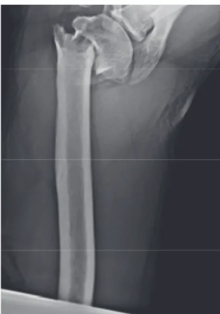
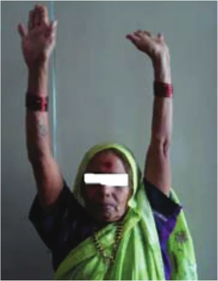
closed reduction under general anaesthesia. Fracture dislocation was reduced under image intensifier con-trol. Immediate immobilization was done using a shoulder arm pouch with immobilizer. Oral analgesics and calcium supplements were given. Physiotherapy in the form of gentle passive range of motion and pendulum exercises was started after 3–4 weeks, once pain was reduced and the patient co-operated. At 6 weeks and 3 and 6 months X-ray shoulder antero- posterior and axillary views were taken (Fig. 3). On clinical and radiological assessment both active and passive shoulder range of motion exercises were started (Figs. 4–6).
Fig. 1 X-ray shoulder joint, A-P view.
Operative management
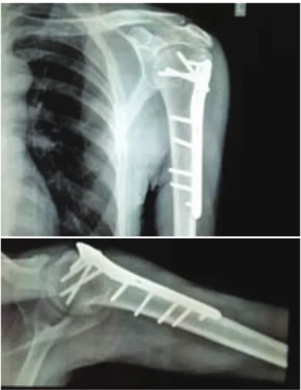
Following pre-operative X-ray shoulder antero-posterior and axillary views (Fig. 7) and baseline investigations, patients were posted for open reduction and internal fixation with locking compression plate.
Surgical technique
A beach-chair position was given to the patient, follow-ing general anaesthesia. The C-arm was positioned prop-erly to view the proximal humerus. Scrubbing, painting and draping of the affected upper limb were done under aseptic precautions. Deltoid-splitting approach was used to expose the proximal humerus. Care was taken not to injure the axillary nerve. When the frac-ture was exposed, the humeral head if dislocated, was relocated first into the glenoid under C-arm guidance. Next, the head was reduced onto the shaft with the help of K-wires and then using them as joysticks reduction was achieved in coronal, sagittal and horizontal planes. Remaining fragments were then reduced with the help of traction sutures placed in the rotator cuff insertions. Once an acceptable reduction was achieved, the lock-ing compression plate was applied at least 1 cm distal to upper end of the greater tubercle and fixed to the
humeral shaft with cortical screws. An aiming device which diverged the screws in the head was temporarily attached to the upper part of the plate. Locking screws were then inserted in the humeral head. After obtaining a stable plate fixation, the K-wires and traction sutures were removed. Suturing was done in layers, followed by sterile dressing.
Post-operative management
Limb was immobilised in a shoulder arm pouch. Immediate post-operative X-rays of shoulder in antero- posterior and axillary views were taken to assess reduction of fracture and stability of fixation (Fig. 8).
Fig. 3 X-ray shoulder joint, A-P view at 6 months.
Fig. 4 Internal rotation.
Fig. 5 Abduction.
Intravenous antibiotics were given for the first 3 days and then shifted to oral antibiotics. Anti-inflammatory, analgesics drugs were also given. Post-operative dress-ings of the surgical wound were done on 2ndand 8th day. Sutures were removed on 12th post-operative day. Mobilisation of affected shoulder was started on the 3rd day with pendulum exercises as per the patient’s toler-ance. At 6 weeks and 3 and 6 months X-ray of the shoul-der in antero-posterior and axillary views were taken (Fig. 9). On clinical and radiological assessment both active and passive shoulder range of motion exercises were started (Figs. 10–12).
Results were then evaluated by the use of Neer’s shoulder score for each case recorded.
OBSERVATION AND RESULTS
Our study included 60 cases of proximal humeral fractures which were managed in our hospital. Thirty patients were treated conservatively and 30 with locking compression plate.
1. Age distribution
The mean age of the patients was 67.15 years. The youngest patient was 51 years old and the oldest patient was 91 years old.
2. Sex distribution
In our study, out of the 60 patients, 38 were male and 22 were female. Male to female ratio was 19:11.
3. Side distribution
In our study, we had 26 patients with right sided proximal humerus fractures and 34 patients with left sided fractures.
4. Distribution according to classification
According to Neer’s classification, 33 patients belonged to the 3 part fracture group out of which 13 were treated conservatively and 20 were treated operatively, 21 patients belonged to the 4 part fracture group out of
Fig. 7 X-ray shoulder joint, A-P and axial views. Fig. 8 X-ray post-operative shoulder joint, A-P and axial views.
Fig. 9 X-ray post-operative shoulder joint, A-P and axial views at
7. Trauma-treatment interval
In our study, the mean duration of trauma-treatment interval was 4. 48 days, with minimum duration of 1 day and maximum duration of 11 days.
8. Follow up period
The mean duration of follow up in our study was 24.27 months with minimum follow up of 24 months and maximum of 27 months.
9. Clinical union
The mean time duration for clinical union in our study was 12.94 weeks, with minimum of 10 weeks and max-imum of 18 weeks.
10. Radiological union
The mean time duration for radiological union in our study was 19.52 weeks, with minimum of 17 weeks and maximum of 23 weeks.
11. Complications
During the follow up period, 6 patients treated conser-vatively and 3 patients treated operatively had shoulder stiffness, 3 patients treated operatively had post-operative infection (5%) and 1 patient treated operatively had implant loosening. Two patients treated conservatively and 2 patients treated operatively had malunion. There were no incidences of non-union or osteonecrosis in our study.
12. Range of motion
At the end of functional recovery, all the patients were assessed for range of motion. They had restriction of abduction, forward flexion, extension and rotation. The mean flexion obtained at the end of our study was 132.63° in the conservative group and 140.3° in the oper-ative group. The mean abduction was 140.53° and 145.4° in the conservative and operative groups, respectively. The mean external rotation was 14.5 and 16.56 in the con-servative and operative groups, respectively (Figs. 7–12).
13. Evaluation of results by Neer’s shoulder score At the end of clinical and radiological union and func-tional recovery, the results were evaluated by Neer’s shoulder score. The mean scores observed were 33.42 units for pain, 23.4 units for function, 16.05 units for
Fig. 10 Internal rotation.
Fig. 11 External rotation.
Fig. 12 Abduction.
which 14 were treated conservatively and 7 were treated operatively and 6 patients belonged to the fracture dislo-cation group out of which 3 were treated conservatively and 3 were treated operatively.
5. Mode of injury
In our study, 31 patients had domestic fall, 26 patients had road traffic accident and 3 patients had assault. 6. Associated injuries
score was 80.94 units (Table 1).
14. Functional outcome
Out of the 60 cases in our study, 8 (13.33%) had excellent functional outcome out of which 3 were treated conser-vatively and 5 were treated operatively, 29 (48.33%) had satisfactory outcome out of which 14 were treated con-servatively and 15 were treated operatively, 19 (31.67%) had unsatisfactory outcome out of which 10 were treated conservatively and 9 were treated operatively and 4 (6.67%) cases were failures out of which 3 were treated conservatively and 1 was treated operatively.
DISCUSSION
Fractures of the proximal humerus are one of the most commonly occurring fractures. Incidence of these frac-tures is 73 per one lakh population, with 75% found in the elderly. About 80–85% of these fractures can be treated conservatively, while the remaining 15–20% which are significantly displaced, require some form of internal fixation15. They have been described two centu-ries back, even before the invention of radiology and have shown various trends in their management. The incidence of proximal humeral fractures has increased in the last few years due to changes in lifestyle and increase in the number of road traffic accidents9,16. The best management for these injuries is still uncertain.Earlier, these fractures were managed by plaster cast techniques, slings and slabs17. But, recent advances in understanding of the anat-omy, development of good surgical skills and availability of better instrumentation have led to various modalities for their treatment18,19.Due to awareness of their com-plexity and complications, these fractures have stimulated a growing interest in finding the optimal treatment. With the aim of getting rapid healing and early restoration of function, open reduction and internal fixation is the preferred modality of treatment8,20.Studies have shown that both non-operative and operative treatments give favourable results. Hence there is uncertainty regarding treatment9,21.An anatomical reduction and good rehabil-itation are predictors of a good functional outcome. In our institution, we have studied 60 patients of proximal humeral fractures, with 30 patients treated conservatively and 30 patients treated with locking compression plate and assessed the outcome using Neer’s shoulder scoring
system. The results of our study were compared with sim-ilar studies performed by other authors.
1. Number of patients
The number of patients is comparable to studies con-ducted by Olerud et al.22,24, Fjalestad et al.23, Boons et al.25, Stableforth et al.26 and Zyto et al.27 (Table 2).
2. Age incidence
In our study, the average age was 67.15 years, with the mean age for conservative group being 68.77 years and that of operative group being 65.53 years. These find-ings are comparable with other similar studies by differ-ent authors as given in Table 3.
3. Sex incidence
The study of literature has revealed predominance of prox-imal humeral fractures in females in an elderly age group. This has been evident in other published studies. In our study, the male to female ratio was 19:11 with 63.4% males as compared to 36.6% females. The higher inci-dence of males in our study can be explained by the higher involvement of males in day-to-day activities and also in road traffic accidents in comparison with females (Table 4).
4. Side involved
Our study included 26 (43.4%) right sided and 34 (56.6%) left sided proximal humeral fractures. Our findings are comparable with the findings of Zyto et al. (Table 5).
Present study 30 vs. 30
Table 3 Age incidence.
Study (conservative vs. operative)Mean age
5. Follow up
In our study, the mean follow up duration was 12.27 months, which is consistent with follow up durations of other similar studies (Table 6).
6. Range of motion
The mean flexion obtained at the end of our study was 132.63°in the conservative group and 140.3° in the oper-ative group. The mean abduction was 140.53° and 145.4° in the conservative and operative groups, respectively. The mean external rotation was 14.5 and 16.56 in the con-servative and operative groups, respectively. These findings are consistent with the results of other studies (Table 7).
7. Complications
In our study of 60 cases, complications were found in 17 cases, of which 8 were present in the conservative group (26.67%) and 9 in the operative group (30%).
These findings are comparable with the complications in other similar studies (Table 8).
Shoulder stiffness was present in 9 cases and they were started with rigorous physiotherapy programme. Two of these cases did not comply with the same and progressed to arthritis and failure outcome.
Post-operative infection was present in 3 cases, all of whom presented early and had superficial infection which subsided with systemic antibiotics and regular sterile dressings.
Malunion was found in 4 cases in our study. But they were within the acceptable range with no significant limitations in the patients activities of daily living.
Implant loosening was present in 1 patient, which appeared at 4 months after the surgery. Patient was advised a revision surgery, but was unwilling for the same. It accounted for failure outcome.
8. Results
Out of the 60 cases in our study, 8 (13.33%) had excellent functional outcome out of which 3 were treated conser-vatively and 5 were treated operatively, 29 (48.33%) had satisfactory outcome out of which 14 were treated con-servatively and 15 were treated operatively, 19 (31.67%) had unsatisfactory outcome out of which 10 were treated conservatively and 9 were treated operatively and 4 (6.67%) cases were failures out of which 3 were treated conservatively and 1 were treated operatively.
The difference in the functional outcomes between two groups by the Chi square test was not statistically significant (P = 0.662).
Our results were similar to the study conducted by Olerud et al.22.In their study, the results obtained using constant score were 61 for operative group versus 58 for conservative group, with a P value of 0.64, and those obtained by DASH score were 26 versus 35 for operative and conservative groups, respectively, with a P value of 0.19, thus showing no statistically significant difference between the two groups.
In the study by Fjalestad et al.23 the mean constant score favoured conservative treatment by 2.4 point with a P value of 0.62, indicating no significant difference between the two groups.
Thus we conclude that both conservative and sur-gical treatment of fractures of the proximal humerus in elderly patients have equally good functional outcomes.
Table 6 Follow up duration.
Study Duration (months)
Olerud et al. 2011 24 Olerud et al. 2011 24 Fjalestad et al. 2012 12 Stableforth 1984 6–48
Present study 12.27
Table 8 Complications.
Study Complications (conservative vs. operative)
Olerud et al. 2011 16.66% vs. 76.67% Olerud et al. 2011 35.71% vs. 37.07% Boons et al. 2012 24% vs. 52% Zyto et al. 1997 25% vs. 40% Present study 26.67% vs. 30%
Table 7 Range of motion.
Study (C vs. O)Flexion Abduction (C vs. O) Ext Rotn (C vs. O)
Olerud et al. 2011 111 vs. 120 106 vs. 114 NA Olerud et al. 2011 95 vs. 93 87 vs. 86 NA Boons et al. 2012 94 vs. 98 87 vs. 77 19 vs. 17
Present study 132.63 vs. 140.3 140.53 vs. 145.4 14.5 vs. 16.56
C: conservative, O: operative.
Table 5 Side involved.
Study Right Left
Zyto et al. 1997 35% 65% Present study 43.4% 56.6%
Table 4 Sex incidence.
Study Male vs. Female
4. Court-Brown CM, Garg A, McQueen MM. The epidemiology of prox-imal humeral fractures. Acta Orthop Scand. 2001;72(4):365–71. 5. Lind T, Krøner K, Jensen J. The epidemiology of fractures of the
proximal humerus. Arch Orthop Trauma Surg. 1989;108(5):285–7. 6. Kristiansen B, Barfod G, Bredesen J, Erin-Madsen J, Grum
B, Horsnaes MW, et al. Epidemiology of proximal humeral fractures. Acta Orthop Scand. 1987;58(1):75–7.
7. Muller ME, Allgovere M, Schneidere R, Willenegger H. Manual of Internal Fixation: Techniques Recommended by AO/ASIF Group, 3rd ed. Berlin: Springer-Verlag; 2002. pp. 438–41. 8. Neer CS. Displaced proximal humeral fractures. II. Treatment of
three-part and four-part displacement. J Bone Joint Surg Am. 1970;52(6):1090–103.
9. Mills HJ, Horne G. Fractures of the proximal humerus in adults. J Trauma. 1985;25(8):801–5.
10. Jacob RP, Kristiansen T, Mayo K, Ganz R, Muller ME. Classification and aspects of treatment of fractures of the proximal humerus. In: Bateman JE, Welsh RP (eds): Surgery of the Shoulder. Philadelphia: BC Decker Inc.; 1984. pp. 330–43.
11. Cofield RH. Comminuted fractures of the proximal humerus. Clin Orthop Relat Res. 1988;230:49–57.
12. Szyszkowitz R, Seggl W, Schleifer P, Cundy PJ. Proximal humeral fractures: management techniques and expected results. Clin Orthop Relat Res. 1993;(292):13–25.
13. Knight RA, Mayne JA. Comminuted fractures and fracture-dislocations involving the articular surface of the humeral head. J Bone Joint Surg Am. 1957;39-A(6):1343–1355.
CM, Wakefield AE. Hemiarthroplasty for treatment of proximal humeral fractures. J Bone Joint Surg Am. 2003;85-A(7):1215–23. 20. Kristiansen B, Christensen SW. Plate fixation of proximal humeral
fractures. Acta Orthop Scand. 1986;57(4):320–23.
21. Horak J, Nilsson BE. Epidemiology of fracture of the upper end of the humerus. Clin Orthop Relat Res. 1975;(112):250–53. 22. Olerud P, Ahrengart L, Ponzer S, Saving J, Tidermark J. Internal
fixation versus nonoperative treatment of displaced 3-part proximal humeral fractures in elderly patients: a randomized controlled trial. J Shoulder Elbow Surg. 2011;20(5):747–55. 23. Fjalestad T, Hole MØ, Hovden IA, Blücher J, Strømsøe K. Surgical
treatment with an angular stable plate for complex displaced proximal humeral fractures in elderly patients: a randomized controlled trial. J Orthop Trauma. 2012;26(2):98–106.
24. Olerud P, Ahrengart L, Ponzer S, Saving J, Tidermark J. Hemiarthroplasty versus non-operative treatment of displaced 4-part proximal humeral fractures in elderly patients: a randomized controlled trial. J Shoulder Elbow Surg. 2011;20(7):1025–33. 25. Boons HW, Goosen JH, van Grinsven S, van Susante JL, van Loon
CJ. Hemiarthroplasty for humeral four-part fractures for patients 65 years and older: a randomized controlled trial. Clin Orthop Relat Res. 2012;470(12):3483–91.
26. Stableforth PG. Four-part fractures of the neck of the humerus. J Bone Joint Surg Br. 1984;66(1):104–8.