Blood on the Shroud of Turin:
An Immunological Review
©2012 Kelly P. KearseAll Rights Reserved
The Shroud of Turin is an approx-imately 14 feet by 3.5 feet linen cloth that bears the image of a man that has been beaten, scourged, and crucified. Here, the scientific evidence for the presence of human blood on the Shroud is evaluated, with particular emphasis on the methodology used to support various conclusions, including blood typing and species classification. The data are specifically considered within the context of immunology as many of the experimental techniques used to study the bloodstains on the Shroud utilize products of the im-mune system as experimental tools in their design. Moreover, the bloodstains themselves represent cellular and fluid components of a once functioning im-mune system and provide clues to their origin.
Introduction
The Shroud of Turin is so named because it is proposed to be the burial cloth, or shroud, that wrapped the body of Jesus following crucifixion; and since 1578, the cloth has resided in the town of Turin, Italy (1). The history of the Shroud from the 14th century to the present has been well documented, including a fire in 1532 which resulted in numerous burn holes, water stains, and subsequent repair patches throughout the cloth (2). In 1898, interest and controversy about the Shroud increased when an amateur photographer, Secondo Pia, noted while developing black-and-whites images of the Shroud that the negatives revealed a strikingly lifelike portrait of a body, with remarkably detailed features, especially around the face (Figure 1).
In the late 1970s, a multidisciplinary team of over twenty scientists, the Shroud of Turin Research Project, or STURP, spent five days examining and documenting the Shroud in great detail, studies which remain among the most referenced. In 1981, in its final report, STURP wrote: "We can conclude for now that the Shroud image is that of a real human form of a scourged, crucified man. It is not the product of an artist. The bloodstains are composed of hemoglobin and also give a positive test for serum albumin. The image is an ongoing mystery and until further chemical studies are made, perhaps by this group of scientists, or perhaps by some scientists in the future, the problem remains unsolved."(3).
Here, the findings from the original scientific proceedings and articles on the blood studies are evaluated, with the intention of striking a balance between engaging readers with varying degrees of scientific background, and at the same time, providing sufficient detail to illustrate the nuances among different immunological techniques and principles. Specific attention is given to the blood typing data and evidence for the blood being of human origin.
Bloodstains on the Shroud
Numerous blood markings are present on the Shroud, which correspond to the wounds of a man that has been tortured and crucified. Bloodstains are evident about the head area, the wrists and the feet (Figure 2), the side, and the back (1,4). Each individual blood wound shows a distinct serum clot retraction ring; such blood halos are only visible
All ©1978 Barrie M. Schwortz Collection, STERA, Inc. Figure 1. Positive and negative images of the Shroud of Turin.
©1978 Barrie M. Schwortz Collection, STERA, Inc.
Figure 2. Bloodstains on the arms and wrist (left); and bottom of the feet (right). under ultraviolet light (5), a detail that a forger is unlikely to have been familiar with. Over ten different chemical tests have established that these markings are indeed bloodstains, and contain specific blood components, including hemoglobin, bilirubin, and albumin (6). These data have been extensively reviewed elsewhere (5, 7-9).
In one of the last interviews Heller and Adler gave on their Shroud blood studies, shortly before Heller’s death, Adler stated that “the most interesting thing is now there is immunological evidence that it is primate blood.” (10). While chemical studies may determine that blood is
indeed present, they cannot distinguish animal blood from human blood or discern between blood of different individuals. Immunological studies provide a level of specificity that chemical data cannot, which is the focus of this review. Those with a scientific back-ground, particularly in the field of immunobiology, may wish to go straight to the sections dealing with specific Shroud studies. For those with a lesser scientific background, numerous illus-trations are included to help clarify the methods described in the text.
The Immune System: Antigen and Antibodies
Molecules that elicit an immune response in the body are referred to as antigens (from the term “antibody generator”). In general, any molecule not expressed in an individual is interpreted as foreign or antigenic, and will stimulate a response (Figure 3, left). Hundreds of millions of different antibodies, or immunoglobulins (Ig), exist within in the immune system; such diversity allows reaction with an untold number of conceivable antigens (11,12). Antibodies may also be generated in animals (for example, mice or rabbits), for use as tools in specific experiments (Figure 3, right). The specificity of a particular antibody is indicated after the word anti-, for example antibodies specific for ragweed antigen would be written as anti-ragweed. The particular species the antibody was produced in is written before the word anti; for example, rabbit anti-ragweed indicates that the antibodies were generated in rabbits.
Structurally, antibody molecules consist of two chains: a smaller chain (the light chain) and a larger one (the heavy chain), (Figure 4 top). Together, the heavy and light chains create an antigen-binding site, or pocket (Figure 4 top).
Figure 3. Stimulation of antibody (im-munoglobulin) production. Hundreds of millions of different antibodies exist in the immune system, each specific for a particular antigen. When antigen enters the body, production of those specific antibodies is greatly increased relative to others that are present. See text for details.
The basic antibody structure contains two identical antigen-binding pockets per molecule, which are specific for a particular antigen (Figure 4, bottom). Five major classes of antibody are produced in the body, only two of which will be highlighted here: Immunoglobulin M (IgM) and Immunoglobulin G (IgG). IgM molecules are produced during an initial immune response and have a pentameric structure (five identical antibody molecules joined together), creating a total of 10 identical antigen-binding sites, (Figure 5), (12). In contrast, IgG molecules are produced during a secondary immune response (upon fur-ther stimulation by antigen), and are dimeric (Figure 5), containing two an-tigen-binding sites, similar to the basic antibody molecule depicted in Figure 4. The formation of immune antibody:anti-gen complexes is the basis for many immunological tests. When intact, antigen-bearing cells are used, antibody is added to establish multiple crosslinks between neighboring cells, resulting in agglutination (clumping).
Figure 4. Basic antibody (immunoglobulin) structure and specificity. Top-Heavy and light chains fit together to create an antigen-binding site (pocket). Bottom-Antigen:antibody binding is specific. Similar to a lock and key interaction, antigen and antibody only react when the binding pocket fits the particular antigen in question.
IgM is ideal for this because of its many antigen-binding sites. Agglutination is useful in the immunological test for blood typing (see below). For antigens not attached to intact cells, immune complexes may be detected microscopically by labeling the antibody with a tag that either fluoresces or changes color.
Immunological tests confirm the presence of (human) blood components on the Shroud
Serum albumin and immunoglobulin (Ig) are two major circulatory proteins that are commonly evaluated to determine the presence of (human) blood in forensic and
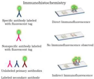
Figure 5. Structure of IgM and IgG classes of antibody. IgM contains ten identical antigen-binding sites; IgG contains two. archaeological samples. In the early 1980s, Heller, Adler, and colleagues used labeled antibody reactive with human albumin to immunologically probe for blood components on the Shroud (5,13). For these studies, anti-albumin antibodies were labeled with a fluorescent tag and added to slides containing sticky tape samples taken from both bloodstained and unstained (control) areas of the Shroud and examined microscopically (Figure 6, top). Any observed fluorescence should result from the labeled antibodies (rabbit anti-human albumin) binding to antigen (albumin) present in the sample (Shroud fibers). Samples taken from bloodstained areas were positive for fluorescence, whereas those obtained from unstained areas were not, suggesting that human blood products were indeed present in marked areas of the cloth. To confirm antigen specificity, the ability of antibody to react with albumin of various species was examined. No reactivity was observed between (anti- human) albumin antibody and albumin synthesized in cow, pig, or horse (10,13). In contrast, chimp antigen (albumin) reacted almost as strongly with labeled antibody as human albumin did, which is not entirely unexpected given that human and chimpanzee albumin differ by only six out of 580 amino acids(14,15).
Figure 6. Immunochemistry techniques. In the direct method (top), antibody containing a fluorescent tag is incubated with antigen immobilized on slides. Nonspecific antibodies serve as a negative control (middle). In the indirect method (bottom), unlabeled antibody is reacted with antigen immobilized on slides, followed by labeled secondary antibody that is specific for the primary antibody. See text for details.
Baima Bollone and colleagues evalu-ated the presence of human Ig on the Shroud using fluorescence-labeled an-tibody techniques, similar to those described above for albumin studies (16-18). Here, samples of linen fibers were obtained by mechanically separating out warp and weft threads from the Shroud. Because differences exist between Ig synthesized in distinct species, when antibody of one species (human), is injected into a different species (for example, rabbit or horse), the host animal will make antibodies directed against the foreign protein (12). Thus, in these studies the injected human antibody serves as an antigen (antibody generator). For these studies, both direct and indirect immunohistochemistry staining methods were used to probe for the presence of human Ig on the Shroud. In the indirect method, unlabeled antibody is first added to the antigen; any unbound antibody is removed by
washing; then a second labeled antibody specific for the first (unlabeled) antibody is then added, forming a type of sandwich (Figure 6, bottom). Taken together, these results showed that fibers from bloodstained areas were positive for staining with anti-human Ig, whereas those from unstained areas were not. Similar findings were also reported by Heller and Adler using direct staining methods (5,10,13). Baima Bollone et al. confirmed and extended the findings that total human Ig was present by also showing that bloodstained areas con-tained human antibody of the IgG class (16-18).
Experimental conclusions and caveats of Shroud blood component studies The above studies demonstrate that bloodstained fibers of the Shroud contain (human) albumin and Ig(G), consistent with the presence of real blood. Although the anti-albumin antibody used was generated against human albumin, cross-reactivity was observed with certain primate species. Regarding the findings that anti-albumin antibody reacted with samples taken from bloodstained areas but not unstained areas, one could argue that fibers from bloodstained areas are nonspecifically adherent for labeled antibody, and might bind other antibodies, regardless of their antigenic specificity. When immunohistochemistry methods are used, it is important to demonstrate that binding is specific (see Figure 6, top and middle), that is, that antibodies generated against an irrelevant antigen do not react. (Please note that if such controls were included during the course of these experiments, it was not apparent when reading the original articles). This is important because intermolecular forces between antigen and antibody may be both specific and nonspecific in nature (12, 19); the degree of nonspecific/specific binding is dependent upon the particular reagents used and the conditions in which the
assays were run. Having said this, it should also be noted that the presence of albumin in bloodstained areas of the Shroud was independently demonstrated by these same investigators using a specific chemical test (Bromcresol Green), (6). Thus, it is reasonable to conclude that the blood component albumin is present in bloodstained areas on the Shroud.
Regarding the studies on (human) Ig by Baima Bollone and coworkers, similar results were obtained with both direct and indirect staining methods: bloodstained fibers were positive for Ig(G), unstained fibers were not. Importantly, these experiments clearly demonstrate that when irrelevant antibodies (with a different antigenic specificity) were used, bloodstained fibers did not react. The absence of binding by irrelevant antibody (Figure 6) was demonstrated for both direct and indirect staining techniques. Thus, it can be concluded that bloodstained fibers specifically reacted with the antisera used in these studies, showing that Ig is present, some fraction of which is IgG. Whether or not other classes of Ig (e.g. IgM) are present remains to be determined.
Although cross-reactivity of anti-human Ig antisera with Ig of other species was not directly examined, the same caveats mentioned in the albumin studies (human vs. nonhuman primates) most likely apply here. Exclusive reactivity with human Ig would have to be verified. Indeed, all four subclasses of IgG (IgG1, IgG2, IgG3, IgG4) present in human serum are also found in chimpanzee serum, as detected with antibodies specific for human IgG (20). Evidence for the human origin of blood on the Shroud is discussed in detail later.
ABO blood groups and blood type Having established that blood components were present on the Shroud, assignment to a specific ABO blood group was next undertaken. Blood typing
studies are perhaps some of the most often misunderstood (and controversial) that have been collected on the Shroud. The notion that “all old blood types as AB” or that “all old blood is degraded to type AB” is misleading and is an oversimplification of the data. Before reviewing specific experiments, a brief overview of blood groups and blood typing methods will be presented.
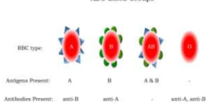
Of the more than twenty different blood antigen groupings that exist, the ABO system is the most important for transfusion and is therefore used as the major classification of an individual’s blood type (21-24). Four fundamental blood types exist: A, B, AB, and O. Persons with type A blood express A molecules, persons with type B blood express B molecules, persons with type AB blood express both A and B molecules, and persons with type O blood express neither A nor B molecules (Figure 7); the O classification originates from the German word “Ohne” which means without (23). In addition to ABO, the designation “positive” or “negative” is often given following a person’s blood group (for example, A positive or O negative). The “+” or “-” refers to expression of the Rh molecule, which is distinct and separate from ABO molecules. Individuals either express Rh molecules (Rh+) or they don’t (Rh-), (22,23).
ABO molecules are carbohydrates (also known as sugars or saccharides), all of which share the same precursor core structure, consisting of a branched carbohydrate chain (Figure 8). In A and B blood types, the core structure receives a further, specific modification that is unique to each blood type (Figure 8). The presence or absence of these specific, terminal carbohydrates (sugars) on the precursor core structure determines the particular blood type (21,25, 26). In red blood cells, ABO carbohydrates are attached mainly to proteins, and to a lesser extent, lipids (fats), both of which
Figure 7. ABO Blood Groups. Depicted are the types of antigens and antibodies present in individuals with A, B, AB, and O blood groups. are anchored in the cell membrane. A single red blood cell expresses almost two million ABO molecules on its surface (23). Although most commonly discussed in association with red blood cells, ABO molecules are actually present on many cell types throughout the body (25-27). Additionally, in many individuals ABO antigens are secreted in bodily fluids such as saliva, serum, sweat, and tears (22,23).
Because molecules not expressed in an individual are interpreted as foreign (antigenic), persons with certain blood types contain in their circulation specific antibodies that react with the carbohydrate antigens they themselves lack. That is, blood type A individuals have naturally occurring anti-B an-tibodies in their serum; type B individuals have anti-A antibodies; type AB individuals have no anti-A or B antibodies; and individuals of type O have both anti-A and anti-B antibodies present (Figure 7). Although not completely understood, it is hypothesized that naturally occurring anti-A and anti-B antibodies in humans originate during development from exposure to antigens present in the environment, for example influenza virus and gram negative bacteria (12, 25, 26, 28, 29). The vast majority of naturally occurring anti-A and anti-B antibodies are of the IgM class,
although anti-A and anti-B specific IgG are also found (25).
Figure 8. Molecular Basis for ABO blood groups. Modification of the core carbohydrate structure by additional sugars to create type A and type B blood molecules. In type O blood, the core structure receives no further modification.
Blood group typing methods
In blood typing, the presence of type A and B antigens on red blood cells is evaluated, called forward typing (Figure 9), as well as the existence of naturally occurring anti-A and anti-B antibodies in the blood (serum), known as reverse typing (Figure 9), (see also Figure 7). Together, forward and reverse typing provide an accurate indication of an individual’s blood type (30). Forward typing methods evaluate the ability of an individual’s red blood cells to react with experimentally prepared A and anti-B antibodies (22, 30, 31). Red blood cells from individuals expressing type A or type B antigens will agglutinate, or clump, in the presence of anti-A, or anti-B antibodies, respectively. Agglutination occurs because crosslinking is established between A/B antigens on one red blood cell to A/B antigens on another, bridged by antibody (Figure 10). The pentameric nature of IgM makes it ideal for use in such experiments; alter-natively, IgG may be used, in which case a secondary antibody is added (anti-IgG),
that ensures sufficient crosslinking (22). Reverse typing methods evaluate whether or not anti-A and anti-B antibodies are present in an individual’s serum by reaction with experimentally prepared red blood cells of known blood type (Figure 11). As discussed previously,
Figure 9. Forward and reverse blood typing methods. In forward typing, the presence (or absence) of RBC surface antigens is evaluated. In reverse typing, the presence (or absence) of serum antibodies directed against RBC surface antigens is evaluated. Expression of RBC surface antigens and serum antibodies is inversely correlated. See text for details.
a predictable complimentary relationship exists between expression of ABO and the presence of anti-A and anti-B antibodies (Figure 7). Because most anti-A, anti-B antibodies present in an individual’s serum are of the IgM class, efficient crosslinking is readily established between adjacent red blood cells (22). For fresh blood, typing tests are fast, inexpensive, and relatively foolproof. When aged blood is involved, however, modifications of the techniques must be employed since red blood cells become dehydrated and eventually rupture with time (32,33). Remarkably, red blood cells have been detected microscopically in ancient samples, including mummies (34-36), prehistoric rock tools (37), and also the Shroud (38, 39). What is most likely being visualized in such samples are aged red blood cell membranes that have
resealed with each other during the preparation procedure, representing reconstituted cells, not red blood cells that have survived intact over large periods of time. Because of their distinctive biconcave, disc-like shape, (resealed) red blood cells may be identified in a variety of ancient materials with relative confidence. Using light and electron microscopy, Hart et al. reported the discovery of human red blood cells in an Egyptian mummy that is over 3,000 years old (40). More recently, atomic force microscopy allowed individual red blood cells to be visualized with remarkable clarity in tissue sections taken from the 5300 year-old Iceman (41).
Figure 10. Forward typing of fresh red blood cells (RBCs). In this example, RBCs from type A (left) or type B (right) individuals are tested for reactivity with (experimentally added) anti-A antibody. RBCs expressing A antigens will readily clump (agglutinate) when anti-A antisera is added, but type B RBCs will not. Agglutination is easily evaluated visually without additional magnification. If similar experiments were performed using anti-B antisera, the results would be exactly opposite.
In general, five different serological techniques may be utilized to study aged samples, four forward typing methods (mixed agglutination test, absorption elution test, absorption inhibition test, and immunohistochemistry test) and the reverse typing method. With the
development of molecular biology techniques in the mid-1980s, DNA testing became feasible, which may be useful for certain aged samples, such as dried bloodstains or ancient mummies (32,42-44). One potential drawback of this technique is that it relies on relatively intact, non-fragmented stretches of DNA being present encoding the particular gene(s) of interest.
Blood typing studies on the Shroud: Forward typing
In a series of studies, Baima Bollone and colleagues evaluated the blood group present in fibers taken from various bloodstained areas, including the
Figure 11. Reverse typing of freshly drawn sera (blood) for the presence of naturally occurring anti-A and anti-B antibodies. In this example, serum from a type B individual is tested for reactivity with (experimentally added) type A RBCs (left) or type B RBCs (right). Agglutination occurs (left) due to the presence of anti-A Ig in serum. Such clumping is easily evaluated visually without the need for magnification. Individuals of a particular blood type express antibodies specific for the carbohydrate antigens they themselves lack.
“belt of blood” and the soles of the left and right feet (16,18,38,39,45), by the mixed agglutination technique. In this method, bloodstained fibers are mixed with experimentally prepared anti-A or anti-B antibodies, and after a period of incubation, unbound antibody is removed
by washing. Only antibodies specific for the particular type A or type B antigen remain bound. Next, experimentally prepared, fresh red blood cells of known blood type are added, which bind to any remaining, free antigen-binding sites on IgM molecules (Figure 12), forming a type of sandwich. When viewed through the microscope, positively stained fibers will appear “coated” with a layer of red blood cells (Figure 12). Unstained (control) fibers and bloodstained fibers were mechanically separated from the Shroud and evaluated together with fiber samples taken from a cloth experimentally daubed with human blood types A, B, AB, and O (to serve as known, positive controls). In addition, fibers from a fabric taken from an Egyptian funerary urn that were “unquestionably stained with traces of human blood” were examined (45). Bloodstained fibers tested positive for both type A and type B antigens. Type B showed slightly more reactivity than type A, “+++” vs. “++”, respectively, relative to the binding strength observed with experimental stains (positive controls), all with a value of “++++”. Both unstained fibers and fibers from the Egyptian urn were negative (45). Taken together, these data demonstrate that the bloodstained fibers from the Shroud contain blood group antigens A and B, assigning a type AB designation (16,18,39,45).
A few years later, these findings were extended with several distinctions in the experimental design: First, unlike previous investigations which utilized polyclonal antisera, this time monoclonal antibodies (produced in mice) were used (Figure 13). Polyclonal antisera consist of the products of multiple (poly) immune cells (clonal), all of which produce unique, yet specific, antibodies that recognize a particular antigen. In contrast, monoclonal antibodies represent the product of a single (mono) immune cell (clonal): all antibody molecules are exactly alike, identical to each other (Figure 13).
Figure 12. Forward typing of bloodstained fibers by the mixed agglutination test. In this example, bloodstained fibers containing type A antigens are evaluated; unstained fibers serve as a negative control. See text for details.
By analogy, polyclonal antibodies are equivalent to having 20 copies of the same letter, but each in a different font; monoclonals on the other hand, are equivalent to having 20 copies of the
same letter, but all in exactly the
Figure 13. Polyclonal and monoclonal antibodies. Polyclonal antibodies represent the products of multiple immune cells (clones), collected in the blood serum of immunized animals. In contrast, monoclonal antibodies are generated from individual cells (clones) se-lected in culture. Monoclonal antibodies are exactly identical, having been produced from a single, individual cell.
same font, all identical to each other (Figure 13). Monoclonal antibodies pro-vide an additional level of specificity not available with polyclonal sera (25,32). Secondly, in addition to using antibodies specific for A antigen and B antigen, in these studies an additional antibody was included, specific for the core structure,
type O antigen. Third, and most importantly, a different forward typing approach was utilized; here, immunohistochemistry techniques were used to evaluate the presence of ABO antigens on bloodstained Shroud fibers, similar to those used in previous studies on albumin and immunoglobulin (Figure 6). These data demonstrated that bloodstained fibers were positive for both A and B antigens, but not O antigens. Evaluation by electron microscopy showed equal intensities of anti-A and anti-B binding to bloodstained fibers (18,38). Unstained fibers failed to react with anti-A, anti-B, or anti-O specific antibodies. Most significantly, even colorless fibers taken from the bed of the stain were negative (18,38). Collectively, these findings are in agreement with the supposition that the blood on the Shroud is type AB. In 1998, Garza-Valdes performed similar studies and reported that bloodstained fibers were positive for B antigens but not O antigens (46); anti-reactivity was not evaluated.
Experimental conclusions and caveats of Shroud forward typing studies
One of the main objections raised in the serological typing of aged blood samples using forward typing methods is the issue of false positives. Indeed, blood type A and B carbohydrate antigens are expressed in many types of organisms (21,26,28, 29,47-49), a concern noted by the original investigators (45). While true that serological studies have been reported in which ancient materials type with a relatively high incidence as AB (50-54), this is certainly not always the case (55-61). As Sokal points out, certain reported errors in serotyping of ancient specimens result from the failure to determine any blood type, not an error in specific blood type determination (59). Thus, while tempting to dismiss serological typing methods of ancient materials in one broad stroke, the preservation of red blood cell antigens
and the extent of contamination in aged material must be evaluated specifically for each case study. The environmental conditions in which ancient materials exist prior to their discovery and analysis are relatively unique to each artifact (37, 62). Moreover, as the samples themselves often do not consist of the same material (rock, bone, tissue, ceramics, mummies, textiles), direct comparisons between systems are somewhat difficult. A further level of complexity arises when you consider that different studies often use different methods (mixed agglutination versus absorption inhibition versus reverse typing). Indeed, in studies by Crainic and colleagues on ABO typing of tissue antigens of Egyptian mummies, using 3 distinct serological methods, results were concordant for only half (7/14) of the samples tested (63). For the other half, results were inconsistent among the different methods and typing was inconclusive. Thus, even within the same study, variation may exist with specific techniques depending on the conditions of the individual artifact under study (63). Clearly, serological evidence is most useful when corroborated by additional experimentation, especially molecular (DNA) analysis of blood group genes (44,64), (see below).
Throughout its history, the Shroud had been handled by untold numbers of people and stored under less than pristine conditions. The extent of the bacterial and fungal biofilm present on the Shroud is unknown (46, 65, 66), but it is reasonable to assume that during the times the samples for previous studies were gathered, the surface was less than aseptic. False positives from antigens present on contaminating bacteria, fungi and insects is the main objection to the validity of blood typing studies done on the Shroud (10,45,49); a logical deduction given the wavering reliability of serological studies on ancient artifacts. An underlying assumption that must
accompany this criticism, however, is that contamination is stringently restricted to bloodstained areas of the cloth. Indeed, colorless fibers taken from the bed of the bloodstain gave no response with anti-A or anti-B antibody (18,38) indicating that any contaminating false positive antigens cannot be widespread. It could be countered that bacterial, fungal antigens would be expected to be concentrated in areas that are relatively enhanced with body fluids and cellular debris. Rationally, however, one would expect that such contamination would also extend to areas immediately adjacent to (e.g. at bed of) bloodstains and (at least slightly) beyond, making it somewhat difficult to reconcile these findings with the simple explanation that contamination is responsible for these results.
Blood typing studies on the Shroud: Reverse typing
Together with forward typing, Baima Bollone and colleagues also evaluated Shroud fibers by reverse typing techniques. For aged samples, this method relies on the assumptions that anti-blood group antibodies survive in dried bloodstains with age, and most importantly, that the three-dimensional conformation of the antigen-binding pocket is sufficiently intact to function correctly (see Figure 4). Because of these potential problems, reverse typing is typically the least utilized method for evaluation of aged blood samples. For these types of studies, bloodstained fibers are reacted with experimentally prepared, fresh red blood cells of known blood type. Coating of fibers with red blood cells is indicative of the presence of anti-blood group antibodies in the sample being studied (Figure 14),(see also Figure 11). When bloodstained Shroud fibers were evaluated by this method, no anti-A or anti-B antibodies were detected, consistent with forward typing results which showed an AB phenotype (see
Figure 7); however, note specific caveats as discussed below.
Experimental conclusions and caveats of Shroud reverse typing studies
The reverse typing studies on the Shroud are very difficult to interpret because they do not distinguish between the possibilities that (i) no A or anti-B antibodies were ever present in the bloodstains (truly type AB); (ii) anti-A or anti-B antibodies were once present in the bloodstains, but over time were denatured and degraded (type A, B, or O); or (iii) anti-A or anti-B antibodies are still present in the bloodstains, but are nonfunctional, the three-dimensional antigen-binding site no longer exists in a working state (type A, B, or O). While proper controls were included to demonstrate that the typing method was operational and specific, these can only be applied to fiber samples experimentally daubed with (fresh) human blood of known type. Species-specific IgG antibodies have been detected in equid and hominid fossils as old as 1.6 million years (67); and previous studies demonstrate the presence of IgG in the Shroud bloodstains (16,17). However, whether or not such antibodies survive in a functional state after an untold number of years remains unresolved. Even if demonstrated in other aged samples, it would still have to be addressed in these studies, there is no one-size-fits all for conclusions of this nature. Given that the expected result for AB blood type is that no naturally occurring anti-A or anti-B would be present (even in freshly obtained samples, see Figure 7), this issue becomes rather circular. Because most naturally present anti-A and anti-B antibodies are of the IgM class (22,25), it would be interesting to evaluate whether (any) IgM antibodies had survived over time and were present in bloodstained Shroud fibers; this is especially relevant as IgM is thought to be less stable than IgG (68). Unfortunately,
Figure 14. Reverse typing of bloodstained fibers. In this example, bloodstained fibers from an individual of B blood type are evaluated, which naturally contain anti-A antibodies; unstained fibers serve as a negative control. See text for details.
previous studies on bloodstained fibers using antibodies for total Ig (16,18) are not that helpful here, as approximately 80% of total serum immunoglobulin is IgG, only 5-10% consists of IgM (11,12). While the presence of IgM in bloodstains would by no means validate the reverse typing results, this would at least determine if some IgM existed. On the other hand, if no IgM was detected, this would cast further doubt on using reverse typing as a confirmatory technique.
Ideally, forward and reverse serological typing methods crosscheck and complement each other, but as is the case with certain aged samples, such as the Shroud, the validity is somewhat in question, particularly the reverse typing results. Thus, it is best concluded that the results suggest that the Shroud bloodstains are type AB as shown only by forward typing methods. Finally, although widely reported on the internet that the blood on the Shroud is AB positive or AB negative, there is no scientific basis for these claims. In previous tests, the condition of the blood was such that analysis of the Rh factor was not feasible (69). Therefore, the expression of Rh antigens on bloodstained fibers of the Shroud remains
to be determined. It is unknown if the blood type is AB positive or AB negative
Evaluation of the human origins of the blood on the Shroud
In their immunological studies on specific blood components (albumin, immunoglobulin) discussed earlier, Adler and colleagues were (appropriately) cautious in concluding that the data only demonstrated that the blood was of primate origin (5,9,10,13). Although other studies imply a human origin for the blood (16-18), cross-reactivity of antisera was not experimentally verified. This is an important point as specificity is the cornerstone of immunology. While many immune sera are advertised in company catalogues as anti-human, that designation only refers to the particular
Figure 15. Cross-reactivity of anti-human specific antibodies. Antibody raised against protein of one species may also react with the same protein from other, similar species. See text for details.
antigen to which the antisera were raised; cross-reactivity must be experimentally determined. Indeed, as shown by Adler & coworkers, antibodies generated against human albumin also reacted with albumin from other species (Figure 15), (see also earlier discussion).
It has been known for quite some time that blood groups of human and great apes are similar, although not completely
interchangeable. Among nonhuman primates, only anthropoid apes (gibbons, orangutans, gorillas, and chimpanzees) express ABO antigens on their red blood cells (70,71). Prosimians, Old World monkeys, and New World monkeys fail to express ABO antigens on their red blood cells, but secrete ABO molecules in saliva and serum (72-74). In a recent study conducted in 2010, monoclonal antibody technology was used together with DNA sequence analysis to assign ABO groups to great apes housed in North American and European zoos as a practical means to assist in blood transfusion situations among these species (75). These studies confirmed that among the anthropoid apes, only orangutans and gibbons express both A and B antigens, similar to humans. Chimpanzees do not express the B antigen and gorillas lack the A molecule (75). Thus, forward typing studies would effectively exclude all nonhuman primates as a possible source of red blood cell membrane AB antigens on the Shroud, except for possibly gibbons and orangutans.
In addition to ABO molecules, approximately thirty other blood group antigens are expressed on the surfaces of human red blood cells. One such group of blood molecules, the M,N, and S antigens, has been examined on blood-stained Shroud fibers using immuno-histochemistry methods. MN antigens are found on red blood cells of all anthropoid apes, but the S antigen is only expressed in humans (76). Bloodstained fibers were positive for expression of M, N, and most importantly, S antigens, ruling out a contribution of (nonhuman) primate blood (77). It is noteworthy to mention that staining of M antigen was “intesa positiva” (intensely positive), compared to “discreta positiva” (fairly good) for N and S antigens (77). The relative differences in antigen staining are somewhat difficult to interpret, however, as individual antibodies will often show differences in reactivity. For example, it
would not be that unusual for three different lots of anti-S antibodies to show some differences in antigen binding; this is particularly the case when polyclonal antisera is used. A more thorough understanding of how much emphasis to place on such differences would require comparing reactivity of anti-N or anti-S antibodies with samples containing a known level of antigen expression, in addition to evaluating several different anti-N or anti-S antibody preparations for reactivity. Given that unstained fibers showed no reactivity, it is valid to conclude that bloodstained fibers contain both N and S antigens. Moreover, since the S antigen has no counterpart in primates or other animals, these results support the conclusion that the blood on the Shroud is of human origin.
Because a fastidious forger might have included the detail of using real blood on the Shroud, a human blood classification does not rule out the possibility of such trickery. However, regarding the issue of blood being painted on the Shroud, Adler once commented, “The blood must have been taken from the exudate of a clot at a certain point in the clotting process. An artist would therefore have needed the exudate from the wounds of a severely tortured man, or baboon, and he would need to take the substance within a 20-minute period after the clotting had begun.” “One would need a constant supply of fresh clot exudates from a traumatically wounded human to paint all the forensically correct images in the proper nonstereo register and then finally paint a serum contraction ring about every wound. Logic suggests that this is something a forger or artesian before the present century would not only not know how to do but even know that it was required (9). A further level of complexity to be considered is that bloodstains are believed to have pre-existed prior to (body) image formation on the cloth, as shown by proteolytic enzyme digestion experiments (5,8).
Blood DNA studies on the Shroud
In the 1990s, Garza-Valdes and colleagues reported the presence of blood remnants on the Shroud that contained DNA segments of human hemoglobin (46). In contrast to hemoglobin proteins, hemoglobin DNA is present in virtually all cells throughout the body, except for, ironically, the vast majority of red blood cells (78,79). In mammals, mature red blood cells are enucleate, they lose their DNA as they develop and exit the bone marrow (12, 79). Thus, the source of hemoglobin DNA in human bloodstains is almost certainly not from red blood cells, but rather the white blood cells of the immune system (lymphocytes, neutro-phils, macrophages) (78,79). Hemoglobin DNA data can only go so far in its conclusion: that human DNA is present on the Shroud. Given the communal nature of the Shroud, displayed and handled over many years by untold numbers of people, it is uncertain that DNA analysis could distinguish endogenous versus contaminating DNA. The DNA on the Shroud is badly fragmented (46), although the extent to which specific chromosome regions survive remains to be determined. Expression of human ABO blood groups is controlled by a single locus in exons 6 and 7 (coding segments) of chromosome 9 (21). If molecular analysis of this region were feasible, such studies would help address previous concerns raised with serological techniques regarding the blood type.
Blood studies on the Sudarium of Oviedo
The Sudarium of Oviedo is a bloodstained fabric of much smaller dimensions than the Shroud, suggested to represent a companion (face) cloth, placed on the body shortly after death (80,81). Bloodstains on the Sudarium show geometric correspondence to those on the Shroud of Turin, consistent with the idea that they wrapped the same
person (80,81). Naturally, there was much interest in verifying the presence of human blood on the Sudarium, and specifically, in comparing the blood type to that found on the Shroud.
In 1985, Baima Bollone and colleagues performed similar experiments on samples taken from the Oviedo cloth, as those described above for the Shroud, concluding that blood group AB antigens were present (82). These findings were confirmed by Goldini et al., in 1993, who reported a series of standard chemical tests, similar to those used by Heller and Adler, to verify that blood is present, including the demonstration of he-matoporphyrin (83). Immunological tests demonstrated the presence of (human) IgG by immunofluorescence; horse anti-human Ig was used for these studies. The same caveats noted for similar studies on serum Ig present in Shroud fibers (human versus non-human primates) apply here.
For blood grouping studies, Goldini used reverse typing methods and a somewhat different forward typing method than described before, the absorption-inhibition test. This method is similar to the mixed agglutination test, except that instead of evaluating antibody binding to fibers, the relative amount of unbound antibody is measured by their ability to agglutinate experimentally added, freshly prepared red blood cells of known blood type (22,84). These studies suggested that type A and type B antigens are present in bloodstained fibers of the Sudarium; findings which were also reported by Villalain, Blanco and Moreno, although background staining with anti-B reagents in “clean” samples was noted (85, 86), possibly confounding the results. Thus, although not as definitely studied as the Shroud, these results suggest that the blood type may be similar. Like the Shroud, DNA has also been isolated from the Sudarium of Oviedo and is badly fragmented (81,82), possibly precluding any
meaningful molecular analysis of ABO gene expression.
Future directions and final conclusions It is unclear what direction the future may hold regarding further investigation into the bloodstains on the Shroud. Blood typing studies could be greatly advanced by distinguishing A and B antigens associated with human red blood cell membranes from those that may exist on other organisms, effectively eliminating arguments of false positives. Kimura and colleagues described an approach for ABO blood grouping of (fresh) human bloodstains using anti-Band 3 antibodies (over 80% of ABO antigens are associated with Band 3 protein) to specifically isolate ABO antigens present in red blood cell membranes, detected by sandwich ELISA technique. This method resulted in accurate typing of bloodstains on fibers only 1-1.5 cm in length (87). Conceivably, such methods might be adaptable to the study of dried, aged bloodstains, like those on the Shroud. Given that enough sensitivity was present, a type of precipita-tion/release/recapture method could be utilized, which has proven very useful in the study of protein and carbohydrate moieties on other immune cell types (88-90). Detection signals might also be greatly amplified by immuno-PCR methodology, which allows as few as 580 antigen molecules to be reproducibly detected (91), an enhancement of 100,000 fold relative to conventional detection techniques. Finally, until 2010 a simple, rapid confirmatory test for distinguishing human versus animal blood was unavailable. Such analysis is now attainable using only a few microliters of blood and is developed for the study of dried bloodstains (92). This methodology evaluates the expression of
the human blood marker glycophorin A, using specifically developed monoclonal antibodies that react exclusively with human blood proteins.
Careful conclusions about the blood on the Shroud (and related artifacts) must rely on multiple approaches that support and confirm each other. It is somewhat precarious to base deductions on a single type of evidence. The use of techniques and methodologies that crosscheck and verify specific findings are instrumental in any type of scientific investigation. Ideally, additional serological analyses with greater sensitivity and DNA studies of specific coding segments (for example, ABO, S, HLA transplantation antigens) could be included among such data on the Shroud in the future.
In summary, the preponderance of current scientific evidence indicates that: (i) there is blood on the Shroud of Turin; (ii) the blood is of primate, i.e. human origin; and (iii) the blood type is most likely AB as determined by forward typing methods, specifically mixed agglutination and immunohistochemistry techniques. Expression of the Rh factor (AB positive or AB negative) remains to be determined.
Acknowledgments
Thank you to the many people who so generously responded to my phone messages and myriads of e-mails. Your time and discussion is very much appreciated.
I would like to specifically thank Barrie Schwortz for his help in making information about the Shroud of Turin readily available and Janice Bennett for providing information about the Sudarium of Oviedo.
I would also like give a very special thanks to Emanuela Marinelli-for her tireless help in scanning & e-mailing numerous original articles related to TS research, and for translating my many questions into “good Italian”. Mil grazie. Thank you to Richard Franklin, Barrie Schwortz, and Tammy Walden for critical reading of the manuscript.
Finally, thank you to my wife Kathy, for everything.
______________________________________ References
1. Wilson, I. The Blood and the Shroud, London: The Free Press (1998).
2. Petrosillo O. and E. Marinelli The Enigma of the Shroud: A Challenge to Science, Malta: Enterprises Group (1996). 3. “A summary of STURP’s Conclusions”,
www.shroud.com/78conclu.htm (1981). 4. Bucklin, R. “The Shroud of Turin: Viewpoint of a Forensic Pathologist”, Shroud Spectrum International 13: 3-8 (1984).
5. Jumper, E.J., et al., “A comprehensive examination of the various stains and images on the Shroud of Turin”, ACS Advances in Chemistry, Archaeological Chemistry III: 205, 447-476 (1984). 6. Heller, J.H. and A. Adler, “A Chemical Investigation of the Shroud of Turin”, Canadian Forensic Society Scientific Journal 14: 81-103 (1981).
7. Ford, D. “The Shroud of Turin’s Blood Images: Blood, or Paint? A History of Scientific Inquiry,” (2000).
http://www.shroud.com/pdfs/ford1.pdf
8. Rodgers, R. A Chemist’s Perspective on the Shroud of Turin. Ed. B. Schwortz, Lulu, Raleigh, NC (2008).
9. Adler, A. “The Orphaned Manuscript, A gathering of Publications on the Shroud of Turin” Shroud Spectrum International Special Issue, 1st Edition, ed. D. Crispino. (2002).
10. Case, T.W. The Shroud of Turin and the C-14 Dating Fiasco: A Scientific Detective Story, Cincinnati, OH: White Horse Press (1996).
11. Kuby, J. Immunology, W.H. Freeman and Company, New York, NY (1991). 12. Janeway, C., et al. Immunobiology, Garland Science Publishing, New York, NY (2005).
13. Heller, J.H. Report on the Shroud of Turin. Boston, MA: Houghton-Mifflin Co. (1983).
14. King, M.C. and A.C. Wilson “Evolution at two levels in humans and
chimpanzees”, Science 188: 107-116 (1975).
15. Wallace, D. G., and Wilson, A. C., “Comparison of frog albumins with those of other vertebrates”, J. molec. Evol., 2: 72–86 (1972).
16. Baima Bollone, P.L., et al., “La
Dimonstrazione della presenza di trace di sangue umano sulla Sindone”, Sindon 30: 5-8 (1981).
17. Baima Bollone, P.L. and A. Gaglio, “Demonstration of blood, aloes and myrrh on the Holy Shroud with
immunofluorescence techniques”, Shroud Spectrum International 13: 3-8 (1984). 18. Baima Bollone, P.L.. “The Forensic Characteristics of the Blood Marks” In The Turin Shroud: Past, Present, and Future, ed. S. Scannerini and P. Savarino [Int. Scientific Symposium, Torino, Mar. 2-5, 2000], 125-135. Torino, Italy: Effata Editrice (2000).
19. Deves, P. Roitt's Essential Immunology,
Wiley-Blackwell, Hoboken, NJ (2006).
20. Black, C.M. et al., “Human markers for IgG2 and IgG4 appear to be on the same molecule in the chimpanzee”, Immunology 72: 94-98 (1991).21. Yamamoto, F. “Review: ABO blood group system-ABH oligosaccharide antigens, anti-A and anti-B, A and B glycosyltransferases, and ABO genes”, Immunohematology 20: 3-22 (2004). 22. Flynn, J.C. Essentials of
Immunohematology, W.B. Saunders, Philadelphia, PA (1998).
23. National Institutes of Health online bookshelf, (2005).
http://www.ncbi.nlm.nih.gov/books/NB K2267/
24. Daniels, G. “Structure and function of red cell surface antigens” ISBT Science Series 1: 3-8 (2006).
25. Storry, J.R., and M.L. Olsson, “The ABO blood group system revisited: a review and update”, Immunohematology 25: 48-59 (2009).
26. Varki, A., et al., Essentials of Glycobiology, Cold Spring Harbor
Laboratory Press, Cold Spring Harbor NY (2009).
27.
Nishi, K., ABO Blood group typing in
forensic autopsies, Japanese J. of Legal
Medicine 59: 111-117 (2005).
28. Spalter, S.H., et al., “Normal human serum contains natural antibodies
reactive with autologous ABO blood group antigens”, Blood 93: 4418-4424 (1999). 29. van Oss, C.J. “Letter to the Editor: Natural versus Regular Antibodies”, The Protein Journal 23: 357 (2004).
30. Stites, et al., Medical Immunology, Appleton & Lange, Stamford, CT (1997). 31. Hillyer, C.D. Blood banking and Transfusion medicine: Basic principles and practice. Churchill Livingstone, Philadelphia, PA (2006).
32. Reid, M.E. “Milestones in laboratory procedures and techniques”,
Immunohematology 25: 39-43 (2009). 33. Baima Bollone, P.L. and A.L. Massaro “Research on extremely minute and ancient traces of blood”, Shroud Spectrum International 11: 2-5 (1984).
34. Reyman, T.A., et al.
“Histopathological Examination of an Egyptian mummy”, Bull. N. Y. Acad. Med. 52: 506-516 (1976).
35. Zimmerman, M.R. “Blood cells preserved in a mummy 2000 years old”, Science 180: 303-304 (1973).
36. Riddle, J.M., et al., “Peripheral blood elements found in an Egyptian mummy: a three-dimensional view”, Science 192: 374-375 (1975).
37. Loy, T.H. “Prehistoric blood residues: Detection on tool surfaces and
identification of species of origin”, Science 220: 1269-1270 (1983).
38. Baima Bollone, P.L. and A. Gaglio, “Ulteriori ricerche sul gruppo delle tracce di sangue umano sulla Sindone”, Sindon 33: 9-14 (1984).
39. Baima Bollone, P.L., Sindon O No, S.E.I. Eds., Torino, IT (1990).
40. Hart, G.D. et al., “Blood group testing of ancient material with particular
reference to the mummy Nahkt”, Transfusion 18: 474-478 (1978). 41. Janko, M. et al., “Preservation of 5300 year old red blood cells in the Iceman”, J. R. Soc. Interface
10.1098/rsif.2012.0174 (2012). 42. Crouse, C. and V. Vincek,
“Identification of ABO alleles on
forensic-type specimens using rapid-ABO
genotyping”, Biotechniques 18: 478-483 (1995).
43. Lin, Z., et al., “Genotyping of ABO blood group system by PCR and RFLP on mummies discovered at Taklamakan desert in 1912”, Nihon Hoigaku Zasshi 50: 336-42, (1996).
44. Hummel, S. “ABO blood group
genotyping of ancient DNA by PCR-RFLP”, Int J. Legal Med 116: 327-333 (2002). 45. Baima Bollone, P.L, et al.,
“Identification of the group of the traces of human blood on the Shroud”, Shroud Spectrum International 6: 3-6 (1983).
46. Garza-Valdes, L., The DNA of God? Doubleday, New York, USA (1999). 47. Hooft, P., et al., “Bloodgroup simulating activity in aerobic
gram-negative oral bacteria cultured from fresh corpses”, Forensic Science International 50: 263-268 (1991).
48. Hart, G.V., et al., “Blood group testing of ancient material”, Masca J. 1: 141-145 (1980).
49. Zugibe, F. The Crucifixion of Jesus: A Forensic Inquiry, M. Evans and Company Inc., New York, NY (2005).
50. Berg, S., et al., “Comparative
methodological contribution and critical observations on the interpretation of blood group determinations of mummies and skeletal remains”, Anthropol Anz 41: 1-19 (1983).
51. Micle, E., et al., “ABO-typing of ancient skeletons from Israel”, Am. J. Phys. Anthrop. 47: 89-91 (1977). 52. Dinic, B. and M. Jankovic, “ABO blood groups in medieval remains (Ras, Novi Pazar, X-XII A.D.”, Journal of Human Evolution 8: 715-718 (1979).
53. Mourant, A.E. The Distribution of the Human Blood Groups, Oxford University Press, Oxford, London (1954).
54. Ellis, L., “Paleoserology” in
Archaeological Method and Theory: An Encyclopedia, Routledge, London (1999). 55. Kellermann, G., “Methodological investigations on the ABO-typing of ancient bones”, Human Genetics 14: 50-55 (1971).
56. Kellermann, G., “Further studies on the ABO-typing of ancient bones”, Human Genetics 14: 232-236 (1972).
57. Matson, G.A., “A procedure for the serological determination of blood-relationship or ancient and modern peoples with special reference to the American Indians”, J. Immunology 30: 459-470 (1936).
58. Connolly, R.C., “Kinship of
Smenkhkare and Tutankhamen affirmed by serological micromethod:
microdetermination of blood group substances in ancient human tissue”, Nature 224: 325-326 (1969).
59. Sokol, R.R., et al., “Spatial autocorrelation of ABO serotypes in Mediaeval cemeteries as an indicator of ethnic and familial structure”, J.
Archaeological Science 14: 615-633 (1987).
60. Thieme, F.P., et al. “A blood typing of human skull fragments from the
Pleistocene”, Am. J. Phys. Anthrop. 14: 437-444 (1956).
61. Malgorzata, K., et al., “A serological and histological study of the Egyptian mummy “Iset Iri Hetes’ from the Ptolemaic period III-I B.C.”, Forensic Science International 99: 229-233 (1999). 62. Thieme, F.P. “Blood typing of aged material”, Science 132: 962-963 (1960). 63. Crainic, K. et al., “ABO Tissue
antigens of Egyptian mummies”, Forensic Science International 43: 113-124 (1989). 64. Thieme, F.P., and C.M. Otten, “The unreliability of blood typing aged bone”, Am J. Phys. Anthrop. 15: 387-397 (1957). 65. Gove, H.E, et al. “Problematic source of organic contamination of linen”, Nuclear Instruments and Methods in Physics Research 123: 504-507 (1997).
66. The Ray Rogers FAQ (2004).
http://www.shroud.com/pdfs/rogers5faq s.pdf
67. Torres, J.M., et al., “Immunoglobulin G in 1.6 million-year-old fossil bones from Venta Micena (Granada, Spain)”, J.
Archaeological Sci. 29: 167-175 (2002). 68. Ruangturakit, S., et al., “Storage stability of dengue IgM and IgG antibodies in whole blood and serum dried on paper filter strips detected by ELISA”, Southeast Asian Trop Med Public Health 25: 560-564 (1994).
69. Personal communication with Emanuela Marinelli who communicated with Professor Baima Bollone, Torino, Italy, 2011.
70. Saitou, N. and F. Yamamoto, “Evolution of primate ABO blood group genes and their homologous genes”, Mol. Biol. Evol. 14: 399-411 (1997).
71. Apoil, P.A., et al., “Evolution of 2fucosyltransferase genes in primates: relation between an intronic Alu-Y element and red cell expression of ABH antigen”, Mol. Biol. Evol. 17: 337-351 (1999).
72. Schneider, H. et al. “ABO blood groups of the capuchin monkey (Cebus apella)”, Rev. Brasil. Genet. VIII: 697-702 (1985).
73. Kermarrec, et al. “Comparison of allele O sequences of the human and non-human primate ABO system”, Immunogenetics 49: 517-526 (1999). 74. Sae-Low, W., and S. Malaivijitnond “The determination of human-ABO blood groups in captive cynomolgus macaques (Macaca fascicularis)”, The Natural
History J. of Chulalongkorn Univ. 3: 55-60 (2003).
75. Gamble, K.C., et al., “Blood groups in the species survival plan, European endangered species program, and managed in situ populations of bonobo (Pan paniscus), common chimpanzee (Pan troglodytes), gorilla (Gorilla ssp.), and orangutan (Pongo pygmaeus ssp.)”, Zoo Biology 29: 1-18 (2010).
76. Blumfeld, O. “Non-human orthologs of the MNS blood group system:”,
http://www.ncbi.nlm.nih.gov/projects/gv /mhc/xslcgi.cgi?cmd=bgmut/orthologs_ mns (2011).
77. Baima Bollone, P.L., et al. “Ricerca degli antigeni M, N ed S nelle trace di sangue sulla Sindone”, Sindon 34: 9-13 (1985).
78. Rodak, B.F., et al., Hematology: Clinical Principles and Applications, Saunders, St. Louis, MO (2007).
79.Gaensslen, R.E., “Forensic Analysis of Biological Evidence”, in Forensic Sciences, vol 1, Wecht, CH ed., Matthew Bender and Col, New York, NY (2000).
80. Bennett, J. Sacred Blood, Sacred Image: The Sudarium of Oviedo, New evidence for the authenticity of the Shroud of Turin, Ignatius Press, San Francisco, CA (2001).
81. Guscin, M. The Oviedo Cloth, Lutterworth Press, United Kingdom (1998).
82. Baima Bollone, Sepoltura del Messia e Sudario Di Oviedo, Societa Editrice
Internazionale, Torino (1997).
83. Goldini, C. “Human Blood on the Sudarium of Oviedo?” in Upinsky A.A. (a cura di), L' Identification Scientifique de l'Homme du Linceul Jesus de Nazareth. Actes du Symposium Scientifique International Rome (1993).
84. Sharma, S. “ABO blood grouping-methods and procedures”
http://www.biotecharticles.com/Others- Article/ABO-Blood-Grouping-Methods-and-Procedures-473.html (2010). 85. Villalain Blanco, J.D. and G.H. Moreno, “El Sudario de Oviedo: Estudio hematolgica forense y geometrico” in El Sudario de Oviedo Hallazgos Recientes, Centro Espanol de Sindologica, Valencia (1998).
86. Villalain Blanco, J.D. “Estudio hematologico forense realizado sobre el “Santo Sudario” de Ovideo” in El Sudario Del Senor: “Sudarium Domini” Actas del I Congreso Internacional sobre El Sudario de Oviedo, Oviedo 29,30 y 31 de Octubre de 1994 (1994).
87. Kimura, A., et al., “ABO blood grouping of bloodstains by sandwich ELISA using monoclonal antibody specific for human red cell band 3”, Int. J. Leg. Med 105: 209-212 (1993).
88. Kearse, K.P., et al. “TCR alpha-CD3 delta epsilon association is the initial step in alpha beta dimer formation in murine T cells and is limiting in immature CD4+ CD8+ thymocytes”, Immunity 2: 391-399 (1995).
89. Kearse, K.P. and A. Singer, “Isolation of immature and mature T cell receptor complexes by lectin affinity chroma-tography”, J Immunol Methods 167: 75-81 (1994).
90. Van Leeuwen, J.E.M., and K.P. Kearse “Deglucosylation of N-linked glycans is an important step in the dissociation of calreticulin-class I-TAP complexes”, Proc Natl. Acad. Sci USA 93: 13997-14001 (1996).
91. Sano, T., et al. “Immuno-PCR: very sensitive antigen detection by means of specific antibody-DNA conjugates”, Science 258: 120-122 (1992).
92. Independent Forensics
http://www.ifitest.com/pdf/BloodValid.p df (2010).