Implementation of global antimicrobial
resistance surveillance system (GLASS) in
patients with bacteremia
Rujipas Sirijatuphat1, Kantarida Sripanidkulchai1, Adhiratha Boonyasiri2, Pinyo Rattanaumpawan1, Orawan Supapueng2, Pattarachai Kiratisin3, Visanu Thamlikitkul1*
1 Department of Medicine, Faculty of Medicine Siriraj Hospital, Mahidol University, Bangkok, Thailand, 2 Department of Research and Development, Faculty of Medicine Siriraj Hospital, Mahidol University,
Bangkok, Thailand, 3 Department of Microbiology, Faculty of Medicine Siriraj Hospital, Mahidol University, Bangkok, Thailand
*visanu.tha@mahidol.ac.th
Abstract
The global antimicrobial resistance surveillance system (GLASS) was launched by the World Health Organization (WHO) in 2015. GLASS is a surveillance system for clinical spec-imens that are sent to microbiology laboratory for clinical purposes. The unique feature of GLASS is that clinical data is combined with microbiological data, and deduplication of the microbiological results is performed. The objective of the study was to determine feasibility and benefit of GLASS for surveillance of blood culture specimens. GLASS was imple-mented at Siriraj Hospital in Bangkok, Thailand using a locally developed web application program (app) to transfer blood culture specimen data, and to enter clinical data of patients with positive blood culture by infection control nurses and physicians via the app installed in their smart phones. The rate of positive blood culture specimens with true infection was 15.2%. Escherichia coli was the most common cause of bacteremia. Secondary bacter-emia, primary bacterbacter-emia, and central line-associated blood stream infection was observed in 61.8%, 30.6%, and 12.6% of cases, respectively. Sepsis was observed in 56.9% of patients. E.coli was significantly more common in community-acquired bacteremia, whereas Klebsiella pneumoniae, Pseudomonas aeruginosa, methicillin-resistant Staphylococcus aureus, and Acinetobacter baumannii were significantly more common in hospital-acquired bacteremia. Hospital-acquired isolates of E.coli, K.pneumoniae, A.baumannii, P.aerugi-nosa, S.aureus and Enterococcus faecium were more resistant to antibiotics than commu-nity-acquired isolates. In-hospital mortality was significantly higher in patients with antibiotic-resistant bacteremia than in patients with antibiotic non-antibiotic-resistant bacteremia (40.5% vs. 28.5%, p<0.001). The patients with antibiotic-resistant bacteremia consumed more resources than those with antibiotic non-resistant bacteremia. Blood culture results com-bined with patient clinical data were shown to have more benefit for surveillance of antimi-crobial resistance, and to be more applicable for developing local antibiotic treatment guidelines for patients suspected of having bacteremia. However, GLASS consumed more time and more resources than the conventional laboratory-based surveillance system. a1111111111 a1111111111 a1111111111 a1111111111 a1111111111 OPEN ACCESS
Citation: Sirijatuphat R, Sripanidkulchai K, Boonyasiri A, Rattanaumpawan P, Supapueng O, Kiratisin P, et al. (2018) Implementation of global antimicrobial resistance surveillance system (GLASS) in patients with bacteremia. PLoS ONE 13 (1): e0190132.https://doi.org/10.1371/journal. pone.0190132
Editor: Giuseppe Vittorio De Socio, Azienda Ospedaliera Universitaria di Perugia, ITALY Received: October 6, 2017
Accepted: December 10, 2017 Published: January 3, 2018
Copyright:©2018 Sirijatuphat et al. This is an open access article distributed under the terms of the
Creative Commons Attribution License, which
permits unrestricted use, distribution, and reproduction in any medium, provided the original author and source are credited.
Data Availability Statement: The data included in this study were collected and retrieved from many sources and these data were linked by Hospital Number (HN) and Admission Number (AN) of the patient. As a result, the data (e.g. cost of hospitalization for each specified patient) are confidential. Qualified and interested researchers may contact the Human Research Protection Unit, Faculty of Medicine Siriraj Hospital, Mahidol University for data access (siethics@mahidol.ac.th,
Introduction
Antimicrobial resistance (AMR) is a continually evolving public health crisis all over the world. The annual AMR burden in Thailand is estimated to be 100,000 new AMR infections, additional 3 million days of hospital stay, and 30,000 deaths [1]. The annual cost of AMR infec-tions in Thailand is estimated to be US$ 200 million for antibiotics and US$ 13,000 million or 0.6% of GDP for total economic loss [1]. Reported estimated AMR burden figures include hos-pital-acquired AMR infections, but they do not include community-acquired AMR infections [2–4]. It is projected that there will be 10 million AMR-related deaths each year and a 3% annual reduction in world GDP by 2050 if effective containment of AMR at a global level is not effectively implemented [5].
AMR surveillance is one of the pillars of the World Health Organization’s (WHO) 2015 global action plan on AMR [6]. Similar to the system at our center, a laboratory-based AMR surveillance system is used at most healthcare facilities. Limitations of conventional AMR surveillance include the following: 1) the nature of the grown organism (causative agent, colonizer, or contaminant) is usually undetermined; 2) the source of infection is usually absent; 3) the type of infection (com-munity-acquired or hospital-acquired infection) is usually unavailable; and, 4) data from the same patient who has the same isolated organisms with same antibiotic susceptibility profiles are often duplicated in the annual report of isolated organisms and their antibiotic susceptibility. As such, the laboratory-based AMR surveillance system model does not provide information regarding the extent of AMR in a given population, and the data produced by this system has limited value for developing antibiotic guidelines for patients with a specific type of infection.
Global antimicrobial resistance surveillance system (GLASS) was launched by WHO in 2015, and the manual for early implementation of GLASS in human infections is available [7]. GLASS is a case finding strategy that evaluates priority specimens that are routinely sent to lab-oratory for clinical purposes. Priority specimen types include blood, urine, feces, and urethral and cervical swabs. The benefit of GLASS is that clinical data is combined with microbiological data, and deduplication of the microbiological results is performed in order to eliminate dupli-cate copies of repeating data. Another potential benefit is that supplementary clinical informa-tion (e.g., morbidity, mortality, and cost), interveninforma-tion outcomes, and potential drivers of AMR can be collected upon availability of resources. GLASS facilitates the monitoring AMR trends and the development of antibiotic guidelines for specific types of infection may be supe-rior to laboratory-based AMR surveillance. However, GLASS may require more time and resources to collect clinical data, and to combine the clinical data with microbiological data.
Stepwise implementation of GLASS began at Siriraj Hospital in June 2016. Surveillance of blood culture specimens was implemented first, followed by feces, sputum, and urine speci-mens. The aim of GLASS implementation was to determine the feasibility and benefit of GLASS at a 2,300-bed tertiary care university hospital in Thailand. We reported herein the results of implementing GLASS for surveillance of blood culture specimens in patients who had their blood culture samples collected for clinical purposes.
Patients and methods
This study protocol was approved by the Siriraj Institutional Review Board (SiIRB), Mahidol University, Thailand. The consent from the study patient was waived by the SiIRB. All blood culture specimens collected from the patients for clinical purposes during July 2016 and Febru-ary 2017 were included. Blood culture specimens were sent to Department of Microbiology and they were processed according the laboratory standard operating procedures using the bioMe´rieux BacT/ALERT 3D Microbial Identification System or the BD BACTEC FX blood culture system. The period of incubation of the blood sample was 5 days before it was
Thailand Telephone: 662 419 2667-72, Fax: 662 411 0162, Line ID: siriraj-irb").
Funding: This work was supported by the Health Systems Research Institute (Thailand),https://
www.hsri.or.th/en. The funder had no role in study
design, data collection and analysis, decision to publish, or preparation of the manuscript. Competing interests: The authors have declared that no competing interests exist.
discarded as negative culture. GLASS was implemented by using a locally developed web appli-cation program (app) to transfer blood culture specimen data, and to enter clinical data of patients with positive blood culture by infection control nurses and physicians at the hospital wards via the app installed in their smart phones. The web application program has 4 parts. Part I contains microbiology and demographic data of all patients who had blood specimens collected for culture. The information on all blood culture specimens are transferred from the laboratory every day, and they are managed by trained back office personnel before the results of positive blood culture specimens are sent to the designated infection control nurses and physicians. Part II contains clinical data of patients with positive blood cultures, including the nature of the isolated organism (infection or contaminant), source or site of infection (primary bacteremia, secondary bacteremia, central line-associated blood stream infection or CLABSI), type of infection (community-acquired or hospital-acquired infection), severity of infection (sepsis or non-sepsis), empirical antibiotics being given on the date of blood specimen collec-tion, specific antibiotics given after the culture results were available, and clinical outcomes at the end of antibiotic treatment. These data were collected from patient medical records or from responsible healthcare personnel, and they were entered into the program by infection control nurses and/or physicians via their smart phones. The average time for entering part II information was 10 minutes. Part III contains antibiotic susceptibility results of the isolated bacteria reported by the laboratory that were managed by trained back office personnel. Part IV contains patient outcome data at hospital discharge and hospitalization costs that were transferred from the hospital database.
Definitions
The isolated bacteria is considered the cause of infection or true bacteremia if the patient has clinical features of infection compatible with the isolated bacteria, has no other causes of those clinical features, and the responsible physician treats such recovered bacteria with antibiotic (s). The isolated bacteria is considered contaminant if the patient has no clinical features of infection or the isolated bacteria usually resides on the skin or environment (e.g.,Bacillusspp., Corynebacteriumspp.,Propionibacteriumspp.), and it is not compatible with causing the infec-tion which the patient may have, or the responsible physician does not treat such recovered bacteria with antibiotic which contains activity against such bacteria, or the infection resolves without receiving antibiotic against such isolated bacteria.
Bacteremia in a patient who is hospitalized more than 2 days at Siriraj Hospital or other hospitals prior to admission to Siriraj Hospital or has healthcare-associated conditions (e.g. prior hospitalization within 3 months, prior use of antibiotic within 90 days, resident of long term care facility, chronic hemodialysis) is defined as hospital-acquired infection (HAI), whereas bacteremia in a patient who is hospitalized at Siriraj Hospital within 2 days and has no healthcare-associated conditions (e.g. prior hospitalization within 3 months, prior use of antibiotic within 90 days, resident of long term care facility, chronic hemodialysis) is defined as community-acquired infection (CAI).
Primary bacteremia is defined as bacteremia with unknown source of the bacteria isolated from blood specimen. Secondary bacteremia is defined as bacteremia in a patient with prior local-ized infection caused by the same bacteria isolated from blood specimen. Central line-associated blood stream infection (CLABSI) is defined as bacteremia in a patient with indwelling central intravascular catheter without other sources of the isolated bacteria from blood specimen.
Sepsis is defined as life-threatening organ dysfunction due to bacteremia with clinical fea-tures of organ dysfunction, such as respiration rate more than 22 per minute, alteration of con-sciousness, systolic blood pressure less than 100 mm Hg.
Concordant empirical antibiotic therapy refers to at least one of the given antibiotics has in vitro activity against the isolated bacteria whereas non-concordant empirical antibiotic therapy refers to none of the given antibiotics has in vitro activity against the isolated bacteria.
The clinical outcomes at the end of antibiotic treatment are classified as cure, superinfection or death. The patient with true bacteremia who received antibiotic (s) is considered cure if all clinical features of infections disappear before or at the end of antibiotic therapy. Superinfec-tion is the infecSuperinfec-tion due to other bacteria from any sites in addiSuperinfec-tion to the bacteria isolated from blood specimen in the patient with true bacteremia and receives antibiotic (s) against the bacteria isolated from blood specimen.
Antibiotic-resistant bacteremia is defined as bacteremia caused by carbapenem-resistant or third-generation cephalosporin-resistantE.coli,K.pneumoniae, and other Enterobacteriaceae; carbapenem-resistantP.aeruginosa; carbapenem-resistantA.baumannii; fluoroquinolone-resistantSalmonellaspp.; methicillin-resistantS.aureus(MRSA); methicillin-resistant coagu-lase-negative staphylococci (MRCNS); vancomycin-resistantE.faecium; and, penicillin-non-susceptibleS.pneumoniae.
Deduplication of the same bacterial isolates is performed for all episodes of bacteremia with recovery of two or more isolates of the same bacteria. Only one isolate of the same bacteria with the identical antibiotic susceptibility profile is included in the analysis of the rate of anti-biotic resistance to each antianti-biotic.
Statistical analysis
The minimum sample size of 865 patients calculated for this study was based on an estimated prevalence of bacteremia of 10%±2% in patients who had blood cultures collected, with a type I error of 5%. Data are presented as number and percentage, mean±standard deviation, or median. Fisher’s exact test orχ2 test was used to compare categorical variables andttest to compare quantitative variables. All statistical analyses were performed using either SPSS Statis-tics or Microsoft Excel. Ap-value of0.05 was considered statistically significant.
Results
There were 8,196 blood culture specimens from 2,825 admissions of 2,393 patients, and 1,611 isolates of organisms recovered from all blood culture specimens during July 2016 and Febru-ary 2017.
The percentage of blood cultures positive for any organisms was 40.2% among 2,393 patients, 39.3% among 2,825 admissions, and 18.7% among 8,196 blood specimens. Organisms isolated from all blood specimens from all admissions of all included patients are shown in Table 1. The most common isolated organism was coagulase-negativeStaphylococcusspp. (CNS), followed byE.coli,K.pneumoniae,S.aureus,A.baumannii, andP.aeruginosa.
The percentage of blood cultures positive for bacteria was 38.8% among 2,393 patients, 37.9% among 2,825 admissions, and 18.0% among 8,196 blood specimens. The causative and contaminant bacteria isolated from all blood specimens from all admissions of all included patients are shown inTable 2. The contamination rate of blood cultures was 3.5% among all blood culture specimens and 18.9% among positive blood culture specimens. The most com-mon contaminant was CNS (86.5%). Acom-mong all isolates of CNS in blood cultures, 84.9% were contaminants and 15.1% were causative bacteria. CNS isolates that were contaminants tended to be more resistant to antibiotics than CNS causing infections. The percentage of blood cul-tures with causative agents was 15.2%, withE.colibeing the most common cause of bacteremia, followed byK.pneumoniae,S.aureus,P.aeruginosa, andA.baumannii.
Secondary bacteremia was observed in 61.8% of infection episodes, with primary bacteremia and CLABSI being observed in 30.6% and 12.6% of infection episodes, respectively. Among 479 episodes of secondary bacteremia, the sources of bacteremia were genitourinary tract (37.2%), respiratory tract (24.6%), gastrointestinal tract (23.0%), and musculoskeletal system (10.6%). Among 98 episodes of CLABSI, CNS (21.4%) was the most common bacteria, followed byS. aureus(20.4%),A.baumannii(14.3%),P.aeruginosa(13.3%),K.pneumoniae(11.2%), Enterococ-cus faecium(9.2%), andE.coli(6.1%). Sepsis was observed in 56.9% of patients.
Comparisons between bacteremia patients with community-acquired infection (CAI) and bacteremia patients with hospital-acquired infection (HAI) are shown inTable 3. Secondary bacteremia was significantly more prevalent in patients with CAI. Patients with HAI were younger and had more septic episodes. Prevalence of bacteremia due toK.pneumoniae,P. aeru-ginosa, andA.baumanniiwas significantly more common in HAI. Overall prevalence of ceftri-axone-resistantE.coliandK.pneumoniaewas 19.3% in CAI, compared with 69.5% in HAI
Table 1. Organisms isolated from all blood specimens from all admissions of all included patients. Type of organism Number of specimens with positive
culture (n = 1,530)
Number of admissions with positive culture (n = 1,109)a
Number of patients with positive culture (n = 963)a Coagulase-negative Staphylococcus spp. 291 (19.0%) 241 (21.7%) 218 (22.6%) Escherichia coli 276 (18.0%) 226 (20.4%) 193 (20.0%) Klebsiella pneumoniae 166 (10.8%) 131 (11.8%) 117 (12.1%) Staphylococcus aureus 149 (9.7%) 95 (8.6%) 89 (9.2%) Acinetobacter baumannii 111 (7.3%) 72 (6.5%) 70 (7.3%) Pseudomonas aeruginosa 105 (6.9%) 78 (7.0%) 75 (7.8%) Yeastsb 51 (3.3%) 37 (3.3%) 34 (3.5%) Proteus mirabilis 39 (2.5%) 33 (3.0%) 30 (3.1%) Enterobacter spp. 38 (2.5%) 21 (1.9%) 21 (2.2%) Enterococcus faecium 38 (2.5%) 31 (2.8%) 31 (3.2%) Enterococcus faecalis 35 (2.3%) 30 (2.7%) 28 (2.9%) Salmonella spp. 28 (1.8%) 21 (1.9%) 20 (2.1%) Stenotrophomonas maltophilia 27 (1.8%) 19 (1.7%) 19 (2.0%) Aeromonas spp. 26 (1.7%) 20 (1.8%) 18 (1.9%) Gram-negative rods, NF 21 (1.4%) 20 (1.8%) 18 (1.9%) Streptococci, group D 21 (1.4%) 20 (1.8%) 14 (1.5%) Coryneform bacteria 19 (1.2%) 17 (1.5%) 17 (1.8%) Streptococci, beta-hemolytic 18 (1.2%) 17 (1.5%) 14 (1.5%) Streptococci, alpha-hemolytic 15 (1.0%) 12 (1.1%) 12 (1.2%) Bacillus spp. 14 (0.9%) 13 (1.2%) 13 (1.3%) Micrococcus spp. 14 (0.9%) 14 (1.3%) 14 (1.5%)
Other Gram-negative bacteriac 72 (4.7%) 49 (4.4%) 46 (4.8%)
Other Gram-positive bacteriad 24 (1.6%) 22 (2.0%) 21 (2.2%)
amay have had more than one positive culture specimen
bC.tropicalis (20); C.albicans (18); C.parapsilosis complex (4); Cryptococcus neoformans (4); C.glabrata (3); C.guilliermondii (1); and, Pseudozyma spp. (1) cPseudomonas spp. (11); Moraxella spp. (9); Acinetobacter spp. (7); Serratia marcescens (7); Achromobacter spp. (6); Vibrio spp. (5); Burkholderia
cepacia (4); Haemophilus spp. (4); Burkholderia pseudomallei (3); Chryseobacterium spp. (3); Citrobacter spp. (3); Pastuerella spp. (2); Capnocytophaga
spp. (1); Klebsiella oxytoca (1); Methylobacterium spp. (1); Plesiomonas shigelloides (1); Proteus vulgaris (1); Providentia rettgeri (1); and, Shewanella spp. (1)
dEnterococcus spp. (8); Streptococcus pneumoniae (7); Streptococcus suis (4); Aerococcus spp. (2); Lactococcus spp. (1); Lactobacillus spp. (1);
Peptostreptococcus spp. (1)
Table 2. Causative and contaminant bacteria isolated from all blood specimens from all admissions of all included patients.
Type of bacteria Causative bacteria Contaminant bacteria
Number of admissions (n = 775)a Number of patients (n = 728)a Number of admissions (n = 251)a Number of patients (n = 237)a Escherichia coli 202 (26.1%) 193 (26.5%) 0 (0%) 0 (0%) Klebsiella pneumoniae 121 (15.6%) 117 (16.1%) 0 (0%) 0 (0%) Staphylococcus aureus 90 (11.6%) 88 (12.1%) 1 (0.4%) 1 (0.4%) Pseudomonas aeruginosa 75 (9.7%) 74 (10.2%) 1 (0.4%) 1 (0.4%) Acinetobacter baumannii 69 (8.9%) 69 (9.5%) 1 (0.4%) 1 (0.4%) Enterococcus faecium 30 (3.9%) 30 (4.1%) 1 (0.4%) 1 (0.4%) Proteus mirabilis 30 (3.9%) 29 (4.0%) 1 (0.4%) 1 (0.4%) Enterococcus faecalis 28 (3.6%) 28 (3.8%) 0 (0%) 0 (0%) Coagulase-negative Staphylococcus spp. 26 (3.4%) 25 (3.4%) 217 (86.5%) 213 (89.9%) Enterobacter spp. 21 (2.7%) 21 (2.9%) 0 (0%) 0 (0%)
Other Gram-negative bacteria 117 (15.1%) 115 (15.8%) 4 (1.6%) 4 (1.7%)
Other Gram-positive bacteria 69 (8.9%) 67 (9.2%) 41 (16.3%) 41 (17.3%)
a
may have had more than one type of bacteria
https://doi.org/10.1371/journal.pone.0190132.t002
Table 3. Comparisons between bacteremia patients with community-acquired infection and bacteremia patients with hospital-acquired infection. Characteristic Community-acquired infection (CAI) Hospital-acquired infection (HAI) p-value
Age (years) Mean±SD 61.6±21.3 50.5±27.2 <0.001 Median 65 57 Male gender 168/314 (53.5%) 183/321 (57.0%) 0.37 Organism Escherichia coli 118/423 (27.9%) 86/373 (23.1%) 0.12 Klebsiella pneumoniae 51/423 (12.1%) 72/373 (19.3%) 0.005 Staphylococcus aureus 50/423 (11.8%) 39/373 (10.4%) 0.54 Pseudomonas aeruginosa 32/423 (7.6%) 44/373 (11.7%) 0.04 Acinetobacter baumannii 12/423 (2.8%) 57/373 (15.3%) <0.001 Clinical features Primary bacteremia 121/423 (28.6%) 119/373 (31.9%) 0.31 Secondary bacteremia 298/423 (70.4%) 181/373 (48.5%) <0.001 Sepsis 216/423 (51.1%) 218/373 (58.4%) 0.04
Empirical antibiotic treatment
Concordant antibiotic therapy 341/431 (79.1%) 240/382 (62.8%) <0.001
Non-concordant antibiotic therapy 90/431 (20.9%) 142/382 (37.2%) <0.001
Length of hospital stay (days)
Mean±SD 17.9±20.0 43.6±41.0 <0.001
Median 13 29
Clinical response at the end of treatment
Response 242/333 (72.7%) 201/349 (57.6%) <0.001
Superimposed infection 43/333 (12.9%) 67/349 (19.2%) 0.03
Death 48/333 (14.4%) 81/349 (23.2%) <0.001
In-hospital mortality 79/333 (23.7%) 141/349 (40.4%) <0.001
(p<0.001). Methicillin-resistantS.aureus(MRSA) was significantly more common in hospital-acquiredS.aureusbacteremia, when compared with community-acquiredS.aureusbacteremia (43.2%vs. 0%,p<0.001). Concordant empirical antibiotic therapy and clinical response of infections at the end of treatment were significantly more favorable in CAI. Length of hospital stay and in-hospital mortality was significantly higher in HAI than in CAI (bothp<0.001).
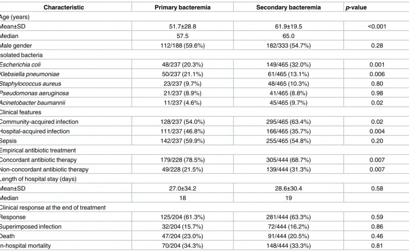
Comparisons between patients with primary bacteremia and secondary bacteremia are shown inTable 4.E.coliandA.baumanniiwere significantly more common in secondary bac-teremia, whereasK.pneumoniaewas significantly more common in primary bacteremia. Pri-mary bacteremia was significantly more prevalent in HAI. Concordant empirical antibiotic therapy was significantly more common in primary bacteremia. Length of hospital stay, clini-cal response of infections at the end of treatment, and in-hospital mortality were comparable between secondary bacteremia and primary bacteremia patients. Patients with CLABSI received more frequent non-concordant empirical antibiotic therapy, and they had a signifi-cantly longer length of hospital stay.
Comparisons between bacteremia patients with and without sepsis are shown inTable 5. K.pneumoniaewas more common in bacteremia patients with sepsis. Clinical response of infections at the end of treatment in bacteremia patients with sepsis was less favorable than in bacteremia patients without sepsis. In-hospital mortality in bacteremia patients with sepsis was significantly higher than in bacteremia patients without sepsis.
Comparisons between patients who received concordant empirical antibiotics and patients who received non-concordant empirical antibiotics are shown inTable 6. Patients with
Table 4. Comparisons between patients with primary bacteremia and patients with secondary bacteremia.
Characteristic Primary bacteremia Secondary bacteremia p-value
Age (years) Mean±SD 51.7±28.8 61.9±19.5 <0.001 Median 57.5 65.0 Male gender 112/188 (59.6%) 182/333 (54.7%) 0.28 Isolated bacteria Escherichia coli 48/237 (20.3%) 149/465 (32.0%) 0.001 Klebsiella pneumoniae 50/237 (21.1%) 61/465 (13.1%) 0.006 Staphylococcus aureus 23/237 (9.7%) 48/465 (10.3%) 0.80 Pseudomonas aeruginosa 21/237 (8.9%) 41/465 (8.8%) 0.98 Acinetobacter baumannii 11/237 (4.6%) 45/465 (9.7%) 0.02 Clinical features Community-acquired infection 128/237 (54.0%) 295/465 (63.4%) 0.02 Hospital-acquired infection 111/237 (46.8%) 166/465 (35.7%) 0.004 Sepsis 142/237 (59.9%) 255/465 (54.8%) 0.20
Empirical antibiotic treatment
Concordant antibiotic therapy 179/228 (78.5%) 305/444 (68.7%) 0.007
Non-concordant antibiotic therapy 49/228 (21.5%) 139/444 (31.3%) 0.007
Length of hospital stay (days)
Mean±SD 27.0±34.2 28.6±30.4 0.58
Median 18 19
Clinical response at the end of treatment
Response 125/204 (61.3%) 281/444 (63.3%) 0.59
Superimposed infection 32/204 (15.7%) 72/444 (16.2%) 0.86
Death 47/204 (23.0%) 91/444 (20.5%) 0.46
In-hospital mortality 70/204 (34.3%) 148/444 (33.3%) 0.81
S.aureusbacteremia, community-acquired infection, and primary bacteremia received concor-dant empirical antibiotics more often; whereas, patients withA.baumanniibacteremia, hospi-tal-acquired bacteremia, and secondary bacteremia received non-concordant empirical antibiotics more often. Patients who received non-concordant empirical antibiotics had a lon-ger length of hospital stay and higher in-hospital mortality than patients who received concor-dant empirical antibiotics.
Duplicate bacterial isolates with identical antibiotic susceptibility profiles for each episode of bacteremia were observed in 80 out of 216 isolates ofE.coli, 45 out of 177 isolates ofK .-pneumoniae, 25 out of 112 isolates ofA.baumannii, 25 out of 108 isolates ofP.aeruginosa, 5 out of 28 isolates ofSalmonellaspp., 7 out of 36 isolates ofE.faecalis, 6 out of 38 isolates ofE. fae-cium, 45 out of 311 isolates of CNS, and 49 out of 149 isolates ofS.aureus. Comparisons of anti-biotic susceptibility profiles between non-duplicate isolates and duplicate isolates of the aforementioned bacteria revealed no significant differences in antibiotic susceptibility between non-duplicate isolates and duplicate isolates.
Comparisons of antibiotic susceptibility of common or important antibiotic-resistant bac-teria between community-acquired and hospital-acquired bacbac-terial isolates are shown in Table 7. Hospital-acquiredE.coliandK.pneumoniaeisolates were more resistant to ceftriaxone than community-acquired isolates. Hospital-acquiredK.pneumoniae,A.baumannii,P. aerugi-nosaisolates were more resistant to meropenem and piperacillin-tazobactam than commu-nity-acquired isolates. None of commucommu-nity-acquiredS.aureusisolates were MRSA whereas
Table 5. Comparisons between bacteremic patients with and without sepsis.
Characteristic Sepsis No sepsis p-value
Age (years) Mean±SD 56.8±23.1 56.3±25.0 0.80 Median 61 61 Male gender 171/321 (53.3%) 145/263 (55.1%) 0.68 Organism Escherichia coli 117/435 (26.9%) 86/361 (23.8%) 0.33 Klebsiella pneumoniae 78/435 (17.9%) 46/361 (12.7%) 0.04 Staphylococcus aureus 51/435 (11.7%) 40/361 (11.1%) 0.82 Pseudomonas aeruginosa 47/435 (10.8%) 29/361 (8.0%) 0.23 Acinetobacter baumannii 43/435 (9.9%) 29/361 (8.0%) 0.39 Clinical features Community-acquired infection 225/435 (51.7%) 198/361 (54.8%) 0.38 Hospital-acquired infection 220/435 (50.6%) 168/361 (46.5%) 0.27 Primary bacteremia 142/435 (32.6%) 99/361 (27.4%) 0.12 Secondary bacteremia 265/435 (60.9%) 214/361 (59.3%) 0.64
Empirical antibiotic treatment
Concordant antibiotic therapy 334/463 (72.1%) 255/367 (69.5%) 0.40
Non-concordant antibiotic therapy 129/463 (27.9%) 112/367 (30.5%) 0.40
Length of hospital stay (days)
Mean±SD 32.5±37.5 29.7±30.0 0.36
Median 22 21
Clinical response at end of treatment
Response 215/377 (57.0%) 213/278 (76.6% <0.001
Superimposed infection 62/377 (16.5%) 44/278 (15.8%) 0.83
Death 100/377 (26.5%) 21/278 (7.6%) <0.001
In-hospital mortality 154/377 (40.9%) 54/278 (19.4%) <0.001
43% of hospital-acquiredS.aureusisolates were MRSA. None of community-acquiredE. fae-cuimisolates were resistant to vancomycin whereas 29% of hospital-acquiredE.faecuimisolates were resistant to vancomycin.
Many bacteria on the list announced by WHO for the antibiotic-resistant bacteria consid-ered posing the greatest threat to human health [8] were observed in this study. Carbapenem-resistantE.coliwas observed in 1.3% ofE.coliisolates; carbapenem-resistantK.pneumoniaein 20.0% ofK.pneumoniaeisolates; carbapenem-resistantP.aeruginosain 27.7% ofP.aeruginosa isolates; and, carbapenem-resistantA.baumanniiin 69.5% ofA.baumanniiisolates. Vancomy-cin-resistant enterococci (VRE) were found in 14.3% ofEnterococcusspp. isolates. All VRE iso-lates wereE.faecium. MRSA was isolated from 19.0% of allS.aureusisolates. MRSA bacteremia was 0% in community-acquiredS.aureusbacteremia, but was 43% in hospital-acquiredS. aureusbacteremia.
Median length of hospital stay in all hospitalized patients with true bacteremia was 17 days. Overall in-hospital mortality of patients with true bacteremia was 33.3%. Mortality was signifi-cantly higher in patients with resistant bacteremia than in patients with antibiotic-non-resistant bacteremia (40.5%vs. 28.5%,p<0.001) as shown inTable 8. The mortality attrib-utable to AMR was 12.0% (95% Confidence Interval 5.7% to 18.1%). Patients with antibiotic-resistantA.baumanniiorE.faeciumbacteremia had the highest mortality (66.7%). Based on these findings from the data collected for eight months, the estimated annual number of deaths was 194 patients with antibiotic-resistant bacteremia, and 171 patients with antibiotic-non-resistant bacteremia.
Table 6. Comparisons between patients who received concordant empirical antibiotic therapy and patients who received non-concordant empiri-cal antibiotic therapy.
Characteristic Concordant empirical therapy* Non-concordant empirical therapy** p-value
Age (years) Mean±SD 55.1±25.5 56.4±26.1 0.57 Median 59 63 Male gender 209/405 (51.6%) 100/161 (62.1%) 0.02 Organism Escherichia coli 152/565 (26.9%) 49/223 (22%) 0.17 Klebsiella pneumoniae 90/565 (15.9%) 31/223 (13.9%) 0.51 Staphylococcus aureus 73/565 (12.9%) 16/223 (7.2%) 0.02 Pseudomonas aeruginosa 46/565 (8.1%) 28/223 (12.6%) 0.05 Acinetobacter baumannii 25/565 (4.4%) 44/223 (19.7%) <0.001 Clinical features Community-acquired infection 342/565 (60.5%) 94/223 (42.3%) <0.001 Hospital-acquired infection 224/565 (39.6%) 139/223 (62.3%) <0.001 Primary bacteremia 186/565 (32.9%) 51/223 (22.9%) 0.006 Secondary bacteremia 323/565 (57.2%) 156/223 (70.0%) <0.001 Sepsis 324/565 (57.3%) 118/223 (52.9%) 0.27
Length of hospital stay (days)
Mean±SD 32.9±38.8 41.3±33.5 0.02
Median 20 31
Clinical response at end of treatment
Response 294/462 (63.6%) 103/223 (46.2%) <0.001
Superimposed infection 78/462 (16.9%) 46/223 (20.6%) 0.23
Death 90/462 (19.5%) 74/223 (33.2%) <0.001
In-hospital mortality 134/462 (29.0%) 110/223 (49.3%) <0.001
Table 7. Percenta ge of antibiotic suscept ibility of common or importan t commun ity-acquire d bacterial isolates (CABI) and hospital-acquire d bacterial isolates (HABI). Bacteria Number of isolates Amoxicillin/ clavulanate Piperacillin/ tazobactam Ceftazidime Ceftriaxone Cefepime Meropenem Amikacin Ciproflo- xacin Colistin Oxacillin Vancomycin Escherichia coli CABI 122 85 99 91 73 94 100 100 58 HABI 94 63 92 36 18 63 100 98 39 Klebsiella pneumoniae CABI 52 94 90 92 98 96 96 98 88 HABI 80 53 56 17 44 59 73 80 49 Acinetobacter baumannii CABI 12 64 64 0 55 70 80 55 100 HABI 75 24 25 0 25 25 34 25 100 Pseudomonas aeruginosa CABI 34 91 91 91 85 97 82 100 HABI 49 67 67 67 63 73 73 98 Staphylococcus aureus CABI 56 95 100 100 HABI 44 57 57 100 Enterococcus faecium CABI 8 100 HABI 24 71 https://doi.o rg/10.1371/j ournal.pone .0190132.t007
The cost of hospitalization for each patient with bacteremia was retrieved from the hospital database of the Computer Unit and the Financial Department of Siriraj Hospital. The annual cost of hospitalizations for patients with bacteremia was estimated from the cost of hospitaliza-tions for patients with bacteremia during the study period for eight months. The estimated total annual cost of hospitalizations for patients with bacteremia was US$ 10,854,132, of which US$ 5,409,816 was spent for patients with antibiotic-non-resistant bacteremia (US$ 8,614/ admission), and US$ 5,444,316 was spent for patients with antibiotic-resistant bacteremia (US $ 15,379/admission). The estimated annual cost of hospitalizations for patients with commu-nity-acquired bacteremia was US$ 2,088,260 (US$ 4,725/admission) and hospital-acquired bacteremia was US$ 8,765,872 (US$ 16,233/admission).
Discussion
The manual for early implementation of GLASS in human infections recommends blood, urine, feces, and urethral and cervical swabs as priority specimens; andE.coli,K.pneumoniae, A.baumannii,S.aureus,S.pneumoniae,Salmonellaspp.,Shigellaspp., andN.gonorrhoeaeas pri-ority bacteria. However, GLASS implementation at Siriraj Hospital included collection of spu-tum because respiratory tract infection is very common infection in hospitalized patients [9, 10]. Furthermore, the interpretation of sputum culture results is challenging, regardless of whether the isolated organism is causative agent, colonizer, or contaminant. We did not include urogenital swabs for gonococcal culture, because these specimens are very uncommon. We also collectedP.aeruginosabecause it is one of the most common causative bacteria, espe-cially in HAI [9,10].
A key feature of GLASS is that patient clinical data and microbiological data are combined. We recognize that many types of relevant patient clinical data are often not included in the information submitted to the laboratory along with the clinical sample. Moreover, new and important patient clinical data will become available after the clinical sample has been sent to the laboratory. As a result, the report of the culture result is usually incomplete and limited in
Table 8. In-hospital mortality of patients with priority pathogens as the cause of bacteremia.
Bacteria Mortality p-value
Antibiotic-resistant bacteriaa Antibiotic-non-resistant bacteria Escherichia coli 23/106 (21.7%) 18/100 (18.0%) 0.51 Klebsiella pneumoniae 23/45 (51.1%) 22/65 (33.8%) 0.07 Other Enterobacteriaceae 3/8 (37.5%) 12/43 (27.9%) 0.58 Pseudomonas aeruginosa 10/21 (47.6%) 19/47 (40.4%) 0.58 Acinetobacter baumannii 38/57 (66.7%) 3/24 (12.5%) <0.001 Salmonella spp. 0/2 (0%) 1/2 (50.0%) 0.25 Staphylococcus aureus 9/16 (56.3%) 13/47 (27.7%) 0.04 Coagulase-negative Staphylococcus spp. 1/14 (7.1%) - -Enterococcus faecium 6/9 (66.7%) 12/20 (60.0%) 0.73 Streptococcus pneumoniae 0/1 (0%) 0/3 (0%)
-Overall in-hospital mortality 113/279 (40.5%) 100/351 (28.5%) <0.001
aCarbapenem-resistant or third-generation cephalosporin-resistant E.coli, K.pneumoniae, and other Enterobacteriaceae; carbapenem-resistant P.aeruginosa; carbapenem-resistant A.baumannii;
fluoroquinolone-resistant Salmonella spp.; MRSA; MRCNS; vancomycin-resistant E.faecium; and, penicillin-non-susceptible S.pneumoniae
value, because it does not include or take into account these important pieces of missing data. Many clinical data were collected from patients in our study using a locally developed user-friendly web application program that could be installed in a smart phone and conveniently used in patient care areas. Supplementary information that was collected included source of infection, severity of infection, empirical and specific antibiotic therapy, clinical outcomes of infection, patient mortality, and cost of hospitalization. This additional data was important to understand the epidemiology of bacteremia at our center, to enhance our ability to develop more appropriate local antibiotic guidelines, and to estimate health and economic burden of bacteremia caused by AMR bacteria.
The reported blood culture contamination rate of 3.5% was higher than the acceptable tar-get rate of less than 3% [11]. CNS was the most common blood culture contaminant, and it accounted for 19% of positive blood culture specimens, but it was less than 32% of CNS among all isolated bacteria in the 2016 annual report of the National Antimicrobial Resistance Surveillance Centre, Thailand [12]. The rate of blood culture contamination is a recommended indicator of health care quality. We, therefore, intend to implement additional measures to reduce the rate of contaminants in blood cultures. Our study revealed that 15.1% of CNS iso-lated from blood specimens were causative bacteria based on patient clinical data. Therefore, information relating to clinical features of patients with positive blood culture for CNS was extremely important for determining if CNS was a causative agent that required antibiotic therapy. This observation emphasized the importance and value of collecting clinical data in addition to demographic data recommended in GLASS manual. CNS that caused infection tended to be less resistant to antibiotics than CNS that was contaminant. This observation sug-gested that bacteria isolated from sputum and urine samples that was colonizer might be more resistant to antibiotics than isolated bacteria that was the cause of infection.
We would have been unable to determine if isolated bacteria was primary bacteremia, sec-ondary bacteremia, or CLABSI unless the clinical features of patients with positive blood cul-tures were taken into account. Classification of these 3 categories of bacteremia was necessary, because the bacteria that caused different types of bacteremia were different, and each type of bacteremia required a different regimen of empirical antibiotic therapy. If all bacteria that cause all types of bacteremia would have been combined and those data were used for develop-ing empirical antibiotic guidelines for patients with bacteremia, many patients with different types of bacteremia would have received inappropriate antibiotic regimens.
Differentiation between bacteremia in CAI and HAI was necessary since the types of causa-tive bacteria, the antibiotic susceptibility profiles of bacteria, and the clinical outcomes of CAI and HAI were significantly different. Important data including hospitalization at other health-care facilities>2 days within 90 days, healthcare-associated conditions, and duration of cur-rent hospitalization>2 days were needed to determine if bacteremia was HAI. We found that if duration from date of hospitalization to date of blood culture collection of2 days was used to classify bacteremia as CAI, at least 10% of patients with HAI would have been classified as CAI. Misclassification of some bacteremic episodes as CAI instead of HAI resulted in signifi-cantly higher prevalence of antibiotic resistance to causative bacteria, such as prevalence of community-acquired MRSA from 0% to 9% and prevalence of ceftriaxone-resistantE.coliand K.pneumoniaefrom 19.3% to 38.5%.
Observations from our study confirmed the results of previous studies thatE.coli,K. pneu-moniae,S.aureus,A.baumannii, andP.aeruginosawere common causative agents of bacter-emia, and that the clinical outcomes of bacteremia due to antibiotic-resistant bacteria were unfavorable [1,9,10,13–19]. However, our study results revealed additional important details about bacteremia, such as the sources of secondary bacteremia, sepsis status in bacteremia patients, and antibiotic susceptibility of community-acquired and hospital-acquired bacterial
blood isolates–all of which will be useful for managing patients with suspected bacteremia in the future.
We found no significant differences in antibiotic susceptibility between non-duplicate iso-lates and duplicate isoiso-lates amongE.coli,K.pneumoniae,Salmonellaspp.,A.baumannii,P. aeru-ginosa,E.faecalis,E.faecium,S.aureus, CNS, andS.pneumoniae. This could be due to the fact that most patients had only one episode of bacteremia, each episode of bacteremia usually had one type of bacteria, and most isolated bacteria were causative agents. It is anticipated that dif-ferences in antibiotic susceptibility between non-duplicate and duplicate isolates of bacteria from other clinical specimens commonly colonized with organisms (e.g., sputum and urine) should be observed similar to our finding of more antibiotic resistance in CNS isolates that were contaminants than CNS isolates that were the cause of infection.
Many metrics of AMR surveillance according to GLASS protocol are presented in our results. However, some recommended metrics could not be computed, such as the number of blood cultures per 100,000 inhabitants. This is because many patients who receive medical care from Siriraj Hospital are not residents of Siriraj Hospital catchment areas. Siriraj Hospital is a national tertiary referral hospital and we receive and treat patients that are referred from across Thailand.
Based on our findings, GLASS was superior to the laboratory-based surveillance for blood culture specimens in patients with bacteremia. Although GLASS consumed more time and resources than the laboratory-based surveillance system, the data derived from GLASS was more useful for developing antibiotic guidelines for patients suspected of having bacteremia. Data derived from GLASS are also valuable for estimating and monitoring the antimicrobial consumption and usage, and health and economic burden of AMR. Furthermore, the results from GLASS can be used to estimate and monitor a drug resistance index [20]. Given that the GLASS that we implemented at Siriraj Hospital exceeded the minimum recommended criteria set forth in GLASS manual, it may be difficult to fully and permanently implement this system in the near term on institution-wide basis. Therefore, we may activate GLASS for one 6-month period every other year because the types of causative agents of infections and their antibiotic susceptibility should not have dramatic changes over a short period of time. Another alterna-tive for ideal implementation of GLASS would require responsible personnel who will send the clinical specimen for culture to provide all relevant patient clinical data along with request for culture of clinical specimen.
Acknowledgments
The authors gratefully acknowledge Ms. Wiyada Arjratanakool, Ms. Sawitta Pramoun, Mr. Nuttapong Srasrisom, Ms. Wanida Cheewathammarat, Mrs. Sukanya Chanboonchuay, and the infection control nurses of Siriraj Hospital for their assistance in managing and collecting data. The authors also thank the Computer Unit and the Financial Department of Siriraj Hos-pital for providing some patients’ outcomes and cost of hosHos-pitalization data.
Author Contributions
Conceptualization: Rujipas Sirijatuphat, Kantarida Sripanidkulchai, Adhiratha Boonyasiri, Pinyo Rattanaumpawan, Pattarachai Kiratisin, Visanu Thamlikitkul.
Data curation: Rujipas Sirijatuphat, Kantarida Sripanidkulchai, Adhiratha Boonyasiri, Pinyo Rattanaumpawan, Orawan Supapueng, Pattarachai Kiratisin, Visanu Thamlikitkul. Formal analysis: Rujipas Sirijatuphat, Kantarida Sripanidkulchai, Orawan Supapueng, Visanu
Funding acquisition: Visanu Thamlikitkul. Project administration: Visanu Thamlikitkul. Supervision: Visanu Thamlikitkul.
Visualization: Visanu Thamlikitkul.
Writing – original draft: Rujipas Sirijatuphat, Kantarida Sripanidkulchai, Visanu Thamlikitkul.
Writing – review & editing: Rujipas Sirijatuphat, Kantarida Sripanidkulchai, Adhiratha Boo-nyasiri, Pinyo Rattanaumpawan, Orawan Supapueng, Pattarachai Kiratisin, Visanu Thamlikitkul.
References
1. Pumart P, Phodha T, Thamlikitkul V, Riewpaiboon A, Prakongsai P, Limwattananon S. Health and eco-nomic impacts of antimicrobial resistance in Thailand. J Health Syst Res. 2012; 6(3): 352–60.
2. Apisarnthanarak A, Kiratisin P, Saifon P, Kitphati R, Dejsirilert S, Mundy LM. Clinical and molecular epi-demiology of community-onset, extended-spectrum beta-lactamase-producing Escherichia coli infec-tions in Thailand: a case-case-control study. Am J Infect Control. 2007; 35(9): 606–12.https://doi.org/ 10.1016/j.ajic.2007.05.008PMID:17980240
3. Suankratay C, Jutivorakool K, Jirajariyavej S. A prospective study of ceftriaxone treatment in acute pyelonephritis caused by extended-spectrum beta-lactamase-producing bacteria. J Med Assoc Thai. 2008; 91(8): 1172–81. PMID:18788687
4. Kanoksil M, Jatapai A, Peacock SJ, Limmathurotsakul D. Epidemiology, Microbiology and Mortality Associated with Community-Acquired Bacteremia in Northeast Thailand: A Multicenter Surveillance Study. PLoS One. 2013;18; 8(1):e54714.https://doi.org/10.1371/journal.pone.0054714PMID: 23349954
5. O’Neill J. Antimicrobial Resistance: Tackling a crisis for the health and wealth of nations. Rev Antimicrob Resist. 2014:1–16.
6. World Health Organization. Global Action Plan on Antimicrobial Resistance, 2015
7. World Health Organization. The Global Antimicrobial Resistance Surveillance System (GLASS), 2015.
8. World Health Organization. List of bacteria for which new antibiotics are urgently needed. February 2017.http://www.who.int/mediacentre/news/releases/2017/bacteria-antibiotics-needed/en/
9. Danchaivijitr S, Judaeng T, Sripalakij S, Naksawas K, Plipat T. Prevalence of nosocomial infection in Thailand 2006. J Med Assoc Thai. 2007; 90(8): 1524–9. PMID:17926980
10. Rongrungruang Y, Sawanpanyalert N, Chomdacha P, Surasarang K, Wiruchkul N, Kachintorn K, Tanti-lipikara P, Danchaivijitr S. Health-care associated infections in Thailand 2011. J Med Assoc Thai. 2013; 96(Suppl 2): S117–23.
11. Hall KK, Lyman JA. Updated review of blood culture contamination. Clin Microbiol Rev. 2006; 19 (4): 788–802.https://doi.org/10.1128/CMR.00062-05PMID:17041144
12. National Antimicrobial Resistance Surveillance Center, Thailand (NARST). Antibiogram 2016. [cited 29 March 2017]. Available fromhttp://narst.dmsc.moph.go.th/
13. Lim C, Takahashi E, Hongsuwan M, Wuthiekanun V, Thamlikitkul V, Hinjoy S, et al. Epidemiology and burden of multidrug-resistant bacterial infection in a developing country. Elife. 2016; 5: e18082.https:// doi.org/10.7554/eLife.18082PMID:27599374
14. Cosgrove SE, Sakoulas G, Perencevich EN, Schwaber MJ, Karchmer AW, Carmeli Y. Comparison of mortality associated with methicillin-resistant and methicillin-susceptible Staphylococcus aureus bacter-emia: a meta-analysis. Clin Infect Dis. 2003; 36(1): 53–9.https://doi.org/10.1086/345476PMID: 12491202
15. Schwaber MJ, Carmeli Y. Mortality and delay in effective therapy associated with extended-spectrum beta-lactamase production in Enterobacteriaceae bacteraemia: a systematic review and meta-analysis. J Antimicrob Chemother. 2007; 60(5): 913–20.https://doi.org/10.1093/jac/dkm318PMID:17848376
16. Leistner R, Sakellariou C, Gu¨rntke S, Kola A, Steinmetz I, Kohler C, Pfeifer Y, Eller C, Gastmeier P, Schwab F. Mortality and molecular epidemiology associated with extended-spectrumβ-lactamase pro-duction in Escherichia coli from bloodstream infection. Infect Drug Resist. 2014; 7: 57–62.https://doi. org/10.2147/IDR.S56984PMID:24648746
17. Lemos EV, de la Hoz FP, Einarson TR, McGhan WF, Quevedo E, Castañeda C, Kawai K. Carbapenem resistance and mortality in patients with Acinetobacter baumannii infection: systematic review and meta-analysis. Clin Microbiol Infect. 2014; 20(5): 416–23.https://doi.org/10.1111/1469-0691.12363 PMID:24131374
18. Zhang Y, Chen XL, Huang AW, Liu SL, Liu WJ, Zhang N, Lu XZ. Mortality attributable to carbapenem-resistant Pseudomonas aeruginosa bacteremia: a meta-analysis of cohort studies. Emerg Microbes Infect. 2016; 5: e27.https://doi.org/10.1038/emi.2016.22PMID:27004762
19. Xu L, Sun X, Ma X. Systematic review and meta-analysis of mortality of patients infected with carbape-nem-resistant Klebsiella pneumoniae. Ann Clin Microbiol Antimicrob. 2017; 16(1): 18.https://doi.org/ 10.1186/s12941-017-0191-3PMID:28356109
20. Laxminarayan R, Klugman KP. Communicating trends in resistance using a drug resistance index. BMJ Open. 2011; 1(2): e000135.https://doi.org/10.1136/bmjopen-2011-000135PMID:22102636