서 론
유두암은 갑상선에서 가장 흔한 암으로 국소 림프절전이의 빈도가 높다 [1-3]. 이러한 림프절전이는 국소재발률과 연관 성이 높고, 이는 치료방법 및 수술범위 결정, 또 수술 후 예후 판정에 중요한 영향을 미친다 [4, 5]. 그러므로 수술 전 평가에 있어 국소 림프절전이의 정확한 예측이 중요하다 [6]. 수술 전 병기결정에서 초음파는 American Thyroid Association guidelines에서 권고하는 가장 유용한 방법이다 [7]. 갑상선암 의 경부 림프절전이를 진단하는 데 초음파는 민감도는 52-84%, 특이도는 95-97%로 보고되었다 [8, 9].갑상선유두암의 경부 림프절전이의 진단:
전산화단층촬영의
조영증강 양상이 림프절전이의 진단에 도움이 되는가?
신현주∙김은경∙문희정 곽진영 연세의대 영상의학교실J Korean Soc Ultrasound Med
2011; 30: 187-191
Received March 22, 2011; Revised July 15, 2011; Accepted July 25, 2011. Address for reprints :
Jin Young Kwak, MD, Department of Radiology, Yonsei University College of Medicine, 134 Shinchon-dong, Seodae-moon-gu, Seoul 120-752, Korea. Tel. 82-2-2228-7400
Fax. 82-2-393-3035 E-mail: docjin@yuhs.ac
Diagnosis of Cervical Metastatic Lymph Nodes in
Papillary Thyroid Carcinoma: Is CT Enhancement Useful
for Diagnosing Lymph Node Metastasis?
Hyun Joo Shin, MD, Eun-Kyung Kim, MD, Hee Jung Moon, MD, Jin Young Kwak, MD
Department of Radiology, Yonsei University College of Medicine
Purpose: We wanted to determine the utility of CT enhancement for diagnosing metastatic lymphadenopathy in patients with papillary thyroid carcinoma (PTC), and especially in the lymph nodes (LNs) of the lateral neck level and that are not suspi-cious for metastasis on ultrasonography (US).
Materials and Methods: Our study population included 34 consecutive LNs of 31 patients (25 females and 6 males, mean age: 46.7 yrs) with PTC and who had no suspicious metastatic lateral cervical LN on preoperative US, but enhancement of the lateral cervical LNs was seen on CT. To objectify the degree of enhancement, the dif-ference of Hounsfield units between the suspicious LN and that of the ipsilateral SCM muscle was calculated. For the node-by-node analysis, marking of the corresponding LN with CT enhancement on the second look US was performed. The final assess-ment was attained by surgical dissection of the marked LNs. The medical records were reviewed for the patients’ age and gender and the size of the LNs.
Results: Among the 34 LNs, 17 LNs were diagnosed as metastasis and 17 were benign. There was no difference in the size of the LNs between two the groups (benign and metastatic). The patients who had metastatic LNs were younger than those patients with benign LNs (p = 0.037). The incidence of metastatic LN was high-er in the male patients than in the female patients (F:M = 38.5%:100%, p = 0.018). There was no statistical difference between the metastatic LNs and benign LNs according to the degree of enhancement (p = 0.953).
Conclusion: The degree of CT enhancement is not feasible to use for diagnosing metastatic LNs in the lateral neck level in patients with PTC.
Key words : Papillary thyroid carcinoma; Computed tomography (CT); Lymph node enhancement
전산화단층촬영 (Computed Tomography, CT)은 지금까지 는 갑상선암 환자의 수술 전 평가에 있어서 관례적으로 시행되 는 검사는 아니었으나, 최근 몇몇 연구에서 수술 전 초음파와 CT가 초음파 단독 시행보다 뛰어난 진단율을 보였다 [10, 11]. CT에서 갑상선유두암의 림프절전이를 시사하는 소견은 석회화, 낭성 또는, 괴사성 변성, 둥근 모양(round shape), 강 한 조영증강이다 [10, 11]. 이 중, 림프절 조영증강은 CT에서 만 평가 가능한 항목으로 이에 대한 객관적인 지표를 제시한 연구는 거의 없다. 기존 연구에서는 갑상선유두암의 수술 전 평가로 시행한 초 음파, CT 결과와 수술 결과를 후향적으로 비교하여 각각의 민 감도, 특이도 등을 분석하였으나, 이번 연구에서는 갑상선유두 암의 림프절전이 진단에 CT의 조영증강이 가지는 유용성 및 CT가 초음파에 더하여 추가적인 의미를 가지고 있는지를 평가 하기 위하여, 초음파에서 전이가 의심되지 않았으나 CT상 조 영증강이 있었던 림프절만을 대상으로 CT의 진단적 가치를 알 아보고자 하였다 [10, 11].
대상 및 방법
환자군 본 병원의 생명윤리위원회가 이 연구에 대하여 승인하였고 검사 시행 전 환자에 대한 사전 동의가 이루어졌다. 2008년 4 월부터 2008년 9월까지 본원에서 갑상선유두암으로 수술 전 병기결정 목적으로 시행한 초음파에서 경부 림프절전이가 의 심되지 않았으나 CT에서 외측 경부림프절의 조영증강이 관찰 되었던 31명의 환자에서 34개의 림프절에 대하여 후향적으로 연구를 진행하였다. 의심되는 림프절 위치는 level Ⅰ (n = 1), level Ⅱ (n = 2), level Ⅲ (n = 20), level Ⅳ (n = 9), level Ⅴ (n = 1), level Ⅵ (n = 1)였고, 장경은 4-17 mm (평균 7.62 mm) 였다. 여자는 25명 (80.6%), 남자는 6명 (19.4%) 이었 으며 평균 연령은 46.7세였다.수술 전 초음파에 의한 림프절 평가
수술 전 병기결정 초음파는 7-15-MHz 선형 탐촉자 (HDI5000; Philips Medical Systems, Bothell, WA) 또는 5-12-MHz 선형 탐촉자(iU22; Philips Medical Systems)를 이 용하였다. 초음파는 11년, 7년, 5년의 경험을 가지고 있는 3명 중 한 명의 영상의학과 전문의 (K.E.K., K.J.Y., K.M.J)가 갑상 선암이 진단된 것을 알고 병기결정의 목적으로 시행하였다. 초 음파상 림프절전이가 의심되는 소견은 주변 근육보다 높은 에 코를 보이거나, 낭성 변성이나 석회화가 있거나, 구형(종경/횡 경 비<1.5)의 모양을 보이는 경우였다. 수술 전 평가를 위해 본원에서는 일상적으로 조영증강 경부 CT (contrast-enhanced CT with an multidetector scanner, SOMATOM Sensation 16 또는 SOMATOM Sensation 64,
Siemens Healthcare, 3 mm reconstructed slice thickness)을 시행하고 있다. 조영제는 90 mL의 iodinated contrast (iopromide, Ultravist 300, Bayer Schering Pharma)를 사용 하였고, 3 mL/s의 속도로 주입되었다. 쇄골하정맥에서 오는 혈 류에 의한 인공허상을 최소화하기 위하여 조영제 주입 직후 생 리식염수를 3 mL/s의 속도로 주입하였다. Scan delay time은 40-60초였다. CT에서 림프절전이가 의심되는 소견은 석회 화, 중심부 괴사 또는 낭성 변성, 주변 근육보다 높은 조영 증강 을 보이는 경우였다. 이 연구에서는 초음파 시행 후 CT를 시행 하여, 초음파에서 전이의심 소견이 없으나 CT에서 다른 의심 스러운 소견없이 단지 조영증강을 보인 병변에 대하여 이 소견 의 진단 정확도를 평가하였다. 정성적으로 조영증강이 의심되 는 림프절을 선택하여, 조영증강의 객관적인 정도를 평가하기 위하여 해당 림프절과 동측 흉쇄유돌근에서 난원형의 region of interests (ROIs)를 그려 이 둘의 차이값을 구하였다. 림프 절의 HU 값에서 근육의 HU 값을 뺀 값을 조영증강정도 (degree of enhancement; DOE)로 생각하였다.
수술 및 병리 보고 초음파에서 전이가 의심되지 않았으나 이후 시행한 CT에서 조영 증강이 있는 림프절에 대하여 다시 한 번 초음파를 시행 하여 그 위치를 수술 직전 경부에 표시하였고, 이를 토대로 수 술 당시 해당 림프절을 절제하였다. 수술을 시행한 림프절의 최종 병리 보고를 위하여, 6th edition AJCC/UICC TNM Classification system을 기준으로 삼았다. 통계 분석 최종진단은 림프절의 병리결과를 토대로 하였고 이들의 초 음파와 전단화단층촬영의 조영증강 소견, 그리고 각 환자의 몇 가지 임상소견과 악성과의 연관성을 알아보았다. 범주형 변수 는 Fisher’s exact test, 연속형 변수는 student t-test를 이용 하여 구하였다. 모든 값에서 p value가 0.05보다 작을 때 그 값 이 통계적으로 유의하다고 보았다. 통계 분석은 SAS software (version 9.1.3; SAS Institute, Cary, NC)를 사용하였다.
결 과
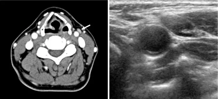
병리결과에서 34개의 림프절 중 17개 (50%)는 갑상선유두 암의 림프절전이가 있고, 나머지 17개 (50%)는 양성이었다 (Fig. 1). CT상에서 전이림프절의 평균 장경은 7.65 mm ± 1.66 mm, 양성림프절은 8.12 mm ± 3.5 mm 이었다 (p value = 0.62). 림프절전이가 있는 환자군은 평균 42.4 세 ± 12.2 세로, 전이가 없는 환자군 (평균 51 세 ± 10.1 세)에 비하여 낮은 연령을 보였다 (p = 0.037). 또한 남자환자 6명의 림프절 에서 모두 전이가 있었고 (100%), 여자는 38%에서 전이가 있 어 남자 환자에게 림프절 전이가 더 많았다 (p = 0.018).조영증강된 림프절과 근육의 HU 차이는, 전이림프절은 평 균 32.53 HU ± 14.66 HU, 양성림프절은 32.18 HU ± 19.49 HU로 둘 간의 유의한 차이는 보이지 않았다 (p = 0.953). 림 프절과 근육의 조영증강 차이가 클수록 전이 가능성이 높은지 알기 위해 구해본 odds ratio는 1.001 (95% 신뢰구간 0.962, 1.042) (p value = 0.951)로 유의한 결과를 보이지 않았다 (Table 1).
고 찰
갑상선유두암은 국소 림프절전이가 흔하고, 재발이 가장 많 이 발생하는 위치는 경부림프절이다 [19, 20]. 반복되는 림프 절전이를 가진 환자는 예후가 나쁘다고 생각하고 있으며 적절 한 림프절절제는 재발 위험을 감소시킨다 [20]. 갑상선유두암 의 림프절전이의 유무에 따라 수술범위 및 방법이 달라지므로 수술 전 림프절 전이의 정확한 평가가 중요하다. 지금까지 초 음파는 갑상선유두암의 림프절전이 평가에 중요한 역할을 하 고 있지만, 검사자 의존도가 높고 검사 가능 범위의 제한이 있 다는 한계가 있다. 초음파상 갑상선유두암의 경부 림프절전이는 내부에 국소 또는 미만성의 고에코 부위가 있거나, 석회화, 낭성 변성, 둥근 모양(종경/횡경 비<1.5), 비정상적 혈류 양상(주변부 혈관 분 포 등)을 띌 때의 다섯 가지 중 하나 이상의 소견을 보일 때 의 심할 수 있다 [10, 12-16]. 림프절의 위치별 비교를 통해 갑상 선유두암의 림프절전이를 평가한 연구들에 따르면, 초음파의 민감도는 52-84%, 특이도는 95-97%로 높았고, 진단정확도 는 76-77%로 보고되었다 [17, 21, 22]. CT상 갑상선유두암의 경부 림프절전이는 내부에 괴사 또는 낭성 변성, 석회화를 동반하거나 주변 근육보다 높은 정도의 조영증강 양상을 보일 때 의심할 수 있다 [10, 11, 17, 18]. 이 러한 소견을 이용한 CT의 림프절전이 평가가 초음파 단독 검 사보다 진단적 가치가 높다는 보고들이 있다 [11, 23]. 이 중,Table 1. Demographics of the Patients Included in This Study Metastatic LN Reactive LN P value Age (years) Mean 42.4 Mean 51
(21-65) (39-75) 0.0373
Gender ratio (M:F) 6:10 0:16 0.0177
Table 2.Characteristics of Included Lymph Nodes
Metastatic LN Reactive LN P value
Size (mm) Mean 7.6 Mean 8.1 0.6195
(6-11) (4-17)
DOE difference (HU) Mean 32.5 Mean 32.2 0.9528 (10-64) (5-64)
Note.─Size : The longest diameter of LN in CT examination, HU : Hounsfield unit
A B
Fig. 1.CT and sonography of the 57-years-old female patient with papillary thyroid cancer.
A.In neck CT scan with contrast administration, about 10 mm sized lymph node in neck level 3 showed enhancement pattern (81 HU, arrow), compared with ipsilateral SCM muscle (57 HU).
B.Follow up sonography for lymph node marking was performed for surgical excision. The final pathology of the lymph node was proven to reactive lymph node.
림프절의 조영증강 양상에 대한 평가는 초음파에서는 가능하 지 않은 CT 고유의 소견으로, 조영증강 양상이 진단에 유용하 다는 몇 가지 연구들이 있으나 이에 대한 정량적 평가를 내린 연구는 없다 [10, 11, 23]. 이 연구는 초음파에서 전이가 의심되지 않는 림프절 중 CT 에서 조영증강의 정도를 객관적으로 측정하여 이 소견이 림프 절전이의 진단에 진단적 가치를 가지는지 알아보고자 하였다. 총 34개의 림프절 중, 초음파에서 전이가 의심되지 않았던 림 프절의 주변근육과의 조영증강 정도 차이는 평균 32.176 HU (±19.485 HU), 전이림프절은 32.529 HU (±14.659 HU)로 둘간의 유의한 차이는 없었고, 이는 CT에서 조영증강 정도를 림프절 전이 예측 소견으로 사용하기 어렵다는 것을 시사하고 있다. 이는 림프절전이 외에도 감염에 따른 반응성 림프절 등 에서도 마찬가지로 조영증강 양상이 나타나기 때문에 조영증 강 양상만으로 여러 질환을 감별하는 것이 어려운 것으로 추측 해볼 수 있다. 갑상선암의 재발과 고위험 인자로는 남성 환자, 종양의 직경이 4 cm보다 큰 경우, 연령이 15세 미만, 45세 이 상일 경우이다 [24]. 이 연구에서 추가적으로 시행한 비교를 통해, 림프절전이는 남성에게 더 많았고 림프절의 직경과 전이 간에는 상관관계가 없는 것으로 나타났으며, 젊은 연령에서 전 이가 더 많은 것으로 나타났다. 이번 연구에는 몇 가지 한계가 있다. 첫째, 초음파상 림프절 전이가 의심되지 않았던 환자 중 CT를 시행하여 림프절 조영 증강이 있었던 환자를 대상으로 하여 실제적으로 모든 림프절 을 대상으로 하지 않아 대상군 설정에 한계가 있다. 둘째, 수술 전 평가를 위하여 총 3명 중 한 명의 영상의학과 전문의가 초음 파 검사를 시행하였으나, 이에 대한 interobserver variability 가 평가되지 않았다. 셋째, 총 32명의 환자, 34개의 림프절을 대상으로 시행하였기 때문에, 더 많은 환자를 대상으로 연구를 진행한다면 CT 소견뿐만 아니라, 환자 연령이나 성별, 림프절 직경에 대해서도 유의한 결과를 얻을 수 있을 것이다. 넷째, 림 프절의 조영증강 양상에 중점을 두었기 때문에, 조영제 주입 양, 속도 또 scan delay time에 따라 결과값이 달라질 수 있다. 그러므로 앞으로 전이림프절의 조영증강 정도 분석을 위해 많 은 수의 환자에 대해 조영제 주입양 및 속도, scan delay time 을 각각 독립적 변수로 지정하여 진행하는 연구도 진행되어야 할 것이다. 이러한 몇 가지 제한점에도 불구하고, 이번 연구에서 CT의 외측 경부림프절 조영증강 정도는 갑상선유두암의 경부 림프 절전이를 진단하는 데에 유용한 도움을 주지 못하였다. 이를 통하여, CT의 외측 경부림프절의 조영증강은 갑상선유두암의 경부 림프절전이를 진단하는 데에 유용하지 않았으며, 림프절 전이의 정확한 진단을 위해 초음파에 추가하여 CT를 시행하여 도 진단정확도의 부가적인 향상을 기대하기는 어렵다는 결론 을 내릴 수 있었다.
요 약
목적: 갑상선유두암의 경부 림프절전이 진단에 있어 초 음파상 전이가 의심되지 않았던 림프절에 대하여 전산화 단층촬영의 조영증강 양상이 가지는 진단적 유용성을 보 고자 하였다. 방법: 갑상선유두암을 진단받은 환자 31명(여성 25명, 남성 6명, 평균 연령 46.7세)에서 초음파상 림프절전이가 의심되지 않았으나 전산화단층촬영상 조영증강 양상이 관 찰된 34개 림프절을 대상으로 하였다. 조영증강 양상의 정 량적 측정을 위해 동측의 흉쇄유돌근과 의심되는 림프절 의 Hounsfield unit 차이를 비교하였다. 각 림프절간의 분 석을 위해 전산화단층촬영상 조영증강이 된 림프절을 다 시 초음파를 시행하여 정확한 위치 표시를 하였고, 이에 대 한 수술적 절제를 통해 병리 결과를 얻었다. 더불어, 각 환 자의 연령, 성별, 림프절 크기에 대한 분석을 더하였다. 결과: 수술 후 34개의 림프절 중 17개는 전이, 17개는 양성 림프절로 진단받았다. 두 환자군간 림프절 크기의 차 이는 없었고 림프절전이가 있는 환자군이 그렇지 않은 환 자군에 비해 연령이 낮았고 (p = 0.037), 남성에게서 림 프절전이가 더 많이 보고되었다 (F:M = 38.5%:100%, p = 0.018). 전산화단층촬영상 림프절 조영증강 양상은 전 이가 있는 림프절과 양성림프절간의 유의한 차이는 없었 다 (p = 0.953). 결론: 갑상선유두암에서 경부 림프절의 전산화단층촬영 상 조영증강 양상은 림프절전이를 예측하는 데에 유용하 지 않았다. References1. Wunderbaldinger P, Harisinghani MG, Hahn PF, et al. Cystic lymph node metastases in papillary thyroid carcino-ma. AJR Am J Roentgenol 2002;178:693-697
2. King AD, Ahuja AT, To EW, Tse GM, Metreweli C. Staging papillary carcinoma of the thyroid: magnetic reso-nance imaging vs ultrasound of the neck. Clin Radiol 2000;55:222-226
3. Lang W, Borrusch H, Bauer L. Occult carcinomas of the thyroid. Evaluation of 1,020 sequential autopsies. Am J Clin Pathol 1988;90:72-76
4. Mazzaferri EL, Jhiang SM. Long-term impact of initial sur-gical and medical therapy on papillary and follicular thy-roid cancer. Am J Med 1994;97:418-428
5. Noguchi S, Murakami N, Yamashita H, Toda M, Kawamoto H. Papillary thyroid carcinoma: modified
radi-cal neck dissection improves prognosis. Arch Surg 1998;133:276-280
6. Marshall CL, Lee JE, Xing Y, et al. Routine pre-operative ultrasonography for papillary thyroid cancer: effects on cervical recurrence. Surgery 2009;146:1063-1072
7. Cooper DS, Doherty GM, Haugen BR, et al. Revised American Thyroid Association management guidelines for patients with thyroid nodules and differentiated thyroid cancer. Thyroid 2009;19:1167-1214
8. Kouvaraki MA, Shapiro SE, Fornage BD, et al. Role of pre-operative ultrasonography in the surgical management of patients with thyroid cancer. Surgery 2003;134:946-954; discussion 954-945
9. Stulak JM, Grant CS, Farley DR, et al. Value of preopera-tive ultrasonography in the surgical management of initial and reoperative papillary thyroid cancer. Arch Surg 2006;141:489-494; discussion 494-486
10. Kim E, Park JS, Son KR, Kim JH, Jeon SJ, Na DG. Preoperative diagnosis of cervical metastatic lymph nodes in papillary thyroid carcinoma: comparison of ultrasound, computed tomography, and combined ultrasound with computed tomography. Thyroid 2008;18:411-418
11. Ahn JE, Lee JH, Yi JS, et al. Diagnostic accuracy of CT and ultrasonography for evaluating metastatic cervical lymph nodes in patients with thyroid cancer. World J Surg 2008; 32:1552-1558
12. Choi JS, Kim J, Kwak JY, Kim MJ, Chang HS, Kim EK. Preoperative staging of papillary thyroid carcinoma: com-parison of ultrasound imaging and CT. AJR Am J Roentgenol 2009;193:871-878
13. Kwak JY, Kim EK, Son EJ, et al. Papillary thyroid carcino-ma carcino-manifested solely as microcalcifications on sonogra-phy. AJR Am J Roentgenol 2007;189:227-231
14. Sohn YM, Kwak JY, Kim EK, Moon HJ, Kim SJ, Kim MJ. Diagnostic approach for evaluation of lymph node metas-tasis from thyroid cancer using ultrasound and fine-needle aspiration biopsy. AJR Am J Roentgenol 2009;194:38-43 15. Miseikyte-Kaubriene E, Trakymas M, Ulys A. Cystic
lymph node metastasis in papillary thyroid carcinoma. Medicina (Kaunas) 2008;44:455-459
16. Langer J, Mandel S. Sonographic imaging of cervical lymph nodes in patients with thyroid cancer. Neuroimaging Clin N Am 2008;18:479-489
17. Jeong HS, Baek CH, Son YI, et al. Integrated 18F-FDG PET/CT for the initial evaluation of cervical node level of patients with papillary thyroid carcinoma: comparison with ultrasound and contrast-enhanced CT. Clin Endocrinol (Oxf) 2006;65:402-407
18. Som PM. Detection of metastasis in cervical lymph nodes: CT and MR criteria and differential diagnosis. AJR Am J Roentgenol 1992;158:961-969
19. Ito Y, Miyauchi A, Jikuzono T, et al. Risk factors contribut-ing to a poor prognosis of papillary thyroid carcinoma: va-lidity of UICC/AJCC TNM classification and stage group-ing. World J Surg 2007;31:838-848
20. Ito Y, Miyauchi A. Lateral lymph node dissection guided by preoperative and intraoperative findings in differentiat-ed thyroid carcinoma. World J Surg 2007;32:729-739 21. Machens A, Holzhausen HJ, Lautenschlager C, Thanh PN,
Dralle H. Enhancement of lymph node metastasis and dis-tant metastasis of thyroid carcinoma. Cancer 2003;98:712-719
22. Soler ZM, Hamilton BE, Schuff KG, Samuels MH, Cohen JI. Utility of computed tomography in the detection of sub-clinical nodal disease in papillary thyroid carcinoma. Archives of otolaryngology--head & neck surgery 2008;134:973-978
23. Park JS, Son KR, Na DG, Kim E, Kim S. Performance of preoperative sonographic staging of papillary thyroid carci-noma based on the sixth edition of the AJCC/UICC TNM classification system. AJR Am J Roentgenol 2009;192:66-72
24. Mazzaferri EL, Kloos RT. Clinical review 128: current ap-proaches to primary therapy for papillary and follicular thyroid cancer. J Clin Endocrinol Metab 2001;86:1447-1463