ORIGINAL PAPER
ÅÑÅÕÍÇÔÉÊÇ ÅÑÃÁÓÉÁ
ARCHIVES OF HELLENIC MEDICINE 2006, 23(5):514–520ÁÑ×ÅÉÁ ÅËËÇÍÉÊÇÓ ÉÁÔÑÉÊÇÓ 2006, 23(5):514–520
...
...
...
The relationship of early atherosclerotic
vascular changes with serum lipoprotein(a)
in predialysis chronic renal failure
and maintenance hemodialysis patients
OBJECTIVE Study of the effect of serum plasma Lp(a) levels on early struc-tural atherosclerotic vascular changes in a group of CRF patients not yet on dialysis and end-stage renal disease patients under regular hemodi-alysis (HD). METHOD This was a cross-sectional study. 29 normal subjects (group one, F=17, M=12), 33 chronic renal failure (CRF) patients not yet on dialysis (group two, F=19, M=14) and 43 HD patients with end-stage renal disease (group three, F=19, M=24). For all patients serum Lp(a) was measured. Carotid intima-media thickness (IMT) was measured and carotid-femoral artery examined for plaque occurrence (plaque score) by B-mode ultrasonography was determined. RESULTS The mean±SD of serum Lp(a) in group one was 42.0±20.0 mg/dL, in the CRF group 57.0±23.0 mg/dL and in the HD group 55.0±16.0 mg/dL. The IMT of group one was 0.84±0.20 mm and the CRF group and HD group 1.30±0.40 mm and 1.10±0.30 mm, respectively. Ninety-three percent of persons of group one had zero plaque score while 39.4% of patients of group two (CRF) and 51.2% of patients in group three (HD) had zero plaque score while 6.8% of subjects in group one, 24.3% in group two and 25.6% of patients in group three had plaque scores of between 1 and 2. For plaque scores of 3 and 4, group one had none, group two had 36.4% and group three had 23.3%. Significant differences were found in IMT group one between group two (P<0.001) and group three (P=0.008), and also be-tween group two and group three (P=0.023). Significant differences in Lp(a) between group one and group two (P=0.016) and group three (P=0.021) were demonstrated. No significant difference in Lp(a) between group two and group three (P>0.05) was found. Significant differences in plaque score between group one and group two (P<0.001) and group three (P=0.020) were found, but not between group two and group three (P>0.05). Positive correlations were found of serum Lp(a) with IMT and plaque score in HD patients. CONCLUSIONS This study showed a positive relationship of Lp(a) with IMT and arterial plaques in HD pa-tients. Lp(a), as a non-traditional factor in the progression of athero-sclerosis, could play an important role in the acceleration of rapid pro-gressive atherosclerosis observed in HD patients, which needs further attention.
Submitted 5.9.2004 Accepted 7.10.2004
H. Nasri,
1A. Baradaran
21Shahrekord University of Medical Sciences, Hajar Medical, Educational and Therapeutic Center, Section of Hemodialysis, Shahrekord, Iran 2Department of Biochemistry, Center of Research and Reference Laboratory of Iran, Hospital Bou-Ali, Tehran, Iran
L
ipoprotein(a) [Lp(a)] when present in high levels in plasma is recognized as an independent risk factor for premature atherosclerotic coronary heart disease.1 Studiesin renal failure have revealed an increase in plasma con-centration of Lp(a).1–3 Elevated plasma Lp(a) levels in
chronic renal failure (CRF) patients have been associat-ed with a frequency distribution of apolipoprotein(a)
[apo(a)] isoforms similar to those found in the general population, which indicates that elevated Lp(a) levels in CRF patients are not genetic in origin.4–6 It has
there-fore been suggested that kidneys play an important role in Lp(a) metabolism, decreasing Lp(a) catabolism or increasing liver production.7–10 Increased Lp(a) levels could
be a contributing factor to the increased incidence of
Key words
Chronic renal failure Hemodialysis Intima-media thickness Lipoprotein(a) Plaque score
Ó÷Ýóç ðñþéìùí áèçñïóêëçñõíôéêþí
áããåéáêþí áëëïéþóåùí
êáé ëéðïðñùôåÀíçò(a) ôïõ ïñïý
óå áóèåíåßò ìå ÷ñoíßá íåöñéêÞ
áíåðÜñêåéá ðñéí êáé ìåôÜ
áðü ôçí Ýíáñîç ôçò ÷ñoíßáò
áéìïêÜèáñóçò
Ðåñßëçøç óôï ôÝëïò ôïõ Üñèñïõatherosclerotic disease observed in CRF and hemodialy-sis (HD) patients.11,12 The early stages of atherosclerosis
are associated with changes in arterial structure. Subtle structural changes such as thickening of the arterial in-tima-media complex thickness (IMT) occur early in the atherosclerotic disease process.12–14 Using B-mode
ultra-sonography (US) for assessing early atherosclerosis is a safe and non-invasive method of studying superficial vascular segments, such as the carotid or femoral ar-tery.11–13 In this way US evaluation of the carotid artery
for IMT can identify patients at risk for cardiovascular disease.12–14 Indeed the carotid arteries are an ideal site
for studying the progression of atherosclerotic lesions from onset to fully developed plaque. Carotid IMT meas-urements are strongly related to the extent of athero-sclerosis in other vascular sites.11–14 Many well-known
and conventional risk factors have been shown to be significantly associated with increased arterial wall thick-ness, consistent with their accepted role in atherogene-sis. Much less is known, however, about the effects of Lp(a) on IMT in CRF and HD patients.15 Therefore, this
study was designed to investigate the effect of plasma Lp(a) levels on early structural atherosclerotic vascular changes in a group of CRF patients not yet on dialysis and end-stage renal disease (ESRD) patients under HD and to explore the correlation of carotid artery IMT and carotid and femoral artery plaques with serum and with the duration of disease.
MATERIAL AND METHOD
This was a cross-sectional study of patients with CRF not yet on dialysis and ESRD patients undergoing maintenance HD treatment. For patient selection exclusion criteria were: Cigarette smoking, body mass index (BMI) of >25, anti-lipid drug treatment, recent myocardial infarction or vascular dis-eases, active or chronic infection and diabetes mellitus. Group one consisted of healthy persons who had no history of hyper-tension or renal disease. Group two consisted of CRF patients not yet on HD and group three of patients who were undergo-ing regular HD because of end-stage renal failure. For labora-tory tests, blood sampling was made after 14 hour overnight fasting. For groups one and two blood samples were taken from the antecubital vein and for group three blood samples were obtained from the venous line of the HD apparatus at the beginning of dialysis. Fasting blood sugar (FBS), Lp(a), triglyceride (Tg), cholesterol (Chol), HDL-cholesterol (HDL-C), LDL-cholesterol (LDL-(HDL-C), BUN, creatinine were measured. Lp(a) was measured by enzyme immunoassay (ELISA) by immuno-biological laboratories (IBL) kit of Hamburg. The oth-er lipids, BUN, creatinine and FBS woth-ere measured by standard
kits. Serum LDL-C was calculated by Friedewald’s formula.16
Creatinine clearance was evaluated from serum creatinine, age and body weight.17 Subjects in group one were interviewed
using a questionnaire prior to consent to ascertain that they were free from any clinical evidence or history of diabetes, cardiac or vascular disease and had no past or current history of hypertension or renal disease. The clinical history of patients in groups two and three was retrieved from the hospital med-ical records. Ninety-eight percent of the patients in groups two and three were hypertensive but taking antihypertensive ther-apy and their blood pressure levels were near normal. Carotid and femoral artery ultrasonography (US) were performed by a single sinologist, who was unaware of the history or laboratory data of the patients. Using a Honda-Hs-2000 Sonograph with 7.5 MHZ linear probe IMT in mm was measured and the carotid and femoral arterial plaques (plaque score) were deter-mined. The procedure was done at the end of the diastolic phase. The sites of measurements were at the distal common carotid artery, the area of bifurcation and the first proximal internal carotid artery and IMT was measured at the plaque free areas. For the examination the subjects were in the supine position with neck hyperextension and rotation of head for facilitation of the procedure. On US, the carotid artery is found to have three different echoes. IMT was defined as the distance from the leading edge of the lumen-intima interface of the far wall to the leading edge of the media-adventitia interface of the far wall. IMT of >0.8 mm was considered abnormal. For statistical analysis the mean of the right and left carotid artery IMT was used. Sonography for plaque was made on the right and left carotid and femoral arteries and scored from 0 (no plaque) to 4 (plaque presence at all four sites), regardless of the number and size of the plaques in each site. Plaque was considered as a local intimal thickness more than 1 mm. For plaque measurement the largest longitude was considered. For statistical analysis descriptive data are expressed as mean±SD and frequency distributions. For comparison between groups ANOVA, Scheffe and chi-square tests were used. For correla-tions, partial correlation test and stepwise regression analysis were used. All statistical analyses were performed using SPSS (version 11.00). Probability (P) was considered significant when P<0.05.
RESULTS
The total number of subjects studied was 105 (F=55, M=50), consisting of 29 (F=17, M=12) normal, healthy subjects (control group, group one), 33 (F=19, M=14) CRF patients not yet on HD (CRF group, group two) and 43 (F=19, M=24) HD patients with end-stage renal disease (HD group, group three). Table 1 shows the characteristics of the subjects. Table 2 shows the mean±SD of the laboratory data of the subjects. Table 3 shows the frequency distribution of the plaque score in the 3 groups.
Mean±SD of the known disease duration in the CRF patients was 36±20 months. The mean±SD of the length of the time the patients had been on HD was 44±31 months. Group one had normal creatinine clearance and the mean±SD of creatinine clearance of group two was 31±18. For the HD group creatinine clearance of <10 mL/min was envisaged. The Lp(a) values in group one, the CRF group and the HD group were 42.0±20.0, 57.0±23.0 and 55.0±16.0 mg/dL, respectively. The mean±SD of IMT in group one was 0.84±0.20 mm, in the group CRF 1.30±0.40 mm and in the HD 1.10±0.30 mm. Ninety-three percent of the subjects of group one had zero plaque score while 39.4% of patients of group two and 51.2% of patients in group three had zero plaque score. Plaque scores of 1–2 were observed in 6.8% of subjects in group one, 24.3% of patients in group two and 25.6% of patients in group three. No subjects in group one had plaque scores of 3–4 but in group two 36.4% and in group three 23.3% had plaque scores of 3–4. All of the plaques were calcified.
By ANOVA testing significant differences in IMT (P<0.001), LDL-C (P<0.001), Chol (P<0.001) and Lp(a) (P=0.006) were observed between the three groups. No significant difference in Tg and HDL-C was found be-tween the three groups (P>0.05). Significant differences in IMT were observed between group one and both group two (P<0.001) and group three (P=0.008), and also between group two and group three (P=0.023) (Scheffe test). Significant differences in Lp(a) were found
Table 1. Frequency distribution of age (years), duration of disease (DD) (months) and creatinine clearance (CLcr) (mL/min) in the three study groups. ÌÔ: Éntima-media index.
Variables Mean±SD Minimum Maximum
Group 1 Age 45±10.4 20 70 DD – – – CLcr 103±4 98 110 IMT 0.84±0.20 0.50 1.20 Group 2 Age 62±14.5 30 88 DD 36±20 2 76 CLcr 31±18 10 70 IMT 1.30±0.40 0.60 2.0 Group 3 Age 47±16.4 15 78 DD 44±31 6 108 CLcr <10 <10 <10 IMT 1.10±0.30 0.50 1.70
DD in group two: Known duration of CRF
DD in group three: The length of the time patients had been on HD
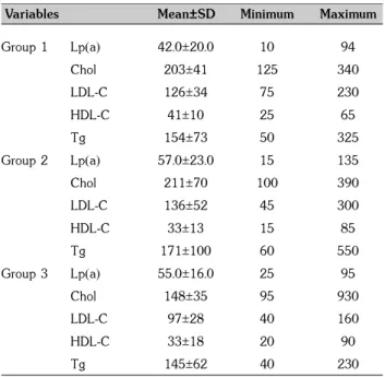
Table 2. Frequency distribution of lipids (mg/dL) in the three study groups. Lp(a): Lipoprotein (a), Chol: Cholesterol, LDL-C: LDL-choles-terol, HDL-C: HDL-cholesLDL-choles-terol, Tg: Triglycerides.
Variables Mean±SD Minimum Maximum
Group 1 Lp(a) 42.0±20.0 10 94 Chol 203±41 125 340 LDL-C 126±34 75 230 HDL-C 41±10 25 65 Tg 154±73 50 325 Group 2 Lp(a) 57.0±23.0 15 135 Chol 211±70 100 390 LDL-C 136±52 45 300 HDL-C 33±13 15 85 Tg 171±100 60 550 Group 3 Lp(a) 55.0±16.0 25 95 Chol 148±35 95 930 LDL-C 97±28 40 160 HDL-C 33±18 20 90 Tg 145±62 40 230
Table 3. Frequency distribution of plaque score in the three study groups.
Plaque score Frequency Percent
Group 1 0 27 93 1 1 3.4 2 1 3.4 3 0 0 4 0 0 Group 2 0 13 39.4 1 5 15.2 2 3 9.1 3 3 9.1 4 9 27.3 Group 3 0 22 51.2 1 3 7 2 8 18.6 3 2 4.7 4 8 18.6
between group one and both group two (P=0.016) and group three (P=0.021) but no significant difference be-tween group two and group three (P=0.962) (Scheffe test). Significant differences of plaque score between the three groups was observed (P=0.02) (chi-square test). The Scheffe test showed significant difference between
Figure 3. Significant positive correlation of plaque score with Lp(a) in the hemodialysis group (group 3) (r=0.375, P=0.008) (partial correlation test after adjustment for age).
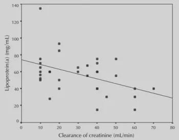
group one and both group two (P<0.001) and group three (P=0.020), but no significant difference in plaque score between group two and group three (P>0.05). There was a significant positive correlation between IMT and age in group one (P=0.035), group two (P=0.017) and group three (P=0.019) (regression analysis with stepwise method). In healthy persons (group one) signif-icant positive correlation of IMT with LDL-C (r=0.350, P=0.03), significant linear inverse correlation of IMT with HDL-C (r=-0.405, P=0.02) and marginal correlation of IMT with Tg (r=0.310, P=0.05) were found. Significant positive correlation was not found of IMT with Lp(a) (r=0.240, P>0.05) or Chol (r=0.260, P>0.05), of IMT with plaque score (r=0.101, P>0.05) or of plaque score with serum Lp(a), LDL-C, HDL-C, Chol, and Tg (P>0.05) (partial correlation test after adjustment for age). In the CRF group, significant positive correlation of IMT with plaque score (r=0.500, P=0.002), but not of IMT and plaque score with serum Lp(a), LDL-C, HDL-C, Chol or Tg (P>0.05) (partial correlation test after adjustment for age, creatinine clearance and known duration of dis-ease). In this group significant linear inverse correlation of creatinine clearance with Lp(a) (r=-0.441, P=0.040) (fig. 1) was seen, but no correlation of IMT and plaque score with creatinine clearance (P>0.05) was found (par-tial correlation test after adjustment for age). In the HD group, significant positive correlation was found of IMT with Lp(a) (r=0.298, P=0.029) (fig. 2), and of plaque score with Lp(a) (r=0.375, P=0.008) (fig. 3). No corre-lation of IMT and plaque score with Chol, LDL-C,
HDL-Figure 1. Significant linear inverse correlation of Lp(a) with creatinine clearance in the chronic renal failure group (group 2) [partial correlation test after adjustment for age (r=-0.441, P=0.06)].
Figure 2. Significant positive correlation of intima-media thickness with serum Lp(a) in the hemodialysis group (group 3) (r=0.298, P=0.029) (partial correlation test after adjustment for age).
C or Tg were found (P>0.05), or of IMT with plaque score (r=0.222, P>0.05) in this group (partial correla-tion test after adjustment for age).
DISCUSSION
The principle findings of this study were, firstly, high-er shigh-erum levels of Lp(a), more thickening of the carotid intima-media complex and more carotid-femoral artery plaque occurrence in patients with CRF, both predialy-sis and those on HD, and secondly, positive correlation
of patients. Studies concerning the effect of Lp(a) on IMT of normal persons have showed various results. Sramek and colleagues in a study on 142 asymptomatic men found no increased IMT in the carotid or femoral artery at high levels of Lp(a) and concluded that Lp(a) levels are not associated with early atherosclerotic vessel wall changes in the carotid or femoral arteries.24 Dentil
and colleagues in a study on 100 elderly subjects (aged 78.5±0.6 years) showed no association between carotid IMT and Lp(a), and concluded that the Lp(a) was unre-lated to the severity of extra-cranial vessel atherosclero-sis.25 Conversely, Baldassarre and colleagues in a study
on 100 type 2 hypercholesterolemic patients showed higher values of carotid IMT in hypercholesterolemic patients with plasma Lp(a) levels >30 mg/dL than in those with lower levels. They concluded that elevated plasma levels of Lp(a) can be considered an additional independent factor associated with carotid artery thick-ening in patients with severe hypercholesterolemia but not in those with moderate hypercholesterolemia or normocholesterolemic subjects.26 Finally, Raitakari and
colleagues in a study on 241 healthy subjects revealed no association between IMT and Lp(a) but significant positive correlation with total cholesterol, LDL-C, LDL/ HDL ratio, age and Tg.1
In renal failure patients the process of accelerated atherosclerosis is frequently observed. As an extraordi-narily high mortality in ESRD patients under HD is due to cardiovascular disease, there is increasing interest in non-traditional atherosclerotic cardiovascular disease risk factors that are prevalent in ESRD, such as Lp(a), which needs to be given more attention because of its effect on the acceleration of rapid progressive atherosclerosis seen in HD patients.
ACKNOWLEDGMENT
We would like to thank Dr M. Mowlaie (sonologist) for carotid-femoral ultrasonographies.
of serum Lp(a) with IMT and plaque score in HD pa-tients only.
Pascasio and colleagues observed a large number of vascular plaques in uremia patients, and concluded that the process of advanced atherosclerosis might be start-ed with the beginning of renal failure. They suggeststart-ed that HD treatment may not be a potential factor in the acceleration of atherosclerosis, and concluded that the progression of atherosclerosis might be related to ather-ogenic factors operative before regular dialysis is in ef-fect.18 Damjanovic and colleagues evaluated the IMT of
45 dialysis patients and found higher mean carotid IMT in HD patients than in a control group with a positive correlation of IMT with certain risk factors for athero-sclerosis (age, duration of dialysis and lipid parameters).19
Correlation of IMT with age and duration of dialysis in HD patients was evaluated by Shoji and Hojs who found no clear relationship of IMT with the duration of HD treatment.20,21 Hojs also, in his study, 28 HD patients,
observed that age was the only significant determinant of the number of plaques, and he concluded that HD patients had advanced atherosclerosis in the carotid ar-teries compared with normal subjects.21 In addition, Hojs
in a more recent study showed no difference in plaque occurrence between 28 HD patients with 28 ESRD patients prior to HD.22 Savage and colleagues in a study
on 24 dialysis patients noted the prevalence of plaque in the carotid and femoral artery and showed the rela-tionship between femoral artery plaque and age of the subjects, and also the correlation of age with carotid artery IMT in HD patients.23 A recent study by Kato
and colleagues showed a significant correlation of IMT with age in 219 HD patients.12 Papagianni and
col-leagues in a study on 112 HD patients showed a posi-tive correlation of plaque score with age.13
The present study showed a positive relationship of Lp(a) with IMT, but no clear relationship between Lp(a) and IMT in the CRF group. No differences were found between the Lp(a) and IMT of the CRF and HD groups
ÐÅÑÉËÇØÇ
...
Ó÷Ýóç ðñþéìùí áèçñïóêëçñõíôéêþí áããåéáêþí áëëïéþóåùí êáé ëéðïðñùôåÀíçò(a)
ôïõ ïñïý óå áóèåíåßò ìå ÷ñïíßá íåöñéêÞ áíåðÜñêåéá ðñéí êáé ìåôÜ áðü ôçí Ýíáñîç
ôçò ÷ñïíßáò áéìïêÜèáñóçò
H. NASRI,1 A. BARADARAN21
Shahrekord University of Medical Sciences, Hajar Medical, Educational and Therapeutic Center,
Section of Hemodialysis, Shahrekord, Iran
2Department of Biochemistry, Center of Research
and Reference Laboratory of Iran, Hospital Bou-Ali, Tehran, Iran
Áñ÷åßá ÅëëçíéêÞò ÉáôñéêÞò 2006, 23(5):514–520
ÓÊÏÐÏÓ Ç ìåëÝôç ôçò åðßäñáóçò ôçò óõãêÝíôñùóçò ôçò Lp(a) óôïí ïñü óôçí åìöÜíéóç ðñþéìùí áèçñï-óêëçñõíôéêþí áããåéáêþí áëëïéþóåùí óå áóèåíåßò ìå ÷ñïíßá íåöñéêÞ áíåðÜñêåéá (×ÍÁ) ðïõ äåí õðïâÜë-ëïíôáé óå ÷ñïíßá áéìïêÜèáñóç (×Á) êáé óå áóèåíåßò ìå ×ÍÁ ôåëéêïý óôáäßïõ ðïõ õðïâÜëõðïâÜë-ëïíôáé óå ×Á. ÕËÉÊÏ–ÌÅÈÏÄÏÓ Ðñüêåéôáé ãéá óõã÷ñïíéêÞ ìåëÝôç, óôçí ïðïßá ðåñéåëÞöèçóáí 29 öõóéïëïãéêÜ Üôïìá (ïìÜäá 1, 17 ãõíáßêåò êáé 12 Üíäñåò), 33 áóèåíåßò ìå ×ÍÁ ðïõ äåí õðïâÜëëïíôáí óå ×Á (ïìÜäá 2, 19 ãõíáßêåò êáé 14 Üíäñåò) êáé 43 áóèåíåßò ìå ×ÍÁ ôåëéêïý óôáäßïõ (ïìÜäá 3, 19 ãõíáßêåò êáé 24 Üíäñåò). Óå üëá ôá Üôïìá ôçò ìåëÝôçò ìåôñÞèçêáí ôá åðßðåäá ôçò Lp(a) óôïí ïñü. Åðßóçò, ìåôñÞèçêå ôï ðÜ÷ïò ôçò Ýóù-ìÝóçò óôéâÜäáò ôùí êáñùôßäùí (ÉÌÔ) êáé åîåôÜóôçêáí ïé êáñùôßäåò êáé ïé ìçñéáßåò áñôçñßåò ãéá ôçí ýðáñîç ðëáêþí (ðñïóäéïñéóìüò âáèìïý ðëÜêáò) ìå B-mode õðåñç÷ïãñáößá. ÁÐÏÔÅËÅÓÌÁÔÁ Ç ìÝóç ôéìÞ (±SD) ôçò óõãêÝíôñùóçò ôçò Lp(a) óôïí ïñü Þôáí óôçí ïìÜäá 1, 42±20 mg/dL, óôçí ïìÜäá 2, 57±23 mg/dL êáé óôçí ïìÜäá 3, 55±16 mg/dL. Áíôéóôïß÷ùò, ôï ÉÌÔ Þôáí 0,84±0,20 mm, 1,30±0,40 mm êáé 1,10±0,30 mm. Ìçäåíéêü âáèìü ðëÜêáò åß÷å ôï 93% ôùí áôüìùí ôçò ïìÜäáò 1, ôï 39,4% ôùí áóèåíþí ôçò ïìÜäáò 2 êáé ôï 51,2% ôùí áóèåíþí ôçò ïìÜäáò 3. Âáèìü ðëÜêáò 1–2 åß÷å ôï 6,8% ôùí áôüìùí ôçò ïìÜäáò 1, ôï 24,3% ôùí áóèåíþí ôçò ïìÜäáò 2 êáé ôï 25,6% ôùí áóèåíþí ôçò ïìÜäáò 3. Âáèìü ðëÜêáò 3–4 äåí åß÷å êáíÝíá áðü ôá Üôïìá ôçò ïìÜäáò 1, åíþ åß÷å ôï 36,4% ôùí áóèåíþí ôçò ïìÜäáò 2 êáé ôï 23,3% ôùí áóèåíþí ôçò ïìÜäáò 3. Ç äéáöïñÜ ôïõ ÉÌÔ ìåôáîý ôçò ïìÜäáò 1 áöåíüò êáé ôùí ïìÜäùí 2 êáé 3 áöåôÝñïõ Þôáí óôáôéóôéêÜ óçìáíôéêÞ (P<0,001 êáé P<0,008, áíôéóôïß÷ùò), êáèþò êáé ìåôáîý ôùí ïìÜäùí 2 êáé 3 (P=0,023). ÓçìáíôéêÝò äéáöïñÝò ùò ðñïò ôá åðßðåäá ôçò Lp(a) óôïí ïñü ðáñáôçñÞèçêáí ìåôáîý ôçò ïìÜäáò 1 áöåíüò êáé ôùí ïìÜäùí 2 (P=0,016) êáé 3 áöåôÝñïõ (P=0,021). Äåí ðáñáôçñÞèçêáí óçìáíôéêÝò äéáöïñÝò ùò ðñïò ôá åðßðåäá ôçò Lp(a) óôïí ïñü ìåôáîý ôùí ïìÜäùí 2 êáé 3 (P>0,05). ÈåôéêÝò óõó÷åôßóåéò ðáñáôçñÞèçêáí ìåôáîý ôùí åðéðÝäùí ôçò Lp(a) áöåíüò êáé ôïõ ÉÌÔ êáé ôïõ âáèìïý ðëÜêáò óôïõò áóèåíåßò ôçò ïìÜäáò 3. ÓÕÌÐÅÑÁÓÌÁÔÁ Ç Lp(a) åíäÝ÷åôáé íá äéáäñáìáôßæåé êÜðïéï óçìáíôéêü ñüëï óôçí åîÝëéîç ôçò áèçñïóêëÞñõíóçò ðïõ ðáñïõóéÜæïõí ïé áóèåíåßò ìå ×ÍÁ, ï ïðïßïò ÷ñÞæåé ðåñáéôÝñù ìåëÝôçò....
ËÝîåéò åõñåôçñßïõ: ÁéìïêÜèáñóç, Âáèìüò ðëÜêáò, ¸óù-ìÝóç óôéâÜäá, ×ñïíßá íåöñéêÞ áíåðÜñêåéá, Lp(a) References1.RAITAKARI OT, ADAMS MR, CELERMAJER DS. Effect of Lp(a) on the early functional and structural changes of atherosclero-sis. Arterioscler Thromb Vasc Biol 1999:990–995
2.OREM A, DEGER O, KULAN K, ÏÍDER E, KIRAN E, UZUNOSMA-NOGLU DET AL. Evaluation of lipoprotein(a) as a risk factor for coronary artery disease in the Ôurkish population. Clin Biochem 1995, 28:171–173
3.MBEWU AD, DURINGTON PN. Lipoprotein(a): Structure and possible involvement in thrombogenesis and atherogenesis.
Atherosclerosis 1990, 85:14
4.KIMAK E, SOLSKI J, JANICKA L, DUMA D, ZAGOJSKA M. Plasma lipoproteins in patients with chronic renal failure. Int Urol Nephrol 1997, 29:597–601
5.GREIBER S, WANNER C. Lipoprotein(a) in nephritic syndrome and end-stage renal disease. Miner Electrol Metab 1997, 23:161–165
6.DIEPLINGER H, LACKNER C, KRONENBERG F, SANDHOLZER C, LHOT-TA K, HOPPICHLER F ET AL. Elevated plasma concentrations of lipoprotein(a) in patients with end-stage renal disease are not related to the size polymorphism of apolipoprotein(a). J Clin Énvest 1993, 91:397–401
7.KRONENBERG F, TRENKWALDER E, LINGENHEL A, FRIEDRICH G, LHOTTA K, SCHOBER M ET AL. Reno-vascular arteriovenous in lipoprotein(a) plasma concentrations suggest removal of Lp(a) from the renal circulation. J Lipid Res 1997, 38:1755–1763
8.MISRA M, REAVELEY DA, COOPER C, BROWN EA, KNIGHT BL, WADE D ET AL. Mechanism for elevated plasma lipoprotein(a) con-centrations in patients on dialysis: Turnover studies. Adv Perit Dial 1998, 14:223–227
9.KODA Y, NISHI S, SUZUKI M, HIRASAWA Y. Lipoprotein(a) is a predictor for cardiovascular mortality of hemodialysis pa-tients. Kidney Int 1999, 71(Suppl):251–253
10.QUASCHNING T, KRANE V, METZGER T, WANNER C. Abnormali-ties in uremic lipoprotein metabolism and its impact on cardiovascular disease. Am J Kidney Dis 2001, 38(Suppl 1):S14–S19
11.RATTASSI M, PUATO M, FAGGIN E, BERTIPAGLIA B, GREGO F, PAU-LETTO P. New markers of accelerated atherosclerosis in end-stage renal disease. J Nephrol 2003, 16:11–20
12.KATO A, TAKAKO T, YUKITAKA M, HIROMISHI K, AKIRA H. Impact of carotid atherosclerosis on long-term mortality in chronic hemodialysis patients. Kidney Int 2003, 64:1472
13.PAPAGIANNI A, KALOVOULOS M, KRIMIZIS D, VAINAS A, BELECHRI AM, ALEXOPOULOS E ET AL. Carotid atherosclerosis is associ-ated with inflammation and endothelial cell adhesion mole-cules in chronic haemodialysis patients. Nephrol Dial Trans-plant 2003,18:113–119
14.BERNADETTE FA, MALLAMACI F, TRIPEPI G, ZOCCALI C. Prognos-tic value of ultrasonographic measurement of carotid inti-ma-media thickness in dialysis patients. J Am Soc Nephrol
2001, 12:2458–2464
15.LONGENECKER JC, CORESH J, MARCOVINA SM, POWE NR, LEVEY AS, GLACULLIF ET AL. Lipoprotein(a) and prevalent cardiovas-cular disease in a dialysis population: The choice for healthy outcomes in caring for ESRD (CHOICE) study. Am J Kidney Dis 2003, 42:108–116
16.FRIEDEWALD WT, LEVY R, FREDRICKSON DS. Estimation of the concentration of low-density lipoprotein cholesterol in plas-ma without use of the preparative ultracentrifuge. Clin Chem
1972, 18:799–802
17.COCKCROFT DW, GAULT MH. Prediction of creatinine clear-ance from serum creatinine. Nephron 1976, 16:31–41
18.PASCASIO L, BLANCO F, GIORGINI A, GALLI G, CORRI G, PANZET-TA G. Echo color doppler imaging of carotid vessels in he-modialysis patients: Evidence of high levels of atheroscle-rotic lesions. Am J Kidney Dis 1996, 28:713–720
19.DAMJANOVIC T, DIMKOVIC N. Dialysis as a risk factor for de-velopment of atherosclerosis. Med Pregl 2003, 56:1–2 20. SHOJI T, EMOTO M, TABATA T, KIMOTO E, SHINOHARA K, MAEKAWA
K ET AL. Advanced atherosclerosis in predialysis patients with chronic renal failure. Kidney Int 2002, 61:2187
21.HOJS R. Carotid intima-media thickness and plaques in he-modialysis patients. Artif Organs 2000, 24:691–695 22.HOJS R, HOJS-FABJANT, BALON BP. Atherosclerosis in patients
with end-stage renal failure prior to initiation of hemodialy-sis. Ren Fail 2003, 25:17–54
23.SAVAGE T, CLARKE AL, GILES M, TOMSON CRV, RAINE AG. Calci-fied plaque is common in the carotid and femoral arteries of dialysis patients without vascular disease. Nephrol Dial Transplant 1998, 13:2004–2012
24.SRAMAK A, REIBER JHC, BAAK-PABLO R, STURK A. Lipoprotein(a) and ultrasonographically determined early atherosclerotic changes in the carotid and femoral artery. J Thromb Hae-most 2003, 1:374–379
25.DENTI L, MARCHING L, PASOLINI G, BAFFONI MT, ABLONI F, VAL-ENTI G. Lipoprotein Lp(a) and cerebrovacsular disease in the elderly: Correlation with the severity of extra-cranial carot-id atherosclerosis assessed by ultrasonography. Acta Biomed Ateneo Parmense 1995, 66:172–183
26.BALDASSARRE D, TREMOLI E, FRANCESCHINI G, MICHELAGNOLI S, SIRTORI CR. Plasma lipoprotein(a) is an independent factor associated with carotid wall thickening in severely but not moderately hypercholesterolemic patients. Stroke 1996, 2716:1044–1049
Corresponding author:
Ç. Nasri, Shahrekord University of Medical Sciences, Hajar Medical, Educational and Therapeutic Center, Section of Hemodialysis, PO Box: 88155-468-Shahrekord-Iran, Shahre-kord, Iran
e-mail: [email protected]