*Corresponding Author; Fax: (+98-2l) 694 9389; E-mail: [email protected]; Abbreviations: Hyperactivation (HA);
Straight-Changes in Motility Parameters of Mouse Spermatozoa in
Response to Different Doses of Progesterone during Course of
Hyperactivation
Majid Katebi
*1, Mansoureh Movahedin
2, Mir Abbas Abdolvahabi
1, Mohammad Akbari
1,
Farid Abolhassani
1, Aligholi Sobhani
1and Fugaku Aoki
31
Dept. of Anatomy, Tehran University of Medical Sciences, Tehran; 2Dept. of Anatomy, Tarbiat Modarres University, Tehran, Iran; 3Graduate School of Frontier Sciences, Dept. of Integrated Biosciences, The University of Tokyo,
KashiwaCampus, Chiba, Japan
Received 3 August 2004; revised 26 December 2004; accepted 3 January 2005
ABSTRACT
The aim of this study was to evaluate changes that occur in motility parameters of progesterone treated mouse spermatozoa during course of hyperactivation. Mouse spermatozoa treated with different doses of progesterone were videotaped after 10 min and 90 min of incubation. For each sperm, one second of movement of the head-midpiece junction was traced from the videotape and for each tracing; seven motility parameters were studied using computer assisted image analysis. For all progesterone treated spermatozoa, motility rate differed significantly from control group after 90 min of incubation. Motility parameters for high doses of progesterone 10 and 100 µg/ml showed hyperactivation occurred during 10 min of incubation. With treatment of 1 µg/ml progesterone, hyperactivated motility pattern of spermatozoa occurred 90 min after incubation similar to the control group showing that low dose of progesterone is unable to induce hyperactivation. In conclusion, progesterone induces hyperactivation in mouse sperm and reduces the motility rate during the time.
Iran. Biomed. J. 9 (2): 73-79, 2005
Keywords: Progesterone, Hyperactivation, Spermatozoa, Mouse
INTRODUCTION
permatozoa are deposited in the female reproductive tract where they undergo several metabolic and structural changes, becoming fertilization-competent. These events are known as sperm capacitation and consist of several biochemical and functional modifications of sperm cells ultimately leading to changes that enable the sperm to undergo both the acrosome reaction and hyperactivation (HA).
HA is a functional change in the sperm movement pattern which usually occurs during the capacitation process [1]. This motility pattern appears to be related to the final stages or completion of capacitation [1]. Early studies described HA as a relatively progressive motion with high-amplitude flagellar bending [2]. However, later studies
referred to non-progressive motility with highly curved, ‘whiplash’ or ‘figure-of-eight’ motion [3]. Experimental evidence gathered from studying populations of mouse spermatozoa showed that HA conferred a direct mechanical advantage on spermatozoa, permitting their penetration through viscous medium [4], which could facilitate passage through oviduct and ovum investments. It has been proposed that HA may improve mammalian sperm migration through the epithelial folds of the oviduct by preventing entrapment and increasing the probability of contact with the cumulus [3].
Several studies have suggested that HA allows spermatozoa to detach from the oviductal epithelium and provides increased thrust for penetration of the cumulus [5, 6].
It has been shown that HA also facilitates penetration of spermatozoa through the oocyte zona
S
pellucida [7]. Consequently, objective measures of HA can serve as biological end-points to evaluate the functional capabilities of spermatozoa.
Blazak et al. [8] determined that quantitative
analysis of sperm motility and velocity is particularly good indicators of deleterious effects in the reproductive system. Among them, the most sensitive measures are the percentage of motile cells, mean sperm swimming speed, and linearity (LIN), as reported for rodents [9] and humans [10].
High concentrations of the steroid are present in the cumulus matrix that surrounds the oocyte which must be necessarily crossed by the sperm to reach the zona pellucida [11]. Progesterone and 17 -hydroxyprogesterone present in the follicular fluid are responsible of most of the biological effects of this fluid on spermatozoa [12]. The effects of progesterone are mediated essentially by three signaling pathway: rapid increase of intracellular Ca2+, efflux of Cl-, stimulation of phospholipases and phosphorylation of sperm proteins [13].
Sperm responsiveness to progesterone is reduced in oligozoospermic and infertile patients [14], suggesting the possibility of predicting sperm fertilizing ability or enhancing fertilization by treatment of spermatozoa in vitro with
progesterone. At present, the appropriate technique of assisted reproduction in the case of male infertility is chosen on the basis of the number of spermatozoa obtained after selection (swim up, mini-Percoll).
In the present study, we have evaluated changes that occur in motility parameters of spermatozoa in response to different doses of progesterone during course of HA as a model to choose high quality spermatozoa.
Computer-assisted image analysis has been used to characterize the motion of mouse epididymal spermatozoa and changes in motion parameters have been interpreted as indicators of effects of progesterone to identify those spermatozoa showing both increased vigour and decreased progression.
MATERIALS AND METHODS
Preparation of sperm. ICR male mice (10-12
weeks) were rapidly killed by cervical dislocation. The cauda epididymis was promptly removed. After removal of blood from epididymal surface with a physiological salt solution, the distal tubules were punctured with a 27-gauge needle in three to five places, and a mass of sperm were squeezed out with forceps into a plastic Petri dish (35 106 mm)
containing 5 ml of mWM medium prewarmed at 37°C, supplemented with 3 mg/ml bovine serum albumin (BSA; fatty acid free Fraction V, Sigma Chemical Inc., Tokyo, Japan), 100U/ml penicillin, and 0.1 mg/ml streptomycin. The spermatozoa were incubated at 37°C under 5% CO2in air.
Treatment of spermatozoa with progesterone.
Progesterone (Sigma Chemical Inc., Tokyo, Japan) was dissolved individually in absolute alcohol (1 mg/ml) and diluted serially in culture medium to obtain a final concentration of 1 µg/ml, 10 µg /ml and 100 µg /ml, and the spermatozoa were cultured for 10 min or 90 min in a CO2incubator.
Sample preparation. Each 14 µl of sperm
suspension was withdrawn to analyze the movement pattern after 10 min, 90 min of incubation. The suspension was diluted with the medium to give a final concentration of around 2106sperm per ml. The suspension was placed on a glass slide prewarmed at 37°C and covered with an 18 24 mm cover slip. The glass slide had been coated with 0.5% PVP (PolyvinylpyrrolidoneK-90), in order to minimize sticking of sperm to glass surfaces. As soon as the sample was prepared, photographs were taken for each analysis.
Experimental design. The experimental groups
were as follows: 1) Control, without progesterone; 2) Exp. 1, with 1 µg/ml progesterone; 3) Exp. 2, with 10 µg/ml progesterone; 4) Exp. 3, with 100 µg/ml progesterone. The groups were studied after 10 and 90 min of incubation.
Analysis for movement pattern of sperm. In the analysis for movement pattern of sperm, photo-graphs were taken in 30 frames per second with FASTCAM-Net high-speed camera (PHOTRON, Tokyo) and the obtained images were recorded using Movie Ruler software (PHOTRON, Tokyo). The analysis was performed using successive 30 frames. An exposure time was 1/2000 second on the phase contrast microscope (Olympus Optical Co., Tokyo, Japan). Immotile or non-progressive sperma-tozoa were omitted from analysis. The sperm motility parameters in this study were as follows:
1) Straight-line velocity (VSL, µm/s) is the straight-line distance between the first and last tracked points, divided by the acquisition time; 2) Curvilinear velocity (VCL, µm/s) is the total distance between adjacent points, divided by the time elapsed; 3) Average path velocity (VAP, µm/s)
is a smoothed path constructed by averaging several neighbouring positions on the track (five points) and joining the averaged positions, which reduced the effect of lateral head displacement; 4) LIN, an index of the straightness (STR) of the path. LIN is derived from the ratio of VSL/VCL multiplied by 100; 5) STR, an index of the departure of the sperm path from a straight line. STR is derived from the ratio of VSL/VAP multiplied by 100; 6) curvilinear progressive ratio (PRC), an inverse index of lateral head displacement. PRC is derived from the ratio of VAP/VCL multiplied by 100 [11].
In order to get motility rate, the video playback option was used. At First, the total number of sperm in a static visual field was determined using the video pause function. The tape was then allowed to run, and sperm that remained in their original position were counted as immotile or non-progressive spermatozoa.
Statistical analysis. Data were analyzed by
One-Way ANOVA followed up with post hoc test (Tukey).
RESULTS
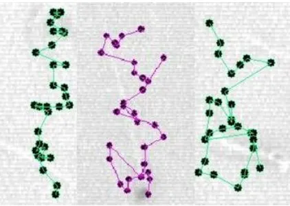
The changes in the motility rate and pattern of movement of spermatozoa under the influence of progesterone were clarified by analyzing the change in movement pattern after incubation for 10 min and 90 min in different doses of progesterone. Change in the motility pattern of mouse spermatozoa during the course of HA is shown in Figure 1.
There was no significant difference in the motility rates after 10 min of incubation for control and experimental groups. But there were significant differences between control with the experimental groups after 90 of min incubation and between experimental groups (P<0.05).
The motility rates of the experimental groups after 90 min of incubation decreased significantly (P<0.05) (Fig. 3a). After 10 min of incubation,
Experimental groups of exp. 2 and exp. 3 and after 90 min of incubation of groups of exp.1 and exp. 2, the track of movement of spermatozoa increased in length of lateral movement and followed a circular trajectory.
The motion parameters (VSL, VCL and VAP) which were studied are shown in Figure 2.
Fig. 1. Definition of changes in motility pattern during capacitation of mouse spermatozoa. Left, incapacitated or fresh sperm; Middle, intermediate sperm; Right, hyperactivated sperm.
The track of lateral movement also changed in length with time. These characteristics of the changes in movement pattern were also shown quantitatively by measuring the parameters such as: VCL, VSL, VAP, LIN, PRC and STR. VCL, an index of swimming speed, increased in the control and exp.1 after 90 min. It means that HA occurred with delay but in the exp.2 and exp.3 HA happened at 10 min and decreased after that (Fig. 3b).
As it was expected VSL, distance between the first and the last tracked points, decreased slightly during 90 min of incubation except the exp.1 which increased insignificantly (Fig. 3c).
Fig. 2. Definition of sperm tracks and related parameters. VCL, curvilinear velocity; VSL, straight-line velocity; VAP, average path velocity. The gray circles show the position of the sperm head in each frame.
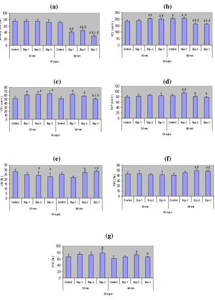
(a) (b)
(c) (d)
(e) (f)
(g)
Fig. 3. Comparison of motility parameters including motility rate (a), VCL (b), VSL (c), VAP (d), LIN (e), PRC (f), STR (g)after treatment of progesterone (0 µg /ml as control, 1 µg /ml as exp. 1, 10 µg /ml as exp. 2 and 100 µg /ml as exp. 3) within time (10 and 90 min). a, significant difference versus control; b, significant difference versus exp. 1; c, significant difference versus exp. 2; d, significant difference versus same group during 10 min.
VAP, obtained by measuring the smooth path of the average of successive 5 points, to reduce the effect of lateral head displacement, increased in the control and exp.1 groups but just significant difference in the exp.1 (Fig. 3d). LIN, an index of path STR, decreased in the control and exp.1 with time, reflecting the time dependent decrease in the radius of the circular swimming path or the increase in the lateral head displacement.
In the exp.2 and 3 as HA accrued during 10 min. LIN increased after 90 min of incubation indicating that progesterone treated spermatozoa loses HA during the 90 min of observation (Fig. 3e).
PRC, an inverse index of lateral head displacement, increased in exp.2 and 3 after 90 min. incubation showing that lateral head displacement is reducing after 90 min. incubation in high doses of progesterone (Fig. 3f). STR, an index of the departure of the sperm path from a straight line, increased in exp.3 after 10 min of incubation (Fig. 3g).
DISCUSSION
Our results showed that the motility rates of mouse spermatozoa treaded with 1 µg/ml , 10 µg/ml and 100 µg/ml progesterone after 90 min of incubation decreased significantly (P<0.05). Other
authors [15] reported low cytotoxic effect of progesterone in human spermatozoa at much higher (3-300 µg/ml) concentrations. Thérien et al. [16]
found that concentrations of progesterone higher than 2 µg/ml provoke a significant decrease of viability in ejaculated bovine spermatozoa at long incubation. Sperm motility appears to be a sensitive parameter of sperm function [17, 18].
The motility of hyperactivated spermatozoa has been analyzed using computer assisted sperm analysis (CASA) in various species, for example mice [19], humans [20], rats [21] and hamster [22]. In CASA analysis, these patterns have been compared in freshly collected and hyperactivated spermatozoa, but have not been examined over time, as HA appears to occur abruptly. However, in elucidating the mechanism of HA, it is important to examine the time-dependent changes in these parameters.
Our study showed that mouse sperm treatment with 10 µg/ml and 100 µg/ml progesterone can induce HA after 10 min incubation. However, treatment of sperm with 1 µg/ml progesterone did not induce HA suggesting that P affects the spermatozoa of mouse in a dose-dependent manner.
The high doses caused HA soon after treatment and the same results obtained by the others [23]. Other studies also failed to show a stimulation of HA by progesterone across a
wide range of progesterone concentrations [24].
Kayet al.[25] demonstrated that no effect upon HA
was detected on exposure of fresh or cryopreserved human spermatozoa to 1ug/ml progesterone. This result indicates that progesterone treated sperm had significantly lower mean straight-line velocities than non-treated sperm (P<0.05); this difference
was reversed after 90 min of incubation. This is very similar to the results previously obtained by Suarez et al. [26]. They were compared swimming
velocities of sperm incubated for 60 min with those of fresh sperm. Their results showed that hyperactivated sperm had significantly lower mean straight-line velocities than fresh sperm. Our results showed that progesterone at 10 and 100 µg/ml after 10 min incubation, significantly increased VCL than non-treated sperm (P<0.05).
After 90 min of incubation, curvilinear progressiveness ratio was increased significantly than non-treated sperm (P<0.05). Suarez et al. [27]
were measured HA of mouse spermatozoa from videotape recordings and compared with those of epididymal sperm incubated for 90 min under capacitating conditions. Their results showed higher VCL and wider-amplitude head movements as measured by PRC. Our result showed higher VCL of untreated spermatozoa after 90 min of incubation than 10 min incubation but there was no significant different in PRC of control groups. The PRC of spermatozoa after 90 min of incubation with 10 µg/ml and 100 µg/ml progesterone was increased significantly (P<0.05). Indicating that progesterone
treated spermatozoa loses the motility pattern of fresh caudal epididymal sperm during the 90 min of observation.
Kobori et al. [28] indicated that prolonged
responses required higher doses of P and their occurrence was enhanced significantly by pre-incubation for 2-4 h as compared with transient responses. Our results showed that with prolongation of time, P treated sperm lost its hyperactivated pattern which was measured using motion parameters and the others have the same results [29]. The reason could be different types of study as Kobori et al. [28] studied the features of
intracellular Ca2+ and we studied the motility parameters. Furthermore, spermatozoa may switch back and forth between transitional and whiplash motion [30] and probably they show HA pattern again in the longer time.
Standardization of methods, such as the one described here, allow accurate and objective identification of hyperactivated spermatozoa. In conclusion, progesterone stimulated HA of mouse spermatozoa in a dose-dependent manner providing a rationale to conduct studies utilizing human spermatozoa to find if the change in motility pattern of spermatozoa in response to progesterone has predicting value of sperm quality. It is necessary to evaluate the correlation between HA pattern of progesterone treated spermatozoa with its fertilization ability during IVF and ICSI until we can have an accurate judgment about HA pattern study as a model to choose high quality spermatozoa.
REFRENCES
1. Katz, D.F., Drobnis, E.Z. and Overstreet, J.W. (1989) Factors regulating mammalian sperm migration though the female reproductive tract and oocyte vestments. Gamete Res. 22: 443-469. 2. Yanagimachi, R. (1970) The movement of golden
hamster spermatozoa before and after capacitation. J. Reprod. Fertil. 23: 193-196.
3. Katz, D.F., Yanagimachi, R. and Dresder, R.D. (1978) Movement characteristics and power output of guinea pig and hamster spermatozoa in relation to activation. J. Reprod. Fertil. 52: 167-172.
4. Suarez, S.S., Katz, D.F. and Owen, D.H. (1991) Evidence for function of hyperactivated motility in sperm. Biol. Reprod. 44: 375-381.
5. Baldi, E., Krausz, C. and Forti, G. (1995) Nongenomic actions of progesterone on human spermatozoa. Trends Endocrinol. Metab. 6: 198-205.
6. Aitken, R.J. (1997) The extragenomic action of progesterone on human spermatozoa. Human Reprod. 12: 38-42.
7. Cancel, M.A., Lobdell, D., Mendola, P. and Perreault, D.S. (2000) Objective evaluation of hyperactivated motility in rat spermatozoa using computer assisted sperm analysis. Human Reprod. 15:1322-1328.
8. Blazak, W.F., Ernst, T.L. and Stewart, BE. (1985) Potential indicators of reproductive toxicity: testicular sperm production and epididymal sperm number, transit time, and motility in Fischer 344 rats. Fundam. Appl. Toxicol. 5:1097-1103.
9. Working, P.K. and Hurtt, M.E. (1987) Computerized video micrographic analysis of rat sperm motility. J. Androl. 8: 330-337.
10. Katz, D.F. and Overstreet, J.W. (1981) Sperm motility assessment by video micrography. Fertil. Steril. 35: 188-193.
11. Neill, J.M. and Olds-Clarke, P. (1987) A
computer-demonstrates that bicarbonate but not bovine serum albumin is required. Gamete Res. 18 (2):121-133. 12. Mahony, M.C. and Gwathmey, T. (1999) Protein
tyrosine phosphorylation during hyperactivated motility of cynomolgus monkey (Macaca fascicularis) Spermatozoa. Biol. Reprod. 60: 1239-1243.
13. Yanagimachi, R. (1969) In vitro capacitation of hamster spermatozoa by follicular fluid. J. Reprod. Fertil. 18: 275-286.
14. Katz, D.F. and Yanagimachi, R. (1981) Movement characteristics of hamster and guinea pig spermatozoa upon attachment to the zona pellucida. Biol. Reprod. 25: 789-791.
15. Parinaud, J., Labal, B. and Vieitez, G. (1992) High progesterone concentrations induce acrosome reaction with a low cytotoxic effect. Fertil. Steril. 58: 599-602.
16. Thérien, I. and Manjunath, P. (2003) Effect on bovine sperm capacitation and acrosome reaction. J. Biol. Reprod. 69: 1408-1415.
17. Aitken, R.J., Best, F.S.M. and Richardson, R.W. (1982) An analysis of sperm quality and sperm function in cases of oligozoospermia. Fertil. Steril. 38: 705-711.
18. Hall, J.L. (1981) Relationship between semen quality and human sperm penetration of zona free hamster ova. Fertil. Steril. 35: 457-463.
19. Neil, J.M. and Olds-Clarke, P. (1987) A computer-assissted assay for mouse sperm hperactivation demonstrates that bicarbonate but not bovine serum albumin is required. Gamete Res. 18: 121-140. 20. Mortimer, S.T. and Swan, M.A. (1995) Kinematics
of capacitating human spermatozoa analysed at 60 Hz. Human Reprod. 10: 873-879.
21. Cancel, A.M., Lobdell, D., Mendola, P. and Perreault, S.D. (2000) Objective evaluation of hyperactivated motility in rat spermatozoa using computer assisted sperm analysis. Human Reprod. 15: 1322-1328.
22. Kinukawa, M., Nagata, M. and Aoki, F. (2003) Changes in flagellar bending during the course of hyperactivation in hamster spermatozoa. Reproduction 125: 43-51.
23. Agarwal, A., Sharma, R.K. and Nelson, D.R. (2003) New semen quality scores developed by principal component analysis of semen characteristics. J. Androl. 24 (3): 343-352.
24. Emiliozzi, C., Cordonier, H. and Guerin, J.F. (1996) Effects of progesterone on human spermatozoa prepared for in vitrofertilization. Int. J. Androl. 19: 39-47.
25. Kay, V.J., Coutts, J.R. and Robertson, L. (1994) Effects of pentoxifylline and progesterone on human sperm capacitation and acrosome reaction. Human Reprod. 9: 2318-2323.
26. Suarez, S.S. and Dai, X. (1992) Hyperactivation enhances mouse sperm capacity for penetrating viscoelastic media. Biol. Reprod. 46: 686-691.
27. Suarez, S.S. and Osman, R.A. (1987) Initiation of hyperactivated flagellar bending in mouse sperm within the female reproductive tract. Biol. Reprod. 36:1191-1198.
28. Kobori, H., Miyazaki, S. and Kuwabara, Y. (2000) Characterization of intracellular (Ca 2+) increase in response to progesterone and cyclic nucleotides in mouse spermatozoa. Biol. Reprod. 63: 113-120.
29. Naser, A. and Mahony, M. (1998) Progesterone increase of intracellular calcium is not a cause of pentoxyfilline-induced hyperactivated motility or acrosome reaction in human sperm. Fertil. Steril. 69 (4): 748-754.
30. Suarez, S.S. (1996) Hyperactivated motility in sperm. J. Androl. 17: 331-335.