R E V I E W
Open Access
Effects of mesenchymal stromal
cell-conditioned media on measures of lung
structure and function: a systematic review
and meta-analysis of preclinical studies
Alvaro Moreira
1*, Rija Naqvi
1, Kristen Hall
1, Chimobi Emukah
1, John Martinez
1, Axel Moreira
2, Evan Dittmar
1,
Sarah Zoretic
1, Mary Evans
1, Delanie Moses
1and Shamimunisa Mustafa
1Abstract
Background:
Lung disease is a leading cause of morbidity and mortality. A breach in the lung alveolar-epithelial
barrier and impairment in lung function are hallmarks of acute and chronic pulmonary illness. This review is part
two of our previous work. In part 1, we demonstrated that CdM is as effective as MSCs in modulating inflammation.
Herein, we investigated the effects of mesenchymal stromal cell (MSC)-conditioned media (CdM) on (i) lung
architecture/function in animal models mimicking human lung disease, and (ii) performed a head-to-head
comparison of CdM to MSCs.
Methods:
Adhering to the animal Systematic Review Centre for Laboratory animal Experimentation protocol, we
conducted a search of English articles in five medical databases. Two independent investigators collected
information regarding lung: alveolarization, vasculogenesis, permeability, histologic injury, compliance, and
measures of right ventricular hypertrophy and right pulmonary pressure. Meta-analysis was performed to generate
random effect size using standardized mean difference with 95% confidence interval.
Results:
A total of 29 studies met inclusion. Lung diseases included bronchopulmonary dysplasia, asthma,
pulmonary hypertension, acute respiratory distress syndrome, chronic obstructive pulmonary disease, and
pulmonary fibrosis. CdM improved all measures of lung structure and function. Moreover, no statistical difference
was observed in any of the lung measures between MSCs and CdM.
Conclusions:
In this meta-analysis of animal models recapitulating human lung disease, CdM improved lung
structure and function and had an effect size comparable to MSCs.
Keywords:
Conditioned media, Mesenchymal stem cell, Lung disease, Animal, Review
© The Author(s). 2020Open AccessThis article is licensed under a Creative Commons Attribution 4.0 International License, which permits use, sharing, adaptation, distribution and reproduction in any medium or format, as long as you give appropriate credit to the original author(s) and the source, provide a link to the Creative Commons licence, and indicate if changes were made. The images or other third party material in this article are included in the article's Creative Commons licence, unless indicated otherwise in a credit line to the material. If material is not included in the article's Creative Commons licence and your intended use is not permitted by statutory regulation or exceeds the permitted use, you will need to obtain permission directly from the copyright holder. To view a copy of this licence, visithttp://creativecommons.org/licenses/by/4.0/. The Creative Commons Public Domain Dedication waiver (http://creativecommons.org/publicdomain/zero/1.0/) applies to the data made available in this article, unless otherwise stated in a credit line to the data.
* Correspondence:[email protected]
1Department of Pediatrics, Division of Neonatology, University of Texas
Background
Pulmonary illness is a leading cause of morbidity and
mortality [
1
]. In children, acute respiratory exacerbations
are a common reason for primary care visits and are
often implicated in hospitalizations [
2
,
3
]. Many of these
pulmonary conditions result in impairments in lung
function that may last into adulthood [
4
,
5
].
Conse-quently, identifying novel therapies for lung disease is
highly warranted.
A unifying theme in many lung diseases includes
in-flammation [
6
–
8
]. While some inflammation is
neces-sary to combat new disease and for proper wound
healing, chronic inflammation may result in altered lung
structure and function. During an acute illness, current
therapies focus on restoring lung function by abating
in-flammation [
9
–
11
]. For instance, glucocorticoids are the
mainstay therapy for reducing inflammation during
acute exacerbations of asthma [
12
]. More recently,
mes-enchymal stromal/stem cells (MSCs) have shown
en-couraging
outcomes
in
animal
models
of
lung
inflammation [
13
–
15
].
MSCs are promising agents as they are easily
har-vested, can be rapidly expanded, and can secrete factors
(exosomes, microvesicles, microRNA) known to reduce
inflammation [
16
–
18
]. The
“
secretome
”
or
“
conditioned
media
”
of MSCs is considered biologically active and can
be easily collected from the surrounding fluid of
propa-gating cells [
19
–
21
]. Remarkably, preclinical studies
sug-gest MSC conditioned media (CdM) may be as
restorative as the MSCs themselves [
22
,
23
]. We
sup-ported this observation in a previous systematic review
and meta-analysis demonstrating that CdM is as
effect-ive as MSCs in modulating inflammation [
24
].
This review is an extension of our previous work. In
this review, we examined the effects of CdM on (i) lung
architecture/function in animal models recapitulating
lung disease and (ii) compare these findings to MSCs.
Given that the therapeutic benefit of MSCs is attributed
to a paracrine fashion, we believed CdM would have
comparable effects to MSCs.
Methods
Overview and literature search
The methods in our review abide to those outlined by
the Systematic Review Centre for Laboratory Animal
Ex-perimentation (SYRCLE) [
25
]. Our protocol was
regis-tered through the Collaborative Approach to
Meta-Analysis and Review of Data from Experimental Studies
(CAMARADES) [
26
]. Details are described in our
previ-ous publication.
We conducted a literature search in five databases
using the following terms: mesenchymal stem
cell-conditioned media, lung disease, and animal. The last
search was performed on March 17th, 2020. Three
independent investigators evaluated titles and abstracts,
followed by full-text review.
Inclusion criteria and outcomes of interest
We included studies administering MSC-CdM to animal
models of acute lung injury or acute respiratory distress
syndrome (ALI/ARDS), asthma, bronchopulmonary
dys-plasia (BPD), chronic obstructive pulmonary disease
(COPD), cystic fibrosis (CF), pneumonia, pulmonary
fi-brosis (PF), and pulmonary hypertension (PH). Refer to
Supplementary File
1
for the list of included studies.
Outcomes of interest
Measures of lung structure and/or function were our
primary endpoint. Lung architecture and function were
assessed under the following categories: alveolarization,
vasculogenesis, right ventricular hypertrophy, fibrosis,
permeability, pulmonary pressures, compliance, and lung
injury. Although the pathogenesis of the included lung
diseases are heterogeneous, we combined all processes
irrespective of disease. This was conducted to obtain a
scoping overview of the impact of CdM on biologic
pro-cesses implicated in lung disease. Subsequently, we
assessed lung structure/function by disease in our
sub-group analysis. Excluded studies were those which did
not provide data concerning our primary outcome of
inflammation.
Data extraction
Three groups of investigators were used (ED and CE;
RN and JM; ME, DM, and SM) to collect data.
Uniform-ity of data was assessed by the primary author. This data
included general study design, animal model
characteris-tics, conditioned media characterischaracteris-tics, and outcomes of
interest.
Data analysis
Results
Study selection
Our literature search resulted in 245 articles. After
re-moving duplicates and viewing the titles and abstracts,
55 articles underwent full-text review. Twenty-nine
arti-cles met inclusion (refer to Supplementary Figure
1
).
Study details
Table
1
summarizes the relevant study characteristics.
Articles included in the review were published between
the years 2009 to 2020. BPD was the most common
ani-mal model (
n
= 8), followed by ALI/ARDS (
n
= 5) and
asthma (
n
= 5). All of the studies used rodents to induce
their lung model.
CdM characteristics
Conditioned media properties are summarized in
Sup-plementary File
3
. Stem cells were most isolated from
bone marrows and cultured in Dulbecco
’
s modified
Ea-gle
’
s medium. Incubation time of the CdM ranged from
24 to 72 h. The volume of CdM administered ranged
from 25
μ
l to 1 ml.
Alveolarization
CdM
: improved alveolarization with an SMD of 1.32
(95% CI 0.99, 1.65; 12 studies; Fig.
1
a) with
moderate heterogeneity (
I
2= 67%;
p
< 0.01).
MSC
: improved alveolarization with an SMD of 1.80
(95% CI 1.52, 2.07; 9 studies; Fig.
1
b) with mild
heterogeneity between groups (
I
2= 36%;
p
= 0.01).
CdM
vs.
MSC
: no significant difference
(Supplementary Figure
2
).
Right ventricular hypertrophy
CdM
: favored CdM over control with an SMD of
−
1.08 (95% CI
−
1.56,
−
0.61); 6 studies; Fig.
2
a) with
significant heterogeneity (
I
2= 70%;
p
< 0.01).
MSC
: favored over the control with an SMD of
−
1.05 (95% CI
−
1.69,
−
0.42; 3 studies, Fig.
2
b) with
significant heterogeneity between groups (
I
2= 71%;
p
< 0.01).
CdM
vs.
MSC
: no significant difference (SMD
−
0.22, 95% CI
−
0.36, 0.16; Supplementary Figure
3
).
Lung fibrosis
CdM
: favored CdM over control with an SMD of
−
1.08 (95% CI
−
1.56,
−
0.61; 6 studies; Fig.
3
a) with
significant heterogeneity (
I
2= 70%;
p
< 0.01).
MSC
: favored MSC over the control with an SMD
of
−
1.99 (95% CI
−
2.93,
−
1.04; 4 studies; Fig.
3
b)
a
b
with significant heterogeneity between groups (
I
2=
90%;
p
< 0.01).
CdM
vs.
MSC
: the comparison between CdM
and MSCs was similar (refer to Supplementary
Figure
4
).
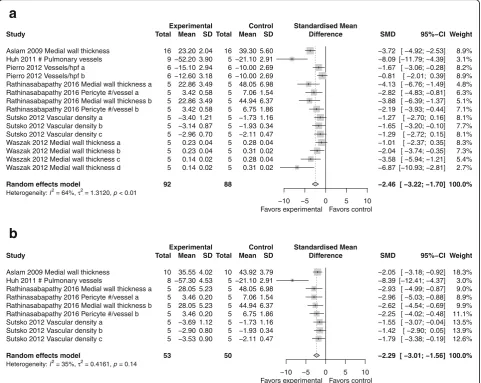
Vasculogenesis
CdM
: superior to control with an SMD of
−
2.46
(95% CI
−
3.22,
−
1.70; 6 studies; Fig.
4
a) with
moderate heterogeneity (
I
2= 76%;
p
< 0.01).
MSC
: superior to control with an SMD of
−
2.29
(95% CI -3.01,
−
1.56; 4 studies; Fig.
4
b) with mild
heterogeneity between groups (
I
2= 35%;
p
= 0.14).
CdM
vs.
MSC
: overall effectiveness between CdM
and MSCs again showed no significant difference
(Supplementary Figure
5
).
Permeability
CdM
: permeability assessment favored CdM over
control with an SMD of
−
0.99 (95% CI
−
1.32,
−
a
b
a
b
0.66; 5 studies; Fig.
5
a) homogeneity that is
non-significant (
I
2= 11.0%;
p
= 0.33).
MSC
: in the evaluation of permeability, the MSC
was favored over the control with an effect size of
−
1.54 (95% CI -2.13,
−
0.95; 4 studies; Fig.
5
b) with
heterogeneity between groups (
I
2= 57.0%;
p
< 0.01).
CdM
vs.
MSC
: equal effectiveness (Supplementary
Figure
6
).
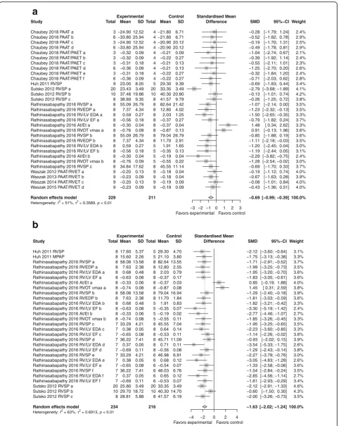
Pulmonary pressures
CdM
: improvement in right ventricular pressures
compared to control with an SMD of
−
0.69 (95% CI
−
0.99,
−
0.39; 5 studies; Fig.
6
a) with moderate
heterogeneity (
I
2= 51%;
p
< 0.01).
MSC
: superior to control with an SMD of
−
1.63
(95% CI
−
2.02,
−
1.24; 3 studies; Fig.
6
b) with
moderate heterogeneity (
I
2= 63%;
p
< 0.01).
CdM
vs.
MSC
: comparable (please refer to
Supplementary Figure
7
).
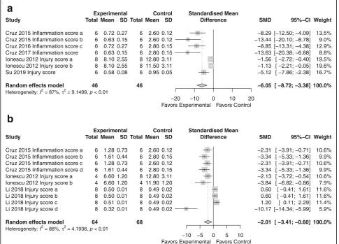
Histologic lung injury
CdM
: improvement in histologic lung injury
compared to control with an SMD of
−
6.05 (95% CI
−
8.72,
−
3.38; 3 studies; Fig.
7
a) with significant
heterogeneity (
I
2= 87%;
p
< 0.01).
MSC
: superior to control with an SMD of
−
2.01
(95% CI -3.41,
−
0.60; 3 studies; Fig.
7
b) with
significant heterogeneity (
I
2= 88%;
p
< 0.01).
CdM
vs.
MSC
: less than 3 studies; comparison not
performed.
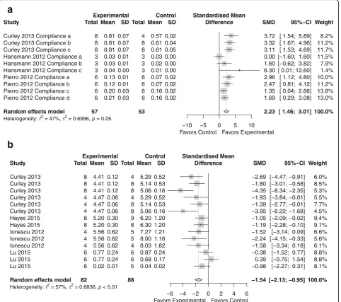
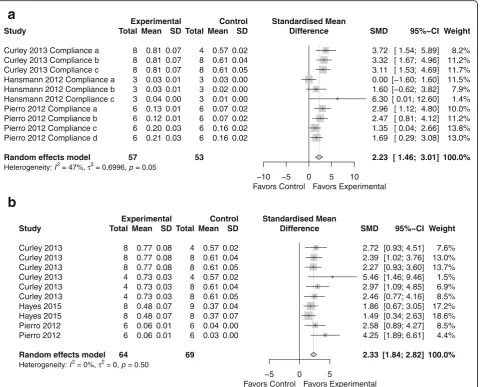
Compliance
CdM
: improvement in lung compliance compared to
control with an SMD of 1.75 (95% CI 0.81, 2.69; 4
a
b
studies; Fig.
8
a) with significant heterogeneity (
I
2=
76%; p < 0.01).
MSC
: improvement in lung compliance compared to
control with an SMD of 2.33 (95% CI 1.84, 2.82;
3 studies; Fig.
8
b) with no heterogeneity (
I
2= 0%;
p
= 0.5).
CdM
vs.
MSC
: not applicable as less than three
studies performed a head-to-head comparison.
All outcomes for lung structure and function combined
CdM
: Supplementary Figure
8
A shows the SMD of
−
1.38 (with 95% CI of
−
1.57,
−
1.19) favoring CdM
over control.
MSC
: Supplementary Figure
8
B shows the SMD of
−
1.66 (with 95% CI of
−
1.91,
−
1.41) favoring MSC
over control.
CdM
vs.
MSC
: no difference was appreciated
between CdM and MSC when all outcomes were
combined (Supplementary Figure
8
C).
Subgroup analysis
Stratification of data was performed by lung disease,
tis-sue source, dose, and route of delivery of CdM.
Evalu-ation was performed if more than 6 studies had data.
Alveolarization
Supplementary Figure
9
A
–
D demonstrates that CdM
had the greatest impact on alveolarization in BPD
a
b
a
b
animal models (SMD 1.67) and when the media was
derived from cord blood (SMD 2.89), given at a dose
of 7
μ
l/g (SMD 2.89), and delivered via the
intraperi-toneal route (SMD 1.56).
RVH
Supplementary Figure
10
A
–
D depicts that CdM
sig-nificantly improved RVH in BPD animal models
(SMD
−
0.93) and only when the media was derived
from adipose tissue (SMD
−
1.05), given at a dose
of 100
μ
l (SMD
−
1.14) and delivered intravenously
(SMD
−
0.86).
Fibrosis
Supplementary Figure
11
A
–
D illustrates that CdM had
the greatest impact in animal models of BPD and PH
(SMD
−
4.1,
−
3.4, respectively) and when the media was
derived from adipose tissue (SMD
−
2.61), given at a
dose of 50
μ
l (SMD
−
4.10) and delivered intravenously
(SMD
−
1.95).
Vascularization
Supplementary Figure
12
A
–
D shows that CdM had the
greatest impact in animal models of COPD (SMD
−
8.09), when the media was derived from adipose tissue
(SMD
−
2.61), given at a dose of 300
μ
l (SMD
−
8.09)
and delivered intravenously (SMD
−
3.65).
Risk of bias
No study was judged as low risk across all ten domains.
Eight studies stated that the allocation selection was
ran-dom. Most studies (
n
= 25) had similar groups at
base-line. Risk of bias was large regarding allocation
concealment, whether authors mention random housing
of animals, and blinding of caregivers or random
selec-tion of outcome. All studies were found to sufficiently
report complete data and being free from other bias.
Refer to Supplementary File
2
[
27
].
Publication bias
Supplementary Figures
13
,
14
,
15
,
16
,
17
,
18
,
19
, and
20
illustrate publication bias through funnel plots. Overall,
a
b
publication bias was low in all the outcomes except for
lung permeability.
Discussion
Preclinical studies reiterate the ability MSCs have on
dampening lung inflammation. This capacity is largely
due to the paracrine secretion of MSC factors
(microve-sicles, exosomes) that provide a basis for future cell-free
therapies for human disease [
28
–
31
]. This is the first
re-view to directly compare the effects of CdM vs MSCs on
lung structure and function in animal models of diverse
lung disease. Overall, we found that CdM improved
measures of alveolarization, right ventricular
hyper-trophy, lung fibrosis, vasculogenesis and permeability.
Furthermore, CdM reduced pulmonary pressures,
ame-liorated histologic lung injury, and increased lung
com-pliance. We found that CdM was comparable to MSCs
in all lung measures evaluated individually and when
combined.
The bioactive factors contained in the CdM of MSCs
have been the focus of multiple studies and review
arti-cles [
32
–
34
]. Congruent with the findings found in this
review, Hansmann et al. show that MSC-CdM,
com-pared to CdM from lung fibroblasts, reversed alveolar
injury, normalized lung function (airway resistance),
and reversed RVH [
35
]. Additionally, the same group
re-cently demonstrated that MSC exosomes (molecular
cargo found within CdM) restored lung architecture,
stimulated pulmonary blood vessel formation, and
mod-ulated lung inflammation [
22
]. In an
E. coli
pneumonia-induced ALI mouse model, MSC microvesicles (also
found in MSC-CdM) reduced lung permeability and
histologic injury score and were equivalent to MSCs
[
36
]. Together, these findings, and those in recent
a
b
reviews, substantiate the results found in this review [
37
,
38
].
This year, Augustine et al. published a network
meta-analysis comparing stem cell and cell-free therapies in
preclinical measures of BPD. MSC-CdM had a similar
effect size to MSCs regarding alveolarization (MSC SMD
1.71 vs. CdM SMD1.68), angiogenesis (SMD 2.24 vs.
1.79), and pulmonary remodeling (1.29 vs. 1.22) [
39
].
Similar to their results, this review showed that CdM
had among the largest impact on measures of
alveolari-zation and vasculogenesis, processes critical for
appro-priate lung healing, development, and function [
40
].
Although vasculogenesis/angiogenesis is an important
process to restore lung function/structure, it can also
en-hance remodeling and thus worsen outcomes in other
lung diseases such as asthma or pulmonary fibrosis [
41
].
In Supplementary Figure
12
A, we demonstrate that this
process improved in BPD, pulmonary hypertension, and
COPD but was not assessed in asthma/pulmonary
fibrosis.
In the study by Hayes et al., they found that MSCs
were superior to CdM in a rodent model of
ventilator-induced lung injury. However, our review suggests that
when you compile the literature, there were no
signifi-cant benefits of using cells over CdM. We cannot
ex-plain why CdM was not comparable in this study;
however, an important challenge that remains in the
field includes the rigorous testing of key variables (tissue
source, dose, route, disease, etc.) that may impact the
quality of CdM [
42
–
44
]. For instance, we found that the
intravenous route provided optimal results. Moreover,
multiple administrations of CdM may augment vascular
development, as seen in the study by Huh et al (
n
= 10
intravenous injections). Conversely, the optimal source
and dose of CdM is dependent on the variable or the lung
disease. This brings to light that it will be incredibly
chal-lenging to find a single CdM product that is ideal for all
lung diseases. Thus, the idea of
“
one-size-fits-all
”
does not
hold true for regenerative cells or products. Illustrating
this concept, Rathinasabapathy et al. showed greater
im-provement in measures of RVH compared to other studies
measuring right ventricular size. Important differences
seen in the study by Rathinasabapathy and colleagues was
that they used a different animal model (PH vs. BPD)
and age of rodents (adult vs. neonatal) [
45
].
As investigators, we should attempt to tease out these
characteristics in order to have the ideal product(s) for
our lung disease of interest. In this way, we may have
translational success in future clinical studies. Refining
these features will take time but will play a vital role in
efficacy. Moreover, pinpointing small and large animal
models of lung disease that will recapitulate what occurs
at the patient bedside is essential if we want to move the
needle in the field [
46
].
The plausibility of using a cell-free product as a
therapeutic agent for lung disease is substantiated by
newly registered human clinical trials. For instance,
NCT04235296
and
NCT04234750
are
evaluating
safety of MSC-CdM in regulating wound
inflamma-tion and promoting wound healing in burn injury.
Another Phase I trial (NCT04134676) plans to study
the therapeutic potential of umbilical cord
tissue-derived stem cell CdM on chronic skin ulcers. Trials
valuing the safety of stem cell CdM constituents
(exo-somes)
are
also
underway
for
ischemic
stroke
(NCT3384433) and ocular conditions (NCT04213248,
NCT03437759).
There are several limitations to our systematic review
and meta-analysis, many of which mirror those
pub-lished in our previous report. We incorporated multiple
animal models of lung disease that have diverse
patho-logic processes resulting in their etiology. Also, most of
the studies lacked methodologic details rendering them
with an unclear risk of bias. Moreover, although
preclin-ical models of lung disease have been helpful in
identify-ing targetable mechanisms/processes, they oftentimes
lack the intricacies of human disease. Thus, meticulous
efficacy studies in large animals may be one approach to
mitigate translational failure in human trials.
Conclusion
This review demonstrates that the administration of
CdM in animal models of lung disease improves lung
architecture and function. When compared to MSCs,
CdM is as efficacious and provides a basis that cell-free
products are a viable option for future studies. However,
mores studies are needed to identify how specific
vari-ables (tissue source, route of delivery, concentration,
etc.) may impact/strengthen their therapeutic potential.
Supplementary information
Supplementary informationaccompanies this paper athttps://doi.org/10. 1186/s13287-020-01900-7.
Additional file 1: Figure S1.Flow diagram demonstrating study selection process.
Additional file 2: Figure S2.Effect size of CdM vs. MSC on lung alveolarization. . Forest plots demonstrate SMD with 95% confidence interval.
Additional file 3: Figure S3.Effect size of CdM on right ventricular hypertrophy. Forest plots demonstrate SMD with 95% confidence interval.
Additional file 4: Figure S4.Effect size of MSC on lung fibrosis. Forest plots demonstrate SMD with 95% confidence interval.
Additional file 5: Figure S5.Effect size of CdM vs. MSC on pulmonary vasculogenesis. Forest plots demonstrate SMD with 95% confidence interval.
Additional file 7: Figure S7.Effect size of CdM vs. MSC on pulmonary pressures. Forest plots demonstrate SMD with 95% confidence interval.
Additional file 8: Figure S8.Effect size of CdM (a), MSCs (b), and CdM vs. MSC (c) on all eight outcomes. Forest plots demonstrate SMD with 95% confidence interval.
Additional file 9: Figure S9.Effect size of CdM on lung alveolarization by disease (a), source (b), dose (c), and route (d). Forest plots
demonstrate SMD with 95% confidence interval.
Additional file 10: Figure S10.Effect size of CdM on right ventricular hypertrophy by disease (a), source (b), dose (c), and route (d). Forest plots demonstrate SMD with 95% confidence interval.
Additional file 11: Figure S11.Effect size of CdM on lung fibrosis by disease (a), source (b), dose (c), and route (d). Forest plots demonstrate SMD with 95% confidence interval.
Additional file 12: Figure S12.Effect size of CdM on pulmonary vascularization by disease (a), source (b), dose (c), and route (d). Forest plots demonstrate SMD with 95% confidence interval.
Additional file 13: Figure S13.Funnel plot assessing for publication bias of CdM on lung alveolarization.
Additional file 14: Figure S14.Funnel plot assessing for publication bias of CdM on right ventricular hypertrophy.
Additional file 15: Figure S15.Funnel plot assessing for publication bias of CdM on lung fibrosis.
Additional file 16: Figure S16.Funnel plot assessing for publication bias of CdM on pulmonary vasculogenesis.
Additional file 17: Figure S17.Funnel plot assessing for publication bias of CdM on lung permeability.
Additional file 18: Figure S18.Funnel plot assessing for publication bias of CdM on pulmonary pressures.
Additional file 19: Figure S19.Funnel plot assessing for publication bias of CdM on histologic lung injury.
Additional file 20: Figure S20.Funnel plot assessing for publication bias of CdM on lung compliance.
Additional file 21: File S1.List of articles included in this review.
Additional file 22: File S2.SYRCLE risk of bias.
Additional file 23: File S3.CdM characteristics.
Abbreviations
ARDS:Acute respiratory distress syndrome; BPD: Bronchopulmonary dysplasia; CAMARADES: Collaborative Approach to Meta-Analysis and Review of Data from Experimental Studies; CdM: Conditioned media; CF: Cystic fibrosis; CI: Confidence interval; MSC: Mesenchymal stem/stromal cell; NCT: National clinical trial; PH: Pulmonary hypertension; SMD: Standardized mean difference; SYRCLE: Systematic Review Centre for Laboratory Animal Experimentation
Acknowledgements
Dr. Robert Cote, Director of the Writing Skills Program at the University of Arizona, Tucson.
Authors’contributions
AM contributed to the design of review, analysis of data, manuscript data, and oversight. RN contributed to the data collection, manuscript writing, and risk of bias. KH contributed to the data collection and manuscript writing. CE contributed to the database search, design of review, assembly of data, and analysis of data. JM contributed to the data collection. AM contributed to the design of review, manuscript writing, and interpretation of data. ED contributed to the database search, design of review, and data collection. SZ contributed to the manuscript writing and interpretation of data. ME contributed to the data collection, risk of bias, and Tables 1 and 2. DM contributed to the data collection and Tables 1 and 2. SM contributed to the data collection and Tables 1 and 2. The author(s) read and approved the final manuscript.
Funding
Supported by NIH NCATS KL2 TR001118, UT Health San Antonio School of Medicine pilot grant, Parker B. Francis Foundation (provided to senior author).
Availability of data and materials
Availability of data and materials will be available through figshare upon publication of the manuscripts.
Ethics approval and consent to participate Not applicable.
Consent for publication
All authors have looked through the manuscript and approved the submission.
Competing interests
The authors declare that they have no competing interests.
Author details
1Department of Pediatrics, Division of Neonatology, University of Texas
Health Science-San Antonio, San Antonio, TX 78229-3900, USA.2Department of Pediatrics, Division of Critical Care, Baylor College of Medicine, Houston, TX, USA.
Received: 12 February 2020 Revised: 19 August 2020 Accepted: 24 August 2020
References
1. Ferkol T, Schraufnagel D. The global burden of respiratory disease. Ann Am
Thorac Soc. 2014;11:404–6. Available from:http://www.atsjournals.org/doi/
abs/10.1513/AnnalsATS.201311-405PS. [cited 2019 Sep 30].
2. Winterstein AG, Choi Y, Cody MH. Association of age with risk of
hospitalization for respiratory syncytial virus in preterm infants with chronic
lung disease. JAMA Pediatr. 2018;172:154–60.
3. Maxwell BG, Nies MK, Ajuba-Iwuji CC, Coulson JD, Romer LH. Trends in
hospitalization for pediatric pulmonary hypertension. Pediatrics. 2015;136:
241–50.
4. Bui DS, Lodge CJ, Burgess JA, Lowe AJ, Perret J, Bui MQ, et al. Childhood
predictors of lung function trajectories and future COPD risk: a prospective cohort study from the first to the sixth decade of life. Lancet Respir Med.
2018;6:535–44.
5. Savran O, Ulrik CS. Early life insults as determinants of chronic obstructive
pulmonary disease in adult life. Int J Chron Obstruct Pulmon Dis. 2018;13: 683–93.https://doi.org/10.2147/COPD.S153555. Published 2018 Feb 26.
6. Zhou-Suckow Z, Duerr J, Hagner M, Agrawal R, Mall MA. Airway mucus,
inflammation and remodeling: emerging links in the pathogenesis of
chronic lung diseases. Cell Tissue Res. 2017;367(3):537–50.
7. Virk H, Arthur G, Bradding P. Mast cells and their activation in lung disease.
Transl Res. 2016;174:60–76.
8. Barnes PJ. Cellular and molecular mechanisms of asthma and COPD. Clin Sci
(Lond). 2017;131(13):1541–58.
9. Robinson D, Humbert M, Buhl R, et al. Revisiting Type high and Type
2-low airway inflammation in asthma: current knowledge and therapeutic
implications. Clin Exp Allergy. 2017;47(2):161–75.
10. Becerra-Díaz M, Wills-Karp M, Heller NM. New perspectives on the
regulation of type II inflammation in asthma. F1000Research. 2017;6:1014.
Available from:http://www.ncbi.nlm.nih.gov/pubmed/28721208. [cited 2019
Jun 11].
11. Dias-Freitas F, Metelo-Coimbra C, Roncon-Albuquerque R Jr. Molecular
mechanisms underlying hyperoxia acute lung injury. Respir Med. 2016;119:
23–8.
12. Barsky EE, Giancola LM, Baxi SN, Gaffin JM. A Practical Approach to Severe
Asthma in Children [published correction appears in Ann Am Thorac Soc.
2018;15(6):767–8.
13. Curley GF, Hayes M, Ansari B, Shaw G, Ryan A, Barry F, et al. Mesenchymal
stem cells enhance recovery and repair following ventilator-induced lung
injury in the rat. Thorax. 2012;67:496–501. Available from:http://www.ncbi.
nlm.nih.gov/pubmed/22106021. [cited 2017 Dec 16].
14. Zhong H, Fan XL, Fang S, Bin LYD, Wen W, Fu QL. Human pluripotent stem
inflammation via TGF-β1-Smad2/Smad3 signaling pathway in mice. Mol
Immunol. 2019;109:51–7.
15. Cui P, Xin H, Yao Y, et al. Human amnion-derived mesenchymal stem cells
alleviate lung injury induced by white smoke inhalation in rats. Stem Cell Res Ther. 2018;9(1):101.
16. Phinney DG, Pittenger MF. Concise review: MSC-derived exosomes for cell
free therapy. Stem Cells. 2017;35:851–8.
17. Vizoso FJ, Eiro N, Cid S, Schneider J, Perez-Fernandez R. Mesenchymal stem
cell secretome: toward cell-free therapeutic strategies in regenerative
medicine. Int J Mol Sci. 2017;18 Available from:http://www.ncbi.nlm.nih.
gov/pubmed/28841158. [cited 2017 Dec 16].
18. Moreira A, Kahlenberg S, Hornsby P. Therapeutic potential of mesenchymal
stem cells for diabetes. J Mol Endocrinol. 2017;59:R109–20. Available from:
http://www.ncbi.nlm.nih.gov/pubmed/28739632. [cited 2017 Nov 30].
19. Zakrzewski W, Dobrzyński M, Szymonowicz M, Rybak Z. Stem cells: past,
present, and future. Stem Cell Res Ther. 2019;10(1):68.
20. Cruz FF, PRM R. Hypoxic preconditioning enhances mesenchymal stromal
cell lung repair capacity. Stem Cell Res Ther. 2015;6:130. Available from:
http://www.ncbi.nlm.nih.gov/pubmed/26169784. [cited 2017 Nov 16].
21. Ferreira JR, Teixeira GQ, Santos SG, Barbosa MA, Almeida-Porada G,
Gonçalves RM. Mesenchymal stromal cell secretome: influencing therapeutic potential by cellular pre-conditioning. Front Immunol. 2018;9: 2837.
22. Willis GR, Fernandez-Gonzalez A, Anastas J, Vitali SH, Liu X, Ericsson M, et al.
Mesenchymal stromal cell exosomes ameliorate experimental bronchopulmonary dysplasia and restore lung function through
macrophage immunomodulation. Am J Respir Crit Care Med. 2018;197:104–
16. Available from:http://www.ncbi.nlm.nih.gov/pubmed/28853608. [cited
2019 Jun 21].
23. van Haaften T, Byrne R, Bonnet S, Rochefort GY, Akabutu J, Bouchentouf M,
et al. Airway delivery of mesenchymal stem cells prevents arrested alveolar growth in neonatal lung injury in rats. Am J Respir Crit Care Med. 2009;180:
1131–42. Available from:http://www.ncbi.nlm.nih.gov/pubmed/19713449.
[cited 2016 Oct 27].
24. Emukah C, Dittmar E, Naqvi R, et al. Mesenchymal stromal cell conditioned
media for lung disease: a systematic review and meta-analysis of preclinical studies. Respir Res. 2019;20(1):239.
25. Zeng X, Zhang Y, Kwong JS, et al. The methodological quality assessment
tools for preclinical and clinical studies, systematic review and metaanalysis, and clinical practice guideline: a systematic review. J Evid Based Med. 2015;
8(1):2–10.
26. Welcome to CAMARADES. Available from:http://www.dcn.ed.ac.uk/
camarades/. [cited 2020 Jan 30].
27. Hooijmans CR, Rovers MM, De Vries RBM, Leenaars M, Ritskes-hoitinga M,
Langendam MW. SYRCLE’s risk of bias tool for animal studies. BMC Med Res
Methodol. 2014;14:1–9.
28. Bruno S, Kholia S, Deregibus MC, Camussi G. The role of extracellular
vesicles as paracrine effectors in stem cell-based therapies. Adv Exp Med
Biol. 2019;1201:175–93.
29. Joo HS, Suh JH, Lee HJ, Bang ES, Lee JM. Current knowledge and future
perspectives on mesenchymal stem cell-derived exosomes as a new therapeutic agent. Int J Mol Sci. 2020;21(3):727. Published 2020 Jan 22.
https://doi.org/10.3390/ijms21030727.
30. Liu C, Wang J, Hu J, Fu B, Mao Z, Zhang H, et al. Extracellular vesicles for
acute kidney injury in preclinical rodent models: a meta-analysis. Stem Cell Res Ther. 2020;11:11.
31. Willis GR, Mitsialis SA, Kourembanas S.“Good things come in small
packages”: application of exosome-based therapeutics in neonatal lung
injury. Pediatr Res. 2018;83:298–307. Available from:http://www.nature.com/
articles/pr2017256. [cited 2019 Jul 12].
32. Madrigal M, Rao KS, Riordan NH. A review of therapeutic effects of
mesenchymal stem cell secretions and induction of secretory modification by different culture methods. J Transl Med. 2014;12:260.
33. Pawitan JA. Prospect of stem cell conditioned medium in regenerative
medicine. Biomed Res Int. 2014;2014:965849. Available from:http://www.
ncbi.nlm.nih.gov/pubmed/25530971. [cited 2019 Jul 20].
34. Augustine S, Avey MT, Harrison B, Locke T, Ghannad M, Moher D, et al.
Mesenchymal stromal cell therapy in bronchopulmonary dysplasia: Systematic review and meta-analysis of preclinical studies. Stem Cells Transl
Med. 2017;6:2079–93. Available from:http://www.ncbi.nlm.nih.gov/
pubmed/29045045. [cited 2019 Mar 22].
35. Hansmann G, Fernandez-Gonzalez A, Aslam M, Vitali SH, Martin T, Mitsialis
SA, et al. Mesenchymal stem cell-mediated reversal of bronchopulmonary
dysplasia and associated pulmonary hypertension. Pulm Circ. 2012;2:170–81.
Available from:http://www.ncbi.nlm.nih.gov/pubmed/22837858. [cited 2018
Jan 18].
36. Zhu YG, Feng XM, Abbott J, Fang XH, Hao Q, Monsel A, et al. Human
mesenchymal stem cell microvesicles for treatment of Escherichia coli
endotoxin-induced acute lung injury in mice. Stem Cells. 2014;32:116–25.
37. Lanyu Z, Feilong H. Emerging role of extracellular vesicles in lung injury and
inflammation. Biomed Pharmacother. 2019;113:108748.
38. Fujita Y, Kadota T, Araya J, Ochiya T, Kuwano K. Clinical application of
mesenchymal stem cell-derived extracellular vesicle-based therapeutics for inflammatory lung diseases. J Clin Med. 2018;7:355.
39. Augustine S, Cheng W, Avey MT, Chan ML, Lingappa SMC, Hutton B, et al.
Are all stem cells equal? Systematic review, evidence map, and meta-analyses of preclinical stem cell-based therapies for bronchopulmonary
dysplasia. Stem Cells Transl Med. 2020;9:158–68. Available from:https://
onlinelibrary.wiley.com/doi/abs/10.1002/sctm.19-0193. [cited 2020 Jan 30].
40. Alvira CM. Aberrant pulmonary vascular growth and remodeling in
bronchopulmonary dysplasia. Front Med. 2016;3:21. Available from:http://
journal.frontiersin.org/Article/10.3389/fmed.2016.00021/abstract. [cited 2018 Nov 8].
41. Kropski J, Richmond B, Gaskill C, Foronjy R, Majika S. Deregulated
angiogenesis in chronic lung diseases: a possible role for lung
mesenchymal progenitor cells (2017 Grover Conference Series). Pulm Circ. 2018;8:2045893217739807.
42. Sensebé L, Gadelorge M, Fleury-Cappellesso S. Production of mesenchymal
stromal/stem cells according to good manufacturing practices: a review.
Stem Cell Res Ther. 2013;4:66. Available from:http://www.ncbi.nlm.nih.gov/
pubmed/23751270. [cited 2017 Jul 11].
43. Galipeau J, Sensébé L. Mesenchymal stromal cells: clinical challenges and
therapeutic opportunities. Cell Stem Cell. 2018;22(6):824–33.
44. Lopes-Pacheco M, Robba C, Rocco P, Pelosi P. Current understanding of the
therapeutic benefits of mesenchymal stem cells in acute respiratory distress
syndrome. Cell Biol Toxicol. 2020;36:83–102.
45. Rathinasabapathy A, Bruce E, Espejo A, et al. Therapeutic potential of
adipose stem cell-derived conditioned medium against pulmonary
hypertension and lung fibrosis. Br J Pharmacol. 2016;173(19):2859–79.
https://doi.org/10.1111/bph.13562.
46. Chamuleau SAJ, van der Naald M, Climent AM, Kraaijeveld AO, Wever KE,
Duncker DJ, et al. Translational research in cardiovascular repair. Circ Res.
2018;122:310–8. Available from:http://www.ncbi.nlm.nih.gov/pubmed/2934
8252. [cited 2019 may 3].
Publisher
’
s Note