Open Access
Hypothesis
Stem cells, senescence, neosis and self-renewal in cancer
Rengaswami Rajaraman*
1, Duane L Guernsey
2, Murali M Rajaraman
3and
Selva R Rajaraman
4Address: 1Department of Medicine, Division of Hematology, Dalhousie University, Halifax NS. B3H 1X5, 2Department of Pathology, Dalhousie University, Halifax NS. B3H 1X5, Canada, 3Nova Scotia Cancer Centre, Department of Radiation Oncology, QEII Health Sciences Center, Dalhousie University, Halifax NS. B3H 1X5, Canada and 4Downtown Clinic, Windsor, ON, N9A 1G5, Canada
Email: Rengaswami Rajaraman* - R.Rajaraman@Dal.Ca; Duane L Guernsey - D.L.Guernsey@Dal.Ca; Murali M Rajaraman - Murali.Rajaraman@cdha.nshealth.ca; Selva R Rajaraman - Silver11@mac.com * Corresponding author
Abstract
We describe the basic tenets of the current concepts of cancer biology, and review the recent advances on the suppressor role of senescence in tumor growth and the breakdown of this barrier during the origin of tumor growth. Senescence phenotype can be induced by (1) telomere attrition-induced senescence at the end of the cellular mitotic life span (MLS*) and (2) also by replication history-independent, accelerated senescence due to inadvertent activation of oncogenes or by exposure of cells to genotoxins. Tumor suppressor genes p53/pRB/p16INK4A and related senescence checkpoints are involved in effecting the onset of senescence. However, senescence as a tumor suppressor mechanism is a leaky process and senescent cells with mutations or epimutations in these genes escape mitotic catastrophe-induced cell death by becoming polyploid cells. These polyploid giant cells, before they die, give rise to several cells with viable genomes via nuclear budding and asymmetric cytokinesis. This mode of cell division has been termed neosis and the immediate neotic offspring the Raju cells. The latter inherit genomic instability and transiently display stem cell properties in that they differentiate into tumor cells and display extended, but, limited MLS, at the end of which they enter senescent phase and can undergo secondary/tertiary neosis to produce the next generation of Raju cells. Neosis is repeated several times during tumor growth in a non-synchronized fashion, is the mode of origin of resistant tumor growth and contributes to tumor cell heterogeneity and continuity. The main event during neosis appears to be the production of mitotically viable daughter genome after epigenetic modulation from the non-viable polyploid genome of neosis mother cell (NMC). This leads to the growth of resistant tumor cells. Since during neosis, spindle checkpoint is not activated, this may give rise to aneuploidy. Thus, tumor cells also are destined to die due to senescence, but may escape senescence due to mutations or epimutations in the senescent checkpoint pathway. A historical review of neosis-like events is presented and implications of neosis in relation to the current dogmas of cancer biology are discussed. Genesis and repetitive re-genesis of Raju cells with transient "stemness" via neosis are of vital importance to the origin and continuous growth of tumors, a process that appears to be common to all types of tumors. We suggest that unlike current anti-mitotic therapy of cancers, anti-neotic therapy would not cause undesirable side effects. We propose a rational hypothesis for the origin and progression of tumors in which neosis plays a major role in the multistep carcinogenesis in different types of cancers. We define cancers as a single disease of uncontrolled neosis due to failure of senescent checkpoint controls.
Published: 08 November 2006
Cancer Cell International 2006, 6:25 doi:10.1186/1475-2867-6-25
Received: 06 October 2006 Accepted: 08 November 2006
This article is available from: http://www.cancerci.com/content/6/1/25
© 2006 Rajaraman et al; licensee BioMed Central Ltd.
Background
Mitosis and meiosis are the classic modes of cell division, which have engaged the attention of the scientists for more than a century. While mitosis involves the symmet-ric division of a diploid, somatic cell to yield two diploid daughter cells identical to the mother cell, meiosis or reduction division yields varying number of haploid daughter cells with chromosomes carrying new combina-tions of alleles due to the cross-over phenomena. While the former is responsible for the somatic growth of multi-cellular organisms from the single celled zygote, the later is involved in sexual reproduction by producing oocytes or sperms, which will reconstitute the diploid somatic cells after fertilization. In both cases, the nuclear envelope is disassembled to facilitate accurate chromosome distri-bution during karyokinesis, and is reassembled at the end of kayrokinesis during the telophase [1]. Errors in chro-mosome segregation during mitosis may cause aneu-ploidy, which will be detrimental to the cell and if viable, the cell will gain genomic instability, possibly leading to cancer growth. In order to assure that the somatic cells faithfully duplicate and distribute the genomic DNA to daughter cells with high fidelity, several checkpoint con-trols have evolved to regulate the mitotic cell cycle pro-gression in order to maintain genomic stability in daughter cells [Reviewed in [2-4]]. In addition, cells have evolved tumor suppressor program consisting of tumor suppressor genes and apoptosis genes.
Recently, the role of senescence as a tumor suppressor program has been demonstrated both in vitro and in vivo, which is considered to be as efficient as the apoptosis pro-gram. Cellular senescence can set in under different cir-cumstances, which fall into two major categories: (1) replication history-dependent telomere attrition-induced senescence and (2) replication history-independent senescence. Senescence phenotype induced by exposure to chemotherapeutic agents fall in the second category. These agents induce accelerated senescence in both nor-mal and tumor cells. Cellular events linking accelerated senescence to mitotic arrest and cell death via mitotic catastrophe, and eventual out growth of aneuploid neo-plastic cells from such senescent populations have not been clearly understood. In this review, (1) we summarize the current concepts of cancer self-renewal, (2) draw attention to recent developments in the understanding of the role of senescence in cancer and (3) describe the novel
mode of cell division termed neosis, which helps cells
bypass senescence and aids tumor growth [5,6]. Further, we discuss the implications of these findings to the cur-rent concepts of cancer biology and propose a neosis-based multistep carcinogenesis hypothesis that provides a more rational explanation of the steps involved in the ori-gin and progression of tumors.
Current concepts in cancer biology
The essentials of the current concepts of cancer growth are three fold: (1) Cancer originates via mitotic division of normal cells with DNA damage [7]; (2) Cancer cells orig-inate from mutant stem cells called cancer stem cells (CSCs), which have the potential to self-propagate by asymmetric division yielding one CSC and one differenti-ating into tissue-specific tumor cell; [8-14] and (3) since CSCs retain the properties of stem cells, they are immortal and have unlimited division potential without being sub-jected to the phenomenon of aging. Emerging new evi-dence hints at a major revision of these current concepts about cancer.
Mitosis and cancer
Current concept of cancer is based on the belief that tumor cells arise after about 13 mitotic divisions of the initiated cell [7]. In addition, cancer cells also multiply by mitotic division. Therefore, the conventional non-surgical cancer treatments, consisting of chemotherapy and radio-therapy target only the mitotic populations of tumor cells. They do not differentiate between proliferating normal and tumor cells. This results in undesirable side effects that limit the level of usable dose-intensity of these treat-ments and restrict their application to only the fittest of patients, this approach results in the growth of resistant tumor cells in the place of originally responsive tumor often in a matter of months by a mechanism that has not been clearly understood. Development of novel strategies to improve current status of cancer therapy will require identification and exploitation of yet unrecognized differ-ences between normal and tumor cells with respect to propagation, evolution and development of resistance to conventional treatments [15,16].
Immortality of cancer cells and limited mitotic life span of diploid somatic cells
The common belief that cancer cells are immortal proba-bly originated from the early attempts at mammalian cell culture in vitro. Gey et al. [17] grew normal epithelial cells and also cultured cells from invasive colon carcinoma. They established one of the oldest cancer cell cultures, HeLa cells with continuous division potential. This resulted in the notion that cells are immortal, while the whole organisms were mortal.
'immortal' [18]. It has been now well established that this Hayflick limit and the phenomenon of senescence are due to the limited mitotic division potential of normal human somatic cells. At the end of their limited mitotic life span (MLS), normal cells enter a permanent non-proliferative senescence phase, characterized by a large, flat morphol-ogy, a high frequency of nuclear abnormalities and often consisted of multinucleate and/or polyploid giant cells (MN/PG cells). Senescent cells display positive stain for senescence associated β-galactosidase at pH 6.0 (SA-β-gal) [19]. Such cells display mitotic crisis and are thought to die by mitotic catastrophe [20]. Onset of replicative senes-cence in human cells is caused by the exhaustion of mitotic potential due to telomere attrition as a function of aging [21-23]. Telomere shortening beyond a certain limit triggers DNA damage response [24-26].
Stem cells and their asymmetric division potential
Stem cells are characterized by (1) the potential to undergo continuous self-renewal and extensive prolifera-tion, (2) the maintenance of a constant pool of the undif-ferentiated stem cells through the life time of the host and (3) the potential to undergo differentiation into multiple cell types upon receiving the appropriate stimulus from the microenvironment. The primordial stem cell is the fer-tilized oocyte or zygote, which gives rise to embryonic, germinal and adult stem cells during development. Embryonic stem cells (ESCs) isolated from the inner cell mass of 5–6 day old mammalian embryos are able to dif-ferentiate into different cell types derived from all three germ layers and are, therefore, considered pluripotent; ESCs undergo symmetric and logarithmic expansion by mitosis, both daughter cells being identical and retaining pluripotency. Germinal stem cells (GSCs) are confined to the gonads. After mitotic amplification, followed by a ter-minal meiotic division, they produce the egg or the sperm for sexual reproduction. During the early embryonic development, as the germ layers are formed from the ESCs, the process of cellular determination and organo-genesis are initiated. The stem cells begin to undergo asymmetric mitotic division, where one daughter cell maintains the stem cell pool specific for different organs and tissues, and the other starts the process of differentia-tion through transit amplifying stages and becomes the progenitor of somatic cells. These are the adult stem cells (ASCs) or resident tissue-specific stem cells that are involved in tissue homeostasis and are capable of tissue regeneration. Thus, during development, the ESCs eventu-ally give rise to about 200 different cell types of the adult organism, as would be expected of their pluripotency [6,27].
Stem cells and Cancer Stem Cells (CSCs)
Studies on the susceptibility of fish and mammalian embryos to tumorigenecity by carcinogens showed that
early embryos in vivo were resistant to carcinogenic agents. They became increasingly susceptible to tumorigenic mutations by carcinogens, after the development of tissue specific adult stem cells or ASCs and the process of orga-nogenesis was initiated [28-31] The following two differ-ent conclusions can be drawn from these data [6]:
(1) Adult stem cells were prone to tumorigenic mutations, while the transit amplifying and differentiating cells were not and the embryonic stem cells were also resistant to tumorigenic mutations due to their innate nature;
(2) Determined adult stem cells (ASCs) and their commit-ted transit amplifying cells and derivatives on the way to differentiation were highly susceptible to tumor initiating mutations, while the embryonic stem cells were relatively resistant.
By this time, the Hayflick's concept of normal somatic cells had limited mitotic division potential [18] and the concept of 'immortality' of tumor cells [17] were well known. It was also demonstrated that stem cells with some mutation might play a role in the clonal origin of some cancers [30-32]. The concept of stem cell origin of
cancers was postulated during the later part of the 19th
with self-renewal potential have been termed "Cancer Stem Cells" (CSCs). Therefore, akin to the ASCs, CSCs are thought to constitute a constant pool of mutated adult stem cells, and by virtue of their asymmetric division potential, are responsible for the continuous growth of tumor tissue, that consists of CSCs and differentiated tumor cells [8,13,14,37].
The concept that resident adult tissue stem cells turn into CSCs with their own specific surface markers has gained wide acceptance. Stem cell origin of cancers has been rec-ognized in hematopoietic malignancies decades ago [8,14,33,38,39] and more recently in solid tumors such as breast tumors, brain tumors [9-13], melanoma [40] and prostate cancer [41], each having its own specific surface markers. The identification of such CSCs as a target for anti-tumor therapy is currently being actively investigated with the aim of using them as specific targets for cancer therapy [11-13]. However, this concept is still controver-sial and implies, but has not yet been unequivocally proven, that CSCs are capable of asymmetric division, i.e., one daughter cell maintaining the CSC pool and the other differentiating into tumor cells [6,42-44]. Recently, the theoretical and technical difficulties of the CSC hypothe-sis have been reported [42]. In addition, it is likely that such tissue specific CSC markers are not unique to CSCs, but may also be shared by normal stem cells and, proba-bly, to a lesser degree by their transit amplifying somatic cell intermediates. Further, since the CSCs are thought to be immortal, this, in turn, implies that they are not subject to aging and senescence, which will inevitably lead to tel-omere attrition due to senescence-checkpoint and rejuve-nation via neosis to turn cancerous (6, Also see below).
Stem cells (ASCs) resemble somatic cells
Weismann [45] popularized the concept of a complete separation in metazoans between a potentially immortal germ line and a mortal soma that transfers the germ line to the next generation and then senesces. This concept has led to the conviction that somatic cells are subject to senescence, while the germ cells are not. However, it should be pointed out that even the germ cells are also subject to aging along with its host [46]; however, they are rejuvenated by sexual reproduction at the beginning of each generation. According to this definition, adult stem cells would also be considered to be somatic cells, since these are probably committed to differentiate, although they might be more primitive compared to transit ampli-fying cells [6,27].
In spite of the obvious difference in the division potentials between ASCs (potential to self-renew and unlimited MLS) and differentiating somatic cells (limited MLS), it is becoming increasingly clear that ASCs may lose division potential as they approach their differentiated state, and
share several properties of the transit amplifying and dif-ferentiating cells. These include: (1) responsiveness of ASCs to both intracellular and extracellular factors to stop cycling and enter differentiation pathways or to enter cycling state and play a role in tissue homeostasis [27,47]; (2) an age-dependent decrease of telomere length in human hematopoietic stem cells (hHSCs), probably con-tributing to a reduction in their proliferation potential [48,49]; (3) limited division potential of adult human mesenchymal stem cells (hMSCs) in vitro that can differ-entiate into multiple cell lineages including bone, carti-lage, adipose and muscle tissues, due to a lack of telomerase [50-52]; (4) the loss of tumor suppressor func-tion of p16Ink4a/p14Arf (or senescence checkpoint con-trols) confers 'immortality' to hMSCs; (5) extension of population doubling of hMSC by transduction of the
tel-omerase gene hTERT in vitro; and (6) the emergence of
some sublines of the cells with extended MLS, loss of con-tact inhibition, anchorage independent growth potential and formation of mesenchymal tumors in 10/10 mice by acquired loss of p16Ink4a/p21, due to epigenetic inactiva-tion by methylainactiva-tion of DBCCRI gene purportedly involved in the onset of senescence [53]
Conditional telomerase expression caused proliferation of hair follicle stem cells in vivo [54], while overexpression of mTERT in basal keratinocytes resulted in increased epi-dermal tumors and increased wound healing in transgenic mice in vivo [55]. These events are similar to the events after telomerase transduction in non-stem somatic cell populations, respectively [6,21-23,48,56-63]. In addition, telomerase is required for retarding the shortening of tel-omeres and to extend the replicative life span of HSCs during serial transplantation [64]. Expression of hTERT in HIV-specific CD8+ T cells showed both an enhanced and sustained capacity to inhibit HIV-1 replication and enhanced antiviral functions accompanied by an increase in proliferative potential and telomere length stabiliza-tion [65]. As in the case of somatic differentiating cells, stem cells also undergo telomere attrition during aging, which is followed by breakage-fusion-bridge cycle, giving rise to self-propagating mechanism for increasing the level of genomic instability via aneuploidy [48,52,63,65-67].
epithelial cells, myoblasts, chondrocytes etc., of various mammalian species including mouse, rat, hamsters and humans; and the resultant transformed cells isolated using the transformed focus assay do form tumors in the appropriate hosts [5,70-73]. Consistent with the concept of origin of tumor growth from the differentiating popu-lation beyond the initiation of determination stage is the fact that almost ~200 different types of human cancers are known, just as many as the number of differentiated cell types [27]. This correlates well with ~200 different micro-RNAs that are known to be involved in the process of dif-ferentiation [74-76]. Recent findings that the microRNA profiles of cancer cells reflect the developmental lineage and differentiation stage of tumors [77] suggest that ASCs and the transit amplifying cells and not simply the 'immortal' ASCs alone are prone to carcinogenic muta-tions. Even differentiated cells may be prone to turn carci-nogenic, when infected with tumor viruses such as acute transforming viruses (Eg. RSV), Human T cell leukemia virus, SV40, human papilloma viruses HPV-5, -8, -16, -18 and -31, human hepatitis virus B, and Burkitt's lymphoma virus etc., since these viruses carry their own transforming genes, that have potential to inactivate the tumor suppres-sor genes required for the onset of senescence and often carry growth promoting genes [78].
Senescence as a tumor suppressor mechanism
Since senescent cells do not respond to growth factors, and display terminal mitotic crisis, the phenomenon of senescence is thought to constitute a tumor suppressor program [79-81] and is considered equivalent to the pro-grammed cell death by apoptosis [82]. The relevance of senescence as a tumor suppressor mechanism has been recently demonstrated unequivocally in different tumor systems in vitro and in vivo [81,83] and constitutes a fail-safe, although not perfect, mechanism to inhibit cancer growth.
While senescent cells do not enter the mitotic cycle even in the presence of growth factors, they are alive and remain metabolically active in culture for several years [84]. This cell cycle arrest is due to the tumor suppressor action of the genes involved in the p53/pRb/p16Ink4 pathway collectively termed the senescence checkpoint control. Abrogation of this pathway by mutation, epige-netic mechanisms or viral inactivation of any one of these genes bypasses senescence checkpoint and the cells grow beyond their normal intrinsic MLS. After an additional 20 – 30 population doublings, the cells enter a terminal mitotic crisis. During this second mitotic crisis period the cells display apoptotic death due to gross chromosomal abnormalities, some rare cells continue to proliferate yielding an 'immortal' cell line, by an unknown mecha-nism. These two mitotic crisis phases in the normal cell life span have been termed M1 and M2 [58,85].
Accelerated premature senescence in the absence of tel-omere attrition can be induced by different non-lethal conditions that cause acute genetic duress in primary cells in vitro and in vivo. Such cells also display similar charac-teristics such as non-responsiveness to growth factors, large, flat cell morphology with nuclear abnormalities, and stain positive for SA-β-gal and senescence associated heterochromatin formation (SAHF) [Reviewed in [19,84]]. DNA-damage induced repair response is acti-vated in such cells, just as in the case of telomere attrition-induced senescent cells. Such acute genetic stress-inducing factors include unfavorable culture conditions [86], or the addition of aberrant oncogenic and mitogenic signals such as activated H-RAS [87]. In addition, as can be expected, chemotherapeutic agents that induce DNA dou-ble strand breaks [88], and mitotic spindle toxins [89,90] are also known to effect accelerated senescent phenotype.
Oncogene-induced premature senescence will terminate a pre-malignant condition before a fully transformed cell can develop from primary cells [80,91] and is associated with the p53, p16INK4a, pRB pathway. Using a mouse
model, in which the oncogene Ras was activated in the
hematopoietic cells of the bone marrow, Braig et al. [92] have demonstrated that cellular senescence phenotype can efficiently block the development of lymphoma. pRB-mediated silencing of growth promoting genes by SAHF was shown to be formed via methylation of histone H3 lysine 9 (H3K9me) [93]. The histone methyltransfearse Suv39h1 protein methylates histones and physically binds with pRB tumor suppressor protein [94,95]. Suv39h1 was shown to be required for oncogene-induced
premature senescence due to the introduction of Ras in
lymphocytes [92]. Proliferation of primary lymphocytes was stalled by a SuV39h1-dependent H3Kme-related senescent growth arrest in response to oncogenic Ras, resulting in the inhibition of the initial step of lymphom-agenesis. In Suv39h1-deficient lymphomas, RB was una-ble to promote senescence [92]. Similarly, using
conditional oncogene K-rasV12 in a mouse model for
human cancer initiation, Collada et al. [96] have shown that both in lung and in pancreas, premalignant tumor tis-sues displayed extensive senescent cells as indicated by the expression of different senescent markers including
p16INK4a, p15INK4b, SA-β-gal, Dec1, DcR2 and SAHF,
while these markers were rare or absent in the same tumors after they turned malignant; this could prove use-ful both in prognoss and diagnosis of cancer. Similarly, chemically induced premalignant skin papillomas with
H-Ras oncogenic mutation displayed senescence marker
in vivo [96].
epithelial cells similar to precancerous lesions in human prostate epithelium. Expression of senescence markers was reported [97] in these lesions, which was associated with inhibition of the development of malignancy. Absence of p53 prevents the senescence response to loss of PTEN and loss of both p53 and PTEN leads to invasive prostate carcinoma in mice [97]. In another study, using a conditional transgenic model with the mitogenic E2F3 transcription factor, Denchi et al., [98] have shown that E2F3 expression induced a burst of initial pituitary hyper-plasia followed by a cessation of cell proliferation accom-panied by expression of senescence markers. Michaloglou et al., [99] have reported that in cultures of human melanocytes and naevi, the benign precursors of malig-nant melanoma, an oncogenic allele BRAF, a protein kinase downstream of Ras, derived from human melano-mas can induce sustained cell cycle arrest and senescence in fibroblasts and melanocytes, accompanied by the expression of p16INK4a and the common senescence marker, SA-β-gal. Congenital naevi in vivo were invariably
positive for both SA-β-gal and spotty induction of
p16INK4a expression, indicating that factors other than p16INK4a may cooperate with the mutant BRAF in bring-ing about senescence phenotype. The senescence pheno-type was not brought about by telomere attrition, supporting the fact that oncogene-induced senescence is a genuine case of protective physiological process. After an initial cell proliferation, which results in the formation of naevi, such lesions typically remain static and benign.
Loss of Ku86 involved in chromosomal metabolism induces early onset of senescence in mice [100]. Telomere fusions responsible for breakage fusion bridge formation can be caused by mutations in the terminal region of tel-omeric DNA [101,102]. It has been recently shown that psychological stress, both perceived and chronic, is signif-icantly associated with higher oxidative stress, lower tel-omerase activity, and shorter telomere length, which are known determinants of cell senescence and longevity, in peripheral blood mononuclear cells from healthy pre-menopausal women [103]
In general, the senescence checkpoint pathway genes such as MAPK [104,105], and overexpression of p53 [reviewed in [106]] and the genes that regulate p53 including ARF
(p14ARF in human or p19ARF in mice), p33ING1 [107],
PML [108-110], nucleoplasmin or NPM [111] and PTEN [97] are all involved in the senescence-induced tumor suppression program [Reviewed in [19,84]].
The above studies indicate that an initial burst of cell divi-sion due to activation of an oncogene expresdivi-sion in pri-mary diploid cells results in the formation of premalignant tumor growth arrest with senescent mor-phology, and such lesions often remain benign without
any further proliferation, until it progresses to a malignant state by additional mutations [112] and epimutations. This type of senescent phenotype appears to be caused by a telomere attrition-independent or proliferative history-independent mechanism and is termed premature or accelerated senescence which is likely to be favored by the absence of telomerase in somatic cells. However, cell types which maintain telomere length due to endogenous
tel-omerase activity also display a senescent phenotype in
vitro. An example is rodent fibroblasts with long telomeres and telomerase expression that can be induced to undergo premature senescence due to culture conditions [85] or after exposure to genotoxins [5]. Human epithelial cells when grown on plastic will undergo senescence, but, when cultured on a feeder layer, they proliferate indefi-nitely without any sign of senescence [113]. These data demonstrate that even in the absence of intrinsic mecha-nism of limited life span due to telomerase expression, such cells will respond to extrinsic factor(s)-induced cumulative damage to DNA by entering premature senes-cence phase.
Induction of accelerated senescence as anti-cancer therapy
It is clear that the senescence phenotype can be induced under different conditions that might cause impediment to normal mitosis creating a mitotic crisis, including: (1) intrinsic ageing induced senescence (M1); (2) prolifera-tive history dependent telomere attrition induced mitotic crisis (M2); (3) spontaneous, cumulative DNA damage induced senescence; (4) oncogene-induced accelerated senescence; and (5) genotoxin-induced premature senes-cence.
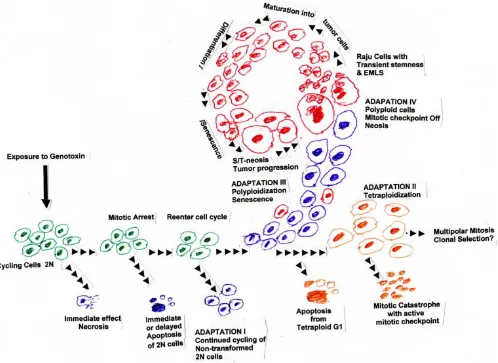
As described above, in primary cells, induction of an accelerated senescent phase acts as an efficient tumor sup-pressor mechanism; and DNA damaging agents or geno-toxins also elicit an accelerated senescence-like phenotype [Reviewed in [114-116]] by activating DNA damage response pathways [88]. Tumor cells in vivo also display the phenomenon of senescence in response to exposure to genotoxins [reviewed in [19,84]]. Such cells may either undergo cytostasis and may never undergo mitosis again, even in the presence of growth factors, or they may die via apoptoss or mitotic catastrophe, Some cells may escape senescence and give rise to resistant tumor growth. (See Fig. 1 for the possible different fates of cells after exposure to genotoxins.)
initia-Fate of cells exposed to genotoxins: Immediate effect of exposure to genotoxins is the arrest of cell cycle progression
Figure 1
tion of the senescent phenotype is accompanied by sup-pression of mitotic genes and overexsup-pression of mitosis inhibitor genes [Reviewed in [84,117]]. Accordingly, te Poele et al., [118] have reported that in the archival breast tumors from patients who had undergone chemotherapy, when sectioned and stained for the senescent marker
SA-β-gal, 41% of the tumors stained positive. In untreated
controls only 10% of the tumors showed sporadic posi-tive staining for SA-β-gal. Prof. E. Sikora, [Personal com-munication] has obsrved that SA-β-gal positive cells can undergo neosis, and tumor cells are apparently defective in senescent checkpoint control(s), and chances are that eventually some of these senescent cells might escape senescence and give rise to resistant tumor growth via neo-sis (see below). Therefore, induction of senescence as an anti-cancer therapy should be approached with caution for the following reasons:
1. Most of the genotoxins are carcinogens.
2. Tumor cells already have mutations in the senescent checkpoint pathway. Therefore, the chances of some cells escaping senescence are very high, especially in advanced tumors.
3. Senescent cells secrete factors that can promote tumor progression [81,119].
4. Senescent cells facilitate tumorigenesis in adjacent cells [119].
5. Ageing senescent cells may accumulate additional mutations due to oxidative damage and escape senescent phase via neosis and may result in recurrence of resistant tumor growth (See below).
6. Since most anti-cancer chemicals are carcinogenic, and the tumor tissue is a mixture of normal and tumor cells, chemotherapeutic drugs may facilitate tumorigenic trans-formation of normal or preneoplastic cells [19].
Therefore, the approach of controlling cancer by inducing senescence in vivo, although tempting, may in the long run increase the chances of resistant tumor growth, or facili-tate origin of new tumors. One has to better understand the molecular events that regulate senescence and the mode of escape from senescence in tumors, the longevity and fate of senescent cells in vivo, before one can design effective anti-cancer treatment strategies based on senes-cence.
What is neosis?
Neosis is the newly defined mode of cell division that occurs only in senescent, polyploid cells, and has not been observed in normal diploid cells. Neosis is a parasexual,
somatic, reduction division displayed by a subset of multinucleate and/or polyploid giant cells or MN/PG cells formed during the spontaneous senescent phase of nor-mal cells at the end of their MLS or genetic stress-induced accelerated senescence phase in tumor cells. It is character-ized by: (1) chromosome distribution to daughter cells via nuclear budding in the presence of an intact nuclear envelope, (2) followed by asymmetric cytokinesis, giving rise to an indefinite number of small, aneuploid, mitoti-cally active cells termed Raju cells (Raju meaning King in Telugu language), after which the polyploid neosis mother cell (NMC) dies. Raju cells display transient stem cell-like properties, and mature into tumor cells with extended MLS, finally arriving at a secondary/tertiary senescent phase. Neosis is interspersed with the extended MLS of Raju cells and their senescent phase and is repeated several times during tumor growth in a progres-sively non-synchronous fashion. Further, neosis is also responsible for the outgrowth of resistant tumor cells after exposure to genotoxins. Neosis appears to be a mode of origin and continuous growth of different tumor types, including hematological malignancies, carcinoma and sarcomas. The significant role of neosis appears to be fine tuning the damaged genome by epigenetic modulation in order to escape death via stress-induced senescence [5,6].
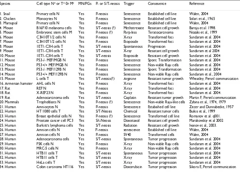
Neosis-like events in the literature (Table 1)
tro-Table 1: Neosis-like events reported in different cell systems of different species. N* = normal cells, T* = transformed or tumor cells, M* = Mutant cells.
Species Cell type N* or T* 0r M* MN/PGs P- or S/T-neosis Trigger Conseuence Reference
1. Snail Primary cells N Yes P-neosis Senescence Established cell line Walen, 2004 2. Chicken Monocytes N Yes P-neosis Senescence Established cell line Solari et al., 1965 3. Marsupial Primary cells N Yes P-neosis Senescence Established cell line Walen, 2004 4. Mouse B16F10 melanoma cells Yes S/T-neosis (?) Methotrexate Resistant cell growth Baroja et al. 1998. 5. Mouse Embryonic stem cells M Yes P-neosis (?) Parp-less Teratocarcinoma Nozaki et al., 1999 6. Mouse C3H10T1/2 cells N Yes P-neosis X-ray Transformed foci Sundaram et al. 2004 7. Mouse C3H10T1/2 cells N Yes P-neosis Etoposide Transformed foci Sundaram et al. 2004 8. Mouse 1ET1-C3H cells T Yes S/T-neosis Spontaneous Progression Sundaram et al. 2004 9. Mouse 1ET1-C3H cells T Yes S/T-neosis X-ray Resistant cell growth Sundaram et al 2004 10. Mouse 1ET1-C3H cells T Yes S/T-neosis Etoposide Resistant cell growth Sundaram et al. 2004 11. Mouse P53-/- MEF/MGB N Yes P-neosis Senescence Spont. Transformation Sundaram et al. 2004 12. Mouse P53+/+ MEF/MGB N Yes P-neosis Senescence Non-viable Raju cells Sundaram et al. 2004 13. Mouse P53-/- MEF/129B N Yes P-neosis Senescence Spont. Transformation Sundaram et al. 2004 14. Mouse P53+/+ MEF/129B N Yes P-neosis Senescence Non-viable Raju cells Sundaram et al., 2004
15. Mouse L cells T Yes S/T-neosis(?) Arginine Resistant tumor growth Wheatley, Persnl communication 16. Armenian hamster AHL cells N Yes P-neosis X-ray Transformed foci Sundaram et al. 2004
17. Rat REF N Yes P-neosis X-ray Transformed foci Sundaram et al. 2004 18. Rat X-REF23 N Yes P-neosis X-ray Transformed foci Sundaram et al. 2004. 19. Rat Adenocarcinoma cells Yes S/T-neosis Cisplatin Resistant tumor growth Martin F, Persnl.communication 20. Mammals Trophoblasts N Yea P-neosis (?) Senescence Non-viable Raju-like cells Zybina et al., 1974, 1979 21. Human Amniocytes N Yes P-neosis Senescence Estabished cell line Zitcer and Dunnabecke, 1957 22. Human HT 1080 cells T Yes S/T-Neosis Chemical Resistant tumor cells Buikis et al., 1999
23. Human Breast epithelial cells N Yes P-neosis (?) Senescence Transformed cell line Romanov et al. q001. 24. Human Prostate cancer cell PC3 Yes S/t-Neosis Doxotaxol Resistant cell growth Marakovskiy et al 2002 25. Human Burkitt's lymphoma cells Yes S/T-neosis Radiation Resistant cell growth Ivanov et al., 2003. 26. Human Amnion cells N Yes P-neosis senescence Established cell line Walen, 2004 27. Human Amnion cells N Yes P-neosis SV40 Transformed cells Walen, 2004 28. Human Adenocarcinoma cells Yes S/T-neosis Spontaneous Tumor progression Sundaram et al. 2004 29. Human FSK cells N Yes P-neosis X-ray Non-viable Raju cells Sundaram et al. 2004 30. Human MRC-5 cells N Yes P-neosis X-ray Non-viable Raju cells Sundaram et al. 2004 31. Human HTB11 cells T Yes S/T-neosis Spontaneous Tumor progression Sundaram et al. 2004 32. Human HTB11 cells T Yes S/T-neosis X-ray Tumor progression Sundaram et al. 2004 33. Human HeLa cells T Yes S/T-neosis X-ray Tumor progression Sundaram et al. 2004 34. Human Colon carcinoma HT116 Yes S/T-neosis Doxorubicin Tumor progression Sikora E, Persnl communication
phoblast development. It is apparent that such a phenom-enon is involved in the origin of tumor cells, due to inadvertent expression of neotic gene(s) during DNA
damage-induced loss of p53 or related senescent
check-point genes.
Solari et al., [126] have reported continuous production of small mononucleate Raju-like cells by nuclear budding from multinucleate giant polyploid cells formed by fusion
of avian senescent peripheral blood mononuclear cells in
vitro, even in the presence of an inhibitor of mitosis. These cells resembled Raju cells in their morphology and fused again initiating secondary nuclear budding to yield the next generation of Raju-like cells. Although the authors did not discus the relationship of their observations to senescence, immortalization or transformation of cells, these events are very similar to the primary (P-neosis) and secondary/tertiary or S/T-neosis reported by us [5,6]. Walen in a series of articles [127-129] has reported similar nuclear budding giving rise to viable cells with continuous division potential in different cell systems. Romanov et
al., [130] have reported the immortalization of senescent human breast epithelial cells in vitro; although the exact events of this process has not been described; they men-tion a process of micronucleamen-tion, which could be the process of nuclear budding related to neosis [5] or the nuclear fragmentation that occurs during mitotic catastro-phe [131].
cells. Makaravskiy et al., [89] reported that exposure of PC3 prostate cancer cells to docetaxel resulted in growth arrest, multinucleation and giant cell formation, which gave rise to docetaxel-resistant clones; this resistance was associated with transient expression of a β-tubulin iso-form and was independent of P-glycoprotein, bcl2 and bcl-xl expression. The recent literature showing neosis-like events are listed in Table 1. In every case, senescent MN/ PGs yield mononuclear Raju-like cells that display extended MLS.
Neosis bypasses senescence
In 1998, using computerized video time-lapse micros-copy, we started to study the cellular process involved in transformed focus formation [5], which is considered to be the in vitro equivalent of in vivo tumorigenicity [70-72]. This involved: (1) video documentation of the transfor-mation process at the level of individual cells, (2) isola-tion and cloning of the individual transformed foci without being contaminated by surrounding non-trans-formed cells, and (3) study the fate of these individual neotic clones (Raju cells derived from a single neosis mother cell or NMC) by serial cultivation under standard culture conditions and under anchorage independent conditions. These studies suggested that transformed focus formation occurs via the novel process termed neo-sis [5] and not via mitoneo-sis as previously thought.
Exposure of C3H10T1/2 cells to carcinogens induced accelerated formation of large senescent MN/PG cells that are supposed to be permanently arrested from undergoing
mitosis and eventually die via mitotic catastrophe [19,81,137,138]. Around 14–21 days post-exposure to carcinogens, a minor subset of these senescence-like MN/ PGs (hereafter termed the neosis mother cells or NMCs), before they died, produced several small mononuclear cells by karyokinesis via nuclear budding in the presence of the intact nuclear membrane. Each nuclear bud was immediately loaded with genomic DNA and surrounded by a small fragment of the cytoplasm delimited by plasma membrane by asymmetric cytokinesis. Each NMC pro-duced about 10+/- 2 Raju cells. The latter were very small
(~6–8 µm in diameter), had very high N/C ratio and
immediately after birth, resumed symmetric mitotic divi-sion, inherited aneuploidy and genomic instability; they grew in soft agar, and displayed genotype and phenotype different from the mother cell. Presumably, they have reactivated telomerase expression [139-141] that resulted in the extension of their mitotic life span (MLS). Thus neosis and not mitosis is the mode of cell division result-ing in tumorigenesis.
If neosis were the mode of transformation of normal cells into neoplastic cells, the spontaneous transformation of p53-/- mouse embryo fibroblasts should also involve senescent phase followed by neosis. In fact this is what we observed, when we studied the mode of spontaneous transformation of p53-/- cells using a similar approach. The primary cultures of p53-/- MEF/MGB cells entered senescent phase at the end of its diploid mitotic life span (about 6–7 passages), and almost all of these MN/PGs underwent spontaneous neotic division, each giving rise
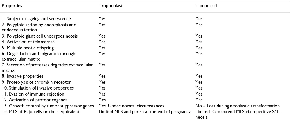
Table 2: Similarities between trophoblast maturation and tumor cell self-renewal
Properties Trophoblast Tumor cell 1. Subject to ageing and senescence Yes Yes 2. Polyploidization by endomitosis and
endoreduplication
Yes Yes
3. Polyploid giant cell undergoes neosis Yes Yes 4. Activation of telomerase Yes Yes 5. Multiple neotic offspring Yes Yes 6. Degradation and migration through
extracellular matrix
Yes Yes
7. Secretion of proteases degrades extracellular matrix
Yes Yes
8. Invasive properties Yes Yes 9. Proteolysis of thrombin receptor Yes Yes 10. Stimulation of invasive properties Yes Yes 11. Evasion of immune rejection Yes Yes 12. Activation of protooncogenes Yes Yes
13. Growth control by tumor suppressor genes Yes. Under normal circumstances No – Lost during neoplastic transformation 14. MLS of Raju cells or their equivalent Limited MLS and perish at the end of pregnancy Limited. Can extend MLS via repetitive
to 50 or more Raju cells, which by repetitive mitosis gave rise to spontaneously transformed cell lines. All the NMCs died within a month leaving the transformed cells in the Petri plate. The isogenic control wild type p53+/+ MEF/ MGB cells underwent neosis yielding fewer number of Raju cells, but these Raju cells died without yielding a transformed cell line, probably due to the presence of p53, which is detrimental to cells with genomic instability [5,6,142] (see additional files 1, 2, 3, 4, 5, 6).
We also observed certain degree of plasticity in neosis, where the nuclear budding during karyokinesis may remain constant, but cytokinesis may follow immediately to form Raju cells sequentially or be delayed to form post-budding synsytium-like multinucleate cell, followed by cytokinesis yielding several Raju cells simultaneously [6].
Raju cells transiently display stem cell properties
Raju cells are defined as the nascent daughter cells of neotic division, before they undergo their first symmetric mitosis. When the extended MLS of individual neotic clones was studied by serial subculturing, around the 20th passage they spontaneously underwent a senescent phase and displayed mitotic crisis. Some of these MN/PGs spon-taneously underwent secondary neosis, each NMC pro-ducing ~10 +/- 2 secondary Raju cells, only to repeat the cycle of EMLS followed by senescence, neosis, and pro-duction of the next generation of Raju cells. [5].
We and others have reported neosis occurring in several human and rodent tumor cell systems [5,6]. In tumor cell cultures, neosis was repeated several times each neotic division interspersed with extended, but limited, MLS of the neotic offspring followed by a senescent phase. Thus, during three years of continuous subculturing, human metastatic neuroblastoma HTB11 cells underwent three episodes of spontaneous senescence followed by S/T-neo-sis and yielded new populations of mitotically active Raju cells in a progressively non-synchronous fashion [5], (see additional files 1, 2, 3, 4, 5, 6). The first and second epi-sodes of mitotic crisis lasted for about 7 to 8 weeks (with medium change twice a week) during which time the cells remained in the senescent phase. The crisis period was succeeded by neosis, giving rise to small, mononucleate Raju cells with extended MLS. However, when HTB11 cells were subjected to carcinogen exposure, the senescent phase lasted for only two weeks before the appearance of Raju cells, reducing the duration of senescent phase and mitotic crisis, indicating that genetic stress was high enough to induce neosis within a shorter duration. This implies that during spontaneous senescence and pro-longed mitotic crisis (up to 7–8 weeks), the cells may slowly be accumulating additional mutations (probably due to oxidative stress) raising the level of genomic insta-bility sufficient to induce spontaneous neosis.
We, therefore, hypothesize that self-renewal of cancer growth is made feasible by the transient re-expression of some stem cell properties in Raju cells. This, probably, occurs after epigenetic modulation of the non-viable
poly-ploid genome of NMC prior to the neotic S phase or SN
phase. Unlike in the mitotic S phase or SM, where DNA is
replicated only once before each division, during the SN
phase, DNA is replicated several times and the newly syn-thesized DNA is preferentially transported to the nuclear bud, followed by asymmetric cytokinesis. Thus, the NMCs display stem cell behavior in that the newly synthesized genomic DNA is asymmetrically segregated to the daugh-ter Raju cells [5,6]. The nascent Raju cell genome often underwent mitotic division even before cellularization while still within the cytoplasm of the NMC [6,133].
Raju cells are unique in that they transiently display cer-tain stem cell-like properties such as extended, but limited MLS, expression of telomerase, and potential to differen-tiate. They entered symmetric mitotic division immedi-ately after they became independent cells. The mitotic derivatives of Raju cells grew larger in size and matured into tumor cells in a couple of days, while undergoing mitosis [5]. We interpret the increase in cell size during the course of mitotic division as an embryonic cell prop-erty that incorporates the G1 phase of the cell cycle [6,143]. As they undergo symmetric mitotic proliferation, they mature into tumor cells and probably undergo defec-tive differentiation and gradually lose stem cell properties. At the end of their extended, but limited MLS, the tumor cells spontaneously entered a senescent phase. Since these cells have already lost some senescent checkpoint path-way gene(s) function, some of these senescent cells stand a good chance of undergoing S/T-neosis, thus contribut-ing to the continuity of tumor cell lineage. Thus, Raju cells behave like committed stem cells immediately after birth and slowly acquire somatic cell properties during the pro-lipherative phase. Given in the box below are some of the stem cell properties of Raju cells and the somatic cell properties of their mitotic derivatives (Table 3).
Mitotic catastrophe and neosis are mutually exclusive with opposite effects
posi-tion of the cell in the mitotic cell cycle, and the severity of the cellular damage. Cells with acute lethal damage may undergo immediate necrosis, or some of them will initiate the process of programmed cell death or apoptosis, which may lead to immediate cell suicide or delayed death by a day or two. In rare occasions, the cells may complete sev-eral mitotic divisions and the whole clone of cells might die simultaneously at a later date. Almost all the surviving cells will stop synthesizing DNA immediately. A major fraction of the surviving cells recover from the shock after several hours of quiescence and reenter cell cycle, proba-bly after repair (or misrepair) of DNA damage. Most of such cells do not show any alterations in their morphol-ogy or growth behavior at least immediately after recovery [Fig. 1].
Of all the different checkpoint controls the most impor-tant one is the mitotic checkpoint or the spindle check-point, which is considered the primary defense against aneuploidy and ensures accurate chromosome segrega-tion in order to produce genetically identical daughter cells [90,146]. During karyokinesis in mitosis and meio-sis, the nuclear envelope is dismantled at which time the spindle checkpoint is activated [146,147]. Often the senescent cells form nuclear envelopes around fragments of the genome, a process termed micronucleation [131,148,149]. Thus, unable to maintain G2 arrest, they enter mitosis and after being arrested for several hours at metaphase, they eventually die without successfully
com-pleting mitosis. This process is known as mitotic catastro-phe or mitotic death [150,151,154,155].
Since most cancer cells are deficient in the tumor suppres-sor function of the p53/pRB/p16INK4a signal transduc-tion pathway, the G1, and G2 checkpoint functransduc-tions are lost. After uncoupling apoptosis from G1 and S, tumor cells with mutant p53 arrive at G2/M interphase, at which time decisions regarding cell survival and death may be initiated. Cells that did not repair the damage to DNA arrive at G2 with point mutations and double strand breaks. Some of the double strand breaks are repaired by homologous recombination and non-homologous end rejoining during the G2 arrest [152,153]. Most of the sur-viving cells with unrepaired chromosomal lesions activate the senescent program via the DNA damage response. Senescent cells are characterized by SA-β-gal expression, SAHF, and are usually very large due to endomitosis and endoreduplication and are generally unable to divide again even in the presence of growth factors. Further, the senescent cells downregulate mitotic genes and upregu-late anti-mitotic genes [19,81,84]. Under these circum-stances, the senescent cancer cells will face inevitable death, unless they adapt and evade cell death, by eliminat-ing the mitotically non-viable genome, and multiply by producing mitotically viable genome in order to be able to maintain the continuity of tumor cell lineage. All these conditions are fulfilled by neosis [5]. In this connection, it is interesting to note that senescent SA-β-gal positive
Table 3: Properties of Raju cells and their mitotic derivatives:
Transient stem cell properties of Raju cells:
1. Short cell cycle duration of nascent Raju cells (before they undergo first mitosis) – an indication of lack of G1 phase? [5, 6]. 2. Reactivation of telomerase conferring extended mitotic life span [139-141].
3. Is it possible to expand Raju cell population without differentiation under proper culture conditions such as EGF or FGF2 [43, 144-148]. 4. Increase in cell size accompanied by increase in cell cycle duration-introduction of G1 phase in the cell cycle [Rajaraman, unpublished; 143]. 5. Resistance to genotoxins – Expression of multidrug resistance genes? [192, 193].
6. Are they transiently expressing tissue stem cell specific surface markers? (e.g., CD34+ for hematopoietic cells [8]; CD133+ for brain cells [12, 13], CD44+, CD33-, LowLin- for breast cells [9-11]; CD20+ for skin cells [40]; CD44+,α 2β 1hi/CD133+ for prostate cancer cells [41]. 7. Are they transiently expressing stem cell specific growth genes? (E.g. Nanog, Oct-4, Wnt, Bmi1 etc.) [188-191]
8. Potential to differentiate, although aberrantly.
Somatic cell properties of mitotic derivatives of Raju cells:
1. Resumption of symmetric mitotic division.
2. Increase in cell size – Introduction of G1 phase in the cell cycle? [143]. 3. Progressive, but, aberrant differentiation.
4. Loss of tissue specific stem cell surface markers due to differentiation during extended mitotic proliferation? 5. Loss of expression of stem cell specific self-renewal genes?
6. Loss of expression of multidrug resistance genes?
7. They are subject to aging and associated senescence brought about by telomere attrition. 8. Therefore, they have limited division potential.
9. Telomere attrition, chromosome breakage-fusion-bridge cycle or genetic stress will result in senescent phase with MN/PG formation, mitotic crisis, and mitotic catastrophe.
cells formed after exposure of human colon carcinoma HT116 cells to doxorubicin underwent nuclear budding and formed Raju cells via neosis [Prof. E. Sikora, personal communication].
We postulated that during such critical time of mitotic cri-sis, the cells may revert back to neocri-sis, which may be an evolutionary throw back just to tide over the crisis [5]. Interestingly, this mode of cell division resembles the asexual reproduction found in parasitic protozoans and protista and has been reported to be an evolutionary phase in sexual reproduction by meiosis in higher organ-isms [153]. However, it has been demonstrated that such a primitive mode of cell division is still being activated during the maturation of extraembryonic tropholast cells during mammalian pregnancy. Thus, it appears that the gene(s) that execute neosis are still functional in the mam-malian genome, which are supposed to be active only dur-ing the trophoblast maturation in the extra embryonic tissue during pregnancy [122,123], and are inadvertently expressed in the absence of certain tumor suppressor
genes (p53?) due to DNA damage.
When tumor cells are exposed to conventional chemo-therapy or radiation chemo-therapy, most of the accelerated senescent cells may eventually die, but some do escape by adapting to the adverse conditions by undergoing poly-ploidization, and karyokinesis via nuclear budding [5,133]. Since mitotic checkpoint or spindle checkpoint is activated only after the dismantling of the nuclear enve-lope [146], polyploidization followed by nuclear budding with an intact nuclear envelope is bound to protect the NMC from cell death due to mitotic checkpoint. There-fore, some of these polyploid cells successfully undergo reduction division or de-polyploidization yielding near diploid daughter Raju cells with transient stem cell prop-erties [5,6].
When spindle checkpoint is defective, a minor fraction of senescent cells escape mitotic catastrophe and become tetraploid due to mitotic slippage or cytokinesis failure [154,155]. Such cells have been shown to undergo multipolar mitosis giving rise to aneuploid cells [154]. However, it is not sure if these aneuploid cells will survive long enough to contribute to the clonal selection in tumor tissue [156]. We have shown that a subpopulation of tetraploid cells can undergo neosis by nuclear budding to give rise to several daughter cells before they die [5]. Since the nuclear envelope is not dismantled during neosis, spindle checkpoint is probably not activated and this might favor successful completion of neosis, often acci-dentally yielding aneuploid neotic offspring, which may survive and proliferate due to loss of genomic stability. During mitotic catastrophe, nuclear envelope is disman-tled and spindle checkpoint is activated, leading to
mitotic catastrophe via micronucleation and death [20,84,148-150]. Thus, mitotic catastrophe and neosis are mutually exclusive phenomena displayed by tumor cells with opposite effects. While the former eliminates the tumor cells with a defective genome, the latter helps adapt them to genomic instability-induced accumulation of genetic and epigenetic alterations by reducing the GI load in the neotic offspring via epigenetic modulation and thus contributing to the continuity of tumor cell lineage.
Neosis and epigenetics in tumor progression
Epigenetic modulation is probably the way the genome alters its behavior in response to the environment [157]. "The genome functions like a highly sensitive organ of the cell that monitors its own activities and corrects common errors, senses unusual and unexpected events, and responds to them, often by restructuring itself" [158]. This statement probably accurately fits the behavior of cancer cells. Recent studies have revealed that genetic mutations alone do not lead to cancer and that epigenetics plays a major role in the origin and progression of tumors. Epige-netic alterations – non-DNA sequence-based heritable alterations – have been shown to initiate genomic insta-bility, even before gene mutations enter into the process [159-162]. Epigenetic mutations fall into two main cate-gories: (1) Altered DNA methylation of CpG dinucle-otides, both losses or hypomethylation (results in gene activation) and gains or hypermethylation (results in silencing the gene) and (2) altered patterns of histone modifications such as acetylation or deacetylation of lysine residues, [160-166].
Epigenetic modulation occurs during all stages of tumor growth from the initiation at the progenitor cells through tumor formation and progression [162,163]. During the initiation stages, epigenetic modifications mimic the effect of genetic damage by altering the expression of tumor suppressor genes, thus compromising the tumor suppressor function of the senescence program; for exam-ple, silencing of tumor suppressor genes by promoter DNA hypermethylation and chromatin hypoacetylation, which may affect the expression of diverse genes including p53, RB1, p16INK4A, Von Hippel-Landau tumor suppres-sor (VHL) and MutL protein homologue 1 (MLH1) [109,162,163,168-170]. Global hypomethylation of chromatin and loss of imprinting lead to chromosomal instability and increased tumor incidence both in vitro and in vivo [165-167]; epigenetic activation of R-ras is respon-sible for gastric cancer [168] and cyclinD2 and mapsin activation in pancreatic cancer [169,170].
Burkitt's lymphoma cells with p53 mutant gene [175]. It has been recently demonstrated that exposure of tumor cells to genotoxins results in the translational upregula-tion of cMOS gene [175], which is involved in switching cells from mitosis to meiosis II and forcing the cell to undergo reduction division [172]. Constitutively expressed in low levels in untreated tumor cells, expres-sion of meiotic coheexpres-sion gene REC8 was also enhanced after irradiation of p53 mutated Namalwa Burkitt's lym-phoma cells, along with other meiosis-specific genes DMC1, STAG3, SYCP1 and SYCP3. Expression of these genes reached a peak level during the mitotic arrest phase and was proportional to the endopolyploid cells [175].
When such defective cells reach senescent phase and undergo neosis, these polyploid NMCs give rise to near-diploid or aneuploid Raju cells; this indicates that neosis must comprise properties of meiosis, mitosis and neosis specific events. During the extended MLS, Raju cells differ-entiate into tumor cells, while also accumulating addi-tional mutaaddi-tional and epimutaaddi-tional alterations. This will increase the GI load, and cells will arrive at the next senes-cent phase due to incomplete differentiation displaying mitotic crisis. The onset of senescence is accompanied by several changes in the gene expression profile of the cells: (1) downregulation of mitotic genes; (2) upregulation of anti-mitotic genes [19,84]. During neosis the following changes in the gene-expression profile must occur: (1) reexpression of the 'immortalizing enzyme' telomerase is obligatory for the extension of mitotic life span, but not sufficient for neoplastic transformation [139-141,176,177]; (2) ectopic expression of meiotic genes col-lectively called cancer/testes (CT) antigens in tumor cells, which are thought to play a role in transformation [171-178], (3) in addition to ectopic expression of stem cell self-renewal genes including microRNAs, notch, oct4, and Bmi1 [77,179-184] and (4) multidrug resistance genes [185,186]. Therefore, the most significant event during neosis appears to be the alteration in gene expression pro-file, which is likely to be brought about by epigenetic modulation. This will in effect reduce the GI load, making it possible to produce mitotically viable genomes of Raju cells from the non-viable polyploid genome. Our data and those of others [5,6,127-129,152,153,175,187] sug-gest that the genome of such polyploid cells, although not mitotically viable, may undergo epigenetic modulation in order to reduce the degree of GI load to yield mitotically active daughter Raju cells with EMLS and thus contribute to the continuity of tumor cell lineage in a non-synchro-nous fashion, while the mitotically non-viable polyploid genome is eliminated by the post-neotic death of NMC. Thus, the genes for neosis which are silent in the genome, and should be active only during the maturation of the extraembryonic trophoblast cells during pregnancy [122,123], are reexpressed in tumor cells in the absence of
p53 or related tumor suppressor genes due to DNA dam-age.
Neosis is repeated several times during the growth of tumors. This implies that global epigenetic modulation occurs throughout the life of the tumors, repetitively at least during each neosis, since continuous proliferation will lead to accumulation of gene mutations and epimu-tations, which may be often detrimental to the cell. There-fore, we propose that during neosis the cell with defective senescence checkpoint control(s) tend to undergo endore-plication/multinucleation and after restructuring the genome followed by multiple rounds of neotic S phase
(SN phase), produces daughter Raju cells with EMLS via
nuclear budding and asymmetric cytokinesis. This decreases the GI load in the Raju cells, while the non-via-ble polyploid genome of the NMC is discarded during its post-neotic demise. In the absence of tumor suppressor gene(s), the genome appears to be highly plastic (and tol-erant to DNA damage) and responds to the damage and never activates the mitotic checkpoint by keeping the nuclear envelope intact and tides over the crisis by pro-ducing several Raju cells with stem cell properties and helps the continuous growth of tumor.
5.6. Neosis as the source of aneuploidy
(1) It has been shown recently that structural and numer-ical chromosome alterations in colon cancer develop through telomere-mediated anaphase bridges and not through mitotic multipolarity. In fact, multipolarity results in uneven chromosome distribution to daughter cells that gives rise to gross genomic changes such as nul-lisomies and non-viable daughter cells, and therefore, rarely contributed to clonal evolution tumor cells [194].
(2) It now seems that all human aneuploidies (cells that have chromosome number other than 46) in normal dip-loid human cell systems that occur during development result in embryonic lethality, except certain combinations of sex chromosomes and hyperdiploid, trisomies 13, 18 and 21, which yield severe birth defects in humans [195,196].
(3) Further, it has been recently shown that chromosome non-disjunction in primary human cells yields tetraploid cells rather than aneuploid cells due to failure of cytokine-sis. This demonstrates that tetraploid cells do not directly give rise of aneuploidy. Therefore, there must be an inter-mediate step between tetraploid cells and the origin of aneuploid progeny of tumor cells [197].
(4) We observed primary neosis that gave rise to trans-formed cells in a binucleate giant cell with a chromosome bridge [5]. The fact that this NMC is binucleate with an isthmus, indicates that this cell has undergone one unsuc-cessful mitotic division attended by cytokinesis failure and has, therefore, an at least tetraploid genome.
(5) Thus, neosis appears to be the intermediate step between tetraploid cells and the origin of aneuploid tumor cells. It has the potential to give rise to aneuploid or near diploid stem cell-like Raju cells via polyploidiza-tion followed by karyokinesis via nuclear budding with-out activating mitotic checkpoint control by keeping the nuclear envelope intact.
(6) Accordingly, p53-/- MN/PG cells spontaneously gave rise to transformed cells via neosis [5] and in p53 null mouse cells cytokinesis failure-generated tetraploid cells promote tumorigenesis in vivo [198].
(7) These cells have extended MLS in the absence of senes-cent checkpoint controls (e.g. P53-/- cells); but they perish in the presence of proper checkpoint controls (eg., p53+/ + cells). Thus, p53+/+ MN/PG mouse cells did not yield viable Raju cells [5,6] and the p53+/+ tetraploid cells did not produce tumors in vivo [198].
(8) In support of this, it is also known that in several human and rodent tumor systems, tetraploidy is the
inter-mediate stage before the genesis of neoplastic growth [155,156,199-203].
Therefore, we propose that senescent cells with tetraploid or higher ploidy genomes have the potential to under go neosis, creating conditions for automatic onset of aneu-ploidy to drive malignancy [67], if all the conditions for survival of the genome are met with in the resultant Raju cells.
Polyploidy can result either due to endomitosis and endoreduplication, or by cell fusion. It has been shown that Mad2- or BubR1-depleted cells (the genes involved in spindle checkpoint control) that do not complete cytoki-nesis remain viable through continued cycles of DNA rep-lication up to at least 32 N [146,205]. In murine melanoma cultures exposed to methotrexate, emergence of resistant clones is preceded by an increase in polyploid cells with DNA content >8c and even >16c, concomitant with a decrease in tetraploid cells and is accompanied by loss of expression of mtotic proliferation markers (PCNA and CDK1) [206]. Even higher ploidy can be attained in certain cell systems [121-124,133]. Polyploidization, in addition to protecting cells from death confers evolvabil-ity by producing cells with non-lethal variations in the genome, upon which the process of natual selection may act [5,207,208]. In addition, since spindle checkpoint control is activated after the dissolution of the nuclear envelope [146], neosis, by performing karyokinesis via nuclear budding without the dissolution of the nuclear envelope, appears to be an adaptation by polyploid cells to escape from spindle checkpoint control-induced mitotic catastrophe. It is known that p53 null cells even-tually develop polyploidy [5,156,197,198] attended by multiple centrosomes.
Often the presence of multiple centrosomes can avoid for-mation of multiple spindle poles by fusion of centro-somes resulting in a complex form of bipolar spindle [154,209]. However, it remains to be elucidated as to how the process of karyokinesis is effected in the presence of nuclear envelope. The question arises if the complex of multiple centrosomes acts like the spindle pole body of budding yeast by physically associating itself with the nuclear envelope and relocating itself back in the cyto-plasm in Raju cells [129] so that they can resume symmet-ric mitosis.
and survive due to loss of checkpoint control(s) and gain of genomic instability. The role of meiotic genes in favor-ing reduction in the number of chromosomes durfavor-ing neo-sis is strongly suspected [175]. Similarly, multiple cycles of DNA replication in the NMC can be made possible by some meiotic gene(s) that are ectopically expressed in tumor cells [171-173][174]. Therefore, we suggest that, in addition to the global epigenetic modulation of the genome discussed above, neosis, rather than multipolar mitosis, is involved in the origin of aneuploid tumor cells.
However, we envisage that once the genesis of tumor cells via neosis is achieved, since these cells have gained genomic instability, further propagation of aneuploidy and the associated chromosomal aberrations including gene duplications, translocations etc will be facilitated by the various mechanisms including centrosome amplifica-tion, mitotic spindle abnormalities, defective attachment of chromatids to kinetchore, telomere dysfunction-induced breakage-fusion-bridge cycle, defective cytokine-sis, mitotic checkpoint defects, among other things [188]. Thus, once the process of aneuploidy is initiated by neo-sis, progressive increase in aneuploidy might play an active role in the on-set of aggressive malignant property in solid tumors, whose survival and proliferation may be favored by loss of genomic stability [66].
The basis for the number of Raju cells produced by each NMC is not known. For example, in p53 MEF/MGB cells we observed that there were multiple giant nuclei present, and often more than one nucleus can produce nuclear buds and yield Raju cells. Number of Raju cells/NMC was around 10+/- 2 in C3H10T1 1/2 cells, ~4 or 5 in HeLa cells, and ~50 or more in p53-/-MGB/MEF cells, but 1 or 2 in p53-/-MEF/129B cells. While it is tempting to postu-late that the number of Raju cells/NMC may depend upon the ploidy of the NMC, this remains to be tested. It is very likely in addition to ploidy, the cellular genetic back-ground may be a determining factor in the number of Raju cells/NMC [5].
Additionally, it is known that cancer cells are living precar-iously at the edge of life [23]. While these cells have shut off spindle checkpoint and escaped mitotic catastrophe, and are on the way to escaping senescence, anything can go wrong in the process of neosis and cells may die at any stage of neosis, a process which we have termed neotic catastrophe. Thus, even at the last phase of neosis the NMC may die without yielding daughter cells, even after they have been successfully formed, for the simple reason that they cannot get out of the mother cells and become independent cells [5].
Limits of extended Mitotic Life Span (MLS) and tumor progression
Neotic clones isolated from C3H10T1/2 cells exposed to etoposide displayed extended MLS, probably due to reex-pression of telomerase [5,6,139-141]. Contrary to the belief that transformed cells are 'immortal,' we observed that at about the 20th passage, the neotic clones entered the next cycle of senescence and underwent secondary neosis yielding the next batch of Raju cells, which contin-ued to multiply [5,6]. Tumor cells undergo spontaneous senescence in vivo and display the senescence marker
SA-β-gal sporadically [118] in a non-synchronous fashion.
This implies that transformation does not confer immor-tality. Instead, this simply results in extension of the MLS of neotic offspring, which would not be feasible if it were not for the rejuvenation process of neosis. In addition, this observation suggests that the 'stemness' of Raju cells was transient and this property is lost during the prolifer-ative phase, probably due to defective differentiation, which leads to the hierarchic nature of the tumor cell pop-ulation. This important aspect of cancer cell mortality would not have been revealed if we did not study the behavior of individual neotic clones [5]. Under normal circumstances, the progenies of different NMCs will be growing either in vitro or in vivo, masking the repetitive renewal or rejuvenation of MLS via neosis, especially since this is occurring in a non-synchronized fashion.
Similarly, the human renal adenocarcinoma ACHN cells displayed rare MN/PG cells that initiated S/T-neosis; while in human metastatic neuroblastoma HTB11 cells, after continuous culture in vitro a high frequency of cells were observed to enter S/T-neosis [5]. Although we do not have any experimental data on the length of the extended MLS in these systems, the fact that these cells also display senescent MN/PGs that act as NMCs implies that cancer cells are not immortal, and that they also undergo sponta-neous senescence due to differentiation and ageing [5] or accelerated senescence after exposure to anti-cancer agents [210]. However, the senescent cells may escape death since these are tumor cells with defective senescent check point control(s) and their growth is rejuvenated incon-spicuously, since neosis is not synchronized.