IJBCP
International Journal of Basic & Clinical Pharmacology
doi: 10.18203/2319-2003.ijbcp20151344Research Article
Determination of hepatoprotective effect of
Mussaenda erythrophylla
in paracetamol induced hepatotoxicity
T. S. Rojin
1*, Sukanya Shetty
2, Rajendra Holla
1INTRODUCTION
Drug-induced liver injury is a major problem facing worldwide even today. Paracetamol is an over the counter drug, and thus the chances of hepatotoxicity are very common. Drug-induced liver disease is a common cause to withdrawn the drugs from the market after it has been introduced into market. Presently almost 900 hepatotoxic drugs are known to cause hepatotoxicity.1 The liver has
its own mechanism to recover from these toxic insults, but in spite of these resiliency liver is continuously being exposed to various toxins, chemicals pollutants, drugs, etc., which can produce hepatotoxicity. In recent years, many researchers have examined the effects of plants used traditionally by herbalists and indigenous healers to treat
the liver diseases. In most cases, research has confirmed by
discovering the mechanisms and mode of action of these
plants as well as reaffirming the therapeutic effectiveness
of certain plants or plant extracts in clinical studies. Many plants have been examined for use in a wide variety of liver disorders.2 Currently, 80% of world population depends
on plant derived medicine for various diseases. Plants are important sources of medicines and presently about 25% of pharmaceutical prescription in the US contain at least one plant-derived ingredient.3 Better therapeutic effect, good
patient compliance and cost effectiveness and less toxicity, are the reasons for choosing a drug from natural origin.4
Mussaenda erythrophylla (ME) is a tropical shrub, native to tropical West Africa. The roots are useful for the treatment of jaundice, cough and also act as an appetizer. The ethyl acetate and methanolic extracts from roots of ME reported having anthelmintic activity against earthworms.5 ME also reported to have diuretic property.
ABSTRACT
Background: Hepatotoxicity may be defined as liver injury caused by drugs and
chemicals. Drug-induced liver injury is a major reason for withdrawing drugs from a market by Food Drug Administration, and it is based on the fact that drug-induced liver injury is responsible for more than 50% of all cases of acute liver failure. Many studies revealed about the hepatotoxic potential of paracetamol. Hence, the present study has undertaken to evaluate the hepatoprotective effect of Mussaenda erythrophylla (ME) in paracetamol induced hepatotoxicity in Wistar albino rats.
Methods: The ethanolic extract ME studied for its hepatoprotective effect on paracetamol induced acute liver damage in Wistar albino rats. The degree of protection was measured using biochemical parameters such as serum glutamate oxalate transaminase (SGOT), serum glutamate pyruvate transaminase (SGPT), total bilirubin (TBL), superoxide dismutase (SOD), glutathione (GSH) peroxidase (GPx), GSH, and ceruloplasmin levels.
Results: Paracetamol treated group had enhanced levels of SGPT, SGOT, TBL (p<0.001) and decreased levels of GSH, SOD, and GPx (p<0.001) when compared with control group. Treatment with silymarin and also 200 mg/kg of MEleaf extract
had significantly (p<0.001) brought down the elevated levels of SGPT, SGOT, and
TBL and an increase in the levels of GSH, SOD, (p<0.01), GPx and ceruloplasmin (p<0.001).
Conclusion: The results showed that ethanolic extract of ME leaf extract possesses
significant hepatoprotective activity.
Keywords: Paracetamol, Mussaenda erythrophylla, Hepatotoxicity
1Department of Pharmacology,
K. S. Hegde Medical Academy, Nitte University, Mangalore, Karnataka, India,
2Department of Biochemistry,
K. S. Hegde Medical Academy, Nitte University, Mangalore, Karnataka, India
Received: 22 September 2015
Accepted: 25 October 2015
*Correspondence to:
T. S. Rojin,
Email: tsrojin@yahoo.co.in
Both the ethanolic and chloroform extract of root of ME showed a dose-dependent increase in urine excretion.6 The
hepatoprotective activity of ethyl acetate and methanol extracts of ME stem against carbon tetrachloride has been reported by Eswaraiah et al. By considering these, the present study has been conducted to evaluate the hepatoprotective effectiveness of leaves of ME against paracetamol induced hepatotoxicity.7
METHODS
Animals
Wistar albino rats of either sex weighing between 150 and 250 g were chosen for the study. They were housed in polypropylene cages with paddy husk bedding under controlled temperature and humidity. The animals were fed with standard pellet diet and water ad-libitum. Institutional Animal Ethics Committee of KSHEMA reference number AEC/15/2010 approved the experimental protocols and procedures employed in this study.
Preparation of plant extract
A weighed quantity of 100 g of ME leaf powder was taken and extracted by 90% of alcohol using soxhlet apparatus. The extract was concentrated by rotary evaporator. The yield of the extract was 15%. The extract was stored in the refrigerator at 4°C and from this stock the extract was diluted freshly according to the need to perform the experiment.
Acute toxicity studies
Wistar albino rats weighed 100-150 g were used for testing acute oral toxicity. It was performed on the basis of OECD guideline no: 423 (OECD, 2001). Overnight fasted animals were administered with ME leaf extract orally as single
dose at five different dose levels of 200, 400, 800, 1600,
and 3200 mg/kg body weight. The rats were observed continuously for 2 hrs for any behavioral changes and
toxicity, and occasionally observed for 4 hrs, finally checked
for overnight mortality. Thereafter, the animals were kept for 14 days and checked for mortality.
Experimental protocol
In this study healthy Wistar rats weighed 200-250 g were used. The animals were randomly allotted into 4 groups of 6 rats each and treated orally as below for 21 days. Group I received distilled water and considered as control. Group II treated as that of control and administered paracetamol 2 g/kg body weight p.o. only on the 21st day. Group III received 200 mg/kg of MEleaf extract for 21 days and administered with 2 g/kg of paracetamol on the 21st day. Group IV received silymarin 50 mg/kg for 21 days and intoxicated with paracetamol as that of Group III. 24 hrs after the intoxication
of paracetamol, all the animals were sacrificed, and samples
were collected for various biochemical analysis.
Biochemical estimations
At the end of drug treatment period, all the animals were
sacrificed by using either. Blood was collected by cardiac
puncture, allowed it to clot for 30 mins, and serum was separated by centrifugation at 3000 rpm for 15 mins. The liver was dissected out, rinsed with water, weighed and homogenized using 0.1 M Tris-HCl buffer of pH 7.5. The resultant homogenate was centrifuged, and the supernatant was collected. The serum as well as liver homogenate was used for determining the biochemical analysis of liver serum marker enzymes as well as oxidative stress parameters like serum glutamate pyruvate transaminase (SGPT), serum glutamic oxaloacetic transaminase (SGOT), total bilirubin (TBL), glutathione (GSH) peroxidase (GPx), glutathione reductase (GSH), superoxide dismutase (SOD), and ceruloplasmin as per standard procedure.
Assessment of hepatoprotective activity
Estimation of aspartate transaminase (AST) and alanine transaminase (Mohun and Cook, 1957)
To 1 ml buffered substrate, 0.05 ml of homogenate dilution was added as an enzyme source, mixed and incubated at 37°C for 5 mins. Then 0.075 ml of aniline-citrate was added. To the blank tube, after the addition of aniline-citrate, 0.05 ml of homogenate was added. The tubes were further maintained for 5 mins at 37°C and added 1 ml of 2,4-dinitrophenylhydrazine reagent, left for 5 mins. Then added 10 ml of 0.4 M NaOH, vortexed and OD was measured at 520 nm after 5 mins. For GPT, same procedure was followed except aniline-citrate addition step.
Total SOD assay
The activity of SOD was assayed according to modified
method of Beauchamp and Fridovich (1971) with NBT at a concentration of 16.8×10⁻⁵ M NBT. Appropriate controls
were taken. The tubes were illuminated uniformly for 5-10 mins at 30°C till the appearance of purple color in
the control tube. One unit of SOD activity was defined as
the amount of enzyme required to cause 50% inhibition of the rate of NBT reduction measured at 560 nm. The values were expressed in units/g liver.
Reduced GSH
-SH groups are measured using 5,51-dithiobis(2-nitrobenzoic acid) (DTNB) method
made up to 900 µl with D/W. After mixing, the tubes were centrifuged at 300 rpm for 5 mins. The supernatant, 600 µl was taken and to this 0.6 ml of 0.3 M Na2HPO4 was added followed by 0.12 ml of DTNB solution. The absorbance was read at 410 nm after 10 mins and within 15 mins.
GPx
-SH groups are measured using DTNB method
Reaction mixture consisted of 0.2 ml of GSH, 0.1 ml EDTA, 0.3 ml buffer, 75 µl each of hydrogen peroxide and sodium azide, 0.2 ml D/W. To this reaction mixture 50 µl enzyme extract was added. After incubation period of 5-min at 30±0.5°C, 0.4 ml of TCA was added. The tube was centrifuged at 3000 rpm for 5 mins. The supernatant, 200 µl was taken carefully without disturbing the pellet and to this; 1.6 ml of 0.3 M Na2HPO4 was added followed by 0. 2 ml of DTNB solution. The absorbance was read at 410 nm after 10 mins and within 15 mins.
Controls were maintained for each sample. Calibration curve was prepared using the GSH solution (0-200 ul).
Ceruloplasmin
To the test add, 1.5 ml of freshly prepared substrate is added followed by 30 µL of serum and incubated at 37°C in water bath. 15 mins after incubation, 0.3 ml of sodium azide is added to stop the oxidation reaction. The intensity of blue color formed is proportional to the amount of ceruloplasmin. The absorbance was read at 546 nm against the blank.
Statistical analysis
Statistical analysis was done by one-way analysis of variance followed by Tukey Kramer multiple group comparison tests performed by Graph Pad Prism software. Analysis between two groups to compare study group and control done by Paired t-test. All values are expressed as mean±standard
deviation. p<0.05 was considered as significant.
RESULTS
Acute toxicity study
There was no mortality among the graded dose groups of animals and not showed any toxicity or behavioral changes at
a dose level of 3200 mg/kg of ME leaf extract. This finding
suggests that ME is safe and non-toxic to rats up to 3200 mg/kg.
Effects of ME leaf extract in paracetamol induced hepatotoxicity
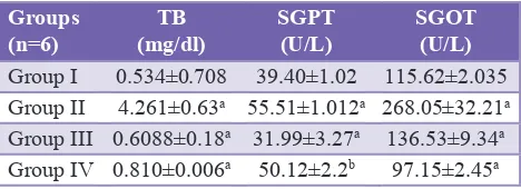
Administration of paracetamol 4 g/kg in Group II resulted
in significant increase in the level of TBL, SGPT, SGOT
(p<0.001) as compared to the normal (Group I). There is
a very significant reduction in the levels of TBL, SGPT,
SGOT in rats which administered with ME+paracetamol (Group III) and silymarin+paracetamol (Group IV) which is summarized in Table 1.
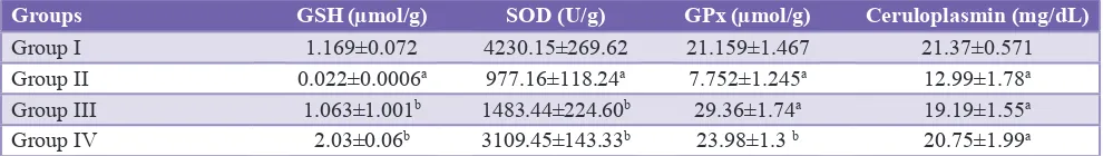
Effect of ME leaf extracts on hepatic oxidative stress parameters
After intoxication with paracetamol in Group II which
caused a significant (p<0.001), reduction of the levels of
GSH, SOD, GPx, and ceruloplasmin when compared with the control group which shows the hepatotoxic potential of paracetamol. After the administration of ME leaf extract with paracetamol (Group III) and silymarin with paracetamol has
shown significant (p<0.01) elevation in the levels of GSH,
SOD, GPx when compared with Group II. Ceruloplasmin
shows a very significant (p<0.001) elevation in Groups III
and IV. This result reveals the hepatoprotective effect of ethanolic extract of ME (Table 2).
DISCUSSION
Paracetamol is one of the common agents which cause hepatotoxicity. The incident was reported to be 10% as per a study conducted in France in 1997-2000.8 Hepatotoxicity
by acetaminophen has been reviewed already. The toxicity was found to be the combination of Phase I and Phase II-induced liver injury, i.e., the formation of both toxic
metabolites as well as inadequate detoxification. Once the
drugs taken into hepatocytes leads to formation of potentially hepatotoxic metabolites like n-acetyl-p-benzoquinoimine, reactive electrophiles, free radicals, etc., by cytochrome
p450 oxidative metabolism (toxification).9 These binds
with cellular membranes alters their functions and lead to hepatocellular necrosis.10 The outcome of Phase I is found
to be an alteration of plasma membrane permeability, disruption of cytoskeleton, mitochondrial dysfunction, loss of intracellular homeostasis. Activation of degradative
[image:3.595.311.545.620.705.2]enzymes and finally apoptosis.11,12
Table 1: Effect of M. erythrophylla leaf extract on biochemical parameters in paracetamol induced
hepatotoxicity.
Groups
(n=6) (mg/dl)TB SGPT (U/L) SGOT (U/L)
Group I 0.534±0.708 39.40±1.02 115.62±2.035 Group II 4.261±0.63a 55.51±1.012a 268.05±32.21a
Group III 0.6088±0.18a 31.99±3.27a 136.53±9.34a
Group IV 0.810±0.006a 50.12±2.2b 97.15±2.45a ap<0.001 when Group II compared with control group. ap<0.001, bp<0.01 when Group III and Group IV compared
In Phase II reaction (detoxification) the toxic drug metabolites NAPQI bind to GSH, glucuronate, or sulfate which leads to formation of non-toxic and excretable hydrophilic products like mercapturic acid. When the dose of paracetamol increases the GSH get exhausted which causes
inadequate detoxification due to inadequate GSH stores.
So overall a series of events beginning from intracellular hepatocyte disruption, cellular necrosis, apoptosis and
immune inflammatory response have been proposed to cause
drug-induced liver injury.13
In the present study to assess hepatoprotective activity the levels of SGPT, SGOT, and TBL has been measured. Marker enzymes are one of the commonest enzymes acting as indicator of hepatic damage. On liver injury, there will be alteration in the plasma membrane integrity and these enzymes will be leaked into the serum. In this study Group II rats were received acute overdose of paracetamol 2 g/kg body weight only on the 21st day which caused a very
significant elevation of hepatic serum markers like SGPT
and SGOT as well as TBL when compared with control group, which could be due to the above-stated mechanism of liver injury. In Group III when the rats received ethanolic extract of ME with paracetamol, the serum marker level has
come down very significantly (p<0.001) when compared
with Group II. This shows the protective effect of ME towardsparacetamol-induced toxicity. The findings on liver
function test are inconsistent with the previous studies. Murugesh et al. has been conducted a study to evaluate hepatoprotective and antioxidant role of Berberis tinctoria
lesch leaves on paracetamol induced hepatic damage in
rats. Their finding shows elevation in the serum marker
enzymes upon paracetamol intoxication and treatment with methanol extract of B. tinctoria at a dose of 150 mg/
kg and 300 mg/kg significantly reduced the elevated levels
of the enzymes.14
Ceruloplasmin is an alpha-2 glycoprotein, synthesized exclusively in the liver. Elevated ceruloplasmin levels are
present in acute infections and various inflammatory states.
Many pathological conditions are accompanied by a marked increase in plasma copper and ceruloplasmin.15 In the present
study paracetamol administration in Group II caused a
significant reduction in the levels of ceruloplasmin. There is a very significant elevation has been observed when the
rats treated with ME. This observation was comparable with silymarin in Group IV.
Protection from free radicals damage is carried out by antioxidant in our body. GPx causes detoxification of organic and inorganic peroxides by using reduced GSH. The regeneration of oxidized GSH is carried out by GSH reductase, in the presence of nicotinamide adenine dinucleotide phosphate-oxidase.16 In enzymatic antioxidant
system SOD was considered to be one of the sensitive index. It diminishes the toxic effects by superoxide anion by scavenging them to form hydrogen peroxide. In the present study administration of paracetamol caused a
significant reduction in the levels of GSH, SOD, and GPx
which gives a clear indication about the paracetamol to cause oxidative stress. Co-administration of ME leaf extract with paracetamol has improved the GSH, SOD and GPx levels. This shows that ME has antioxidant property which can protect the cells from free radical damage. Here, the importance of antioxidants is come to the picture. If free radicals are not inactivated, their chemical reactivity can damage all cellular macromolecules including proteins, carbohydrates, lipids and nucleic acids. Free radical damage to DNA is also implicated in the causation of cancer and its effect on low-density lipoprotein cholesterol is very likely responsible for heart disease.17
The plant products have been used to treat liver diseases because of their antioxidant properties. Most commonly used plants to treat liver diseases like silymarin, glycyrrhiza glabra, reported to have antioxidant constituents.18 Several
investigations have shown that silymarin improved liver function related to hepatocellular necrosis and increased membrane permeability through its antioxidant capacity. The protective effect of silymarin observed in the present study could be attributed to its antioxidant and free radical scavenging properties as reported in earlier studies.19-22
Silymarin prevents the absorption of toxins into the hepatocytes by occupying the binding sites as well as inhibiting many transport proteins at the membrane.23
The antiperoxidative property also has been postulated as the mechanism of protection in iatrogenic and toxic liver diseases.24
An antioxidant is a molecule stable enough to donate an electron to free radical to neutralize it. In a study conducted by Eswaraiah et al., on isolation of phytoconstituents from the stems of ME found to have the presence of
phytosterols, triterpenes, and flavonoids. They have isolated
[image:4.595.51.546.89.159.2]the active constituents from ME stem and it was found
Table 2: Effect of M. erythrophylla leaf extract on biochemical parameters in paracetamol induced hepatotoxicity.
Groups GSH (µmol/g) SOD (U/g) GPx (µmol/g) Ceruloplasmin (mg/dL)
Group I 1.169±0.072 4230.15±269.62 21.159±1.467 21.37±0.571
Group II 0.022±0.0006a 977.16±118.24a 7.752±1.245a 12.99±1.78a
Group III 1.063±1.001b 1483.44±224.60b 29.36±1.74a 19.19±1.55a
Group IV 2.03±0.06b 3109.45±143.33b 23.98±1.3 b 20.75±1.99a
ap<0.001 when Group II compared with Group I. ap<0.001, bp<0.01 when Group III and Group IV compared with Group II (paracetamol
to be β-sitosterol, 5 hydroxy-7, 4’-dimethoxy flavones,
3-isocumaryloxy-cyclopropane-1-oleic acid, 4-hydroxy-3-methoxy cinnamic acid.25 The hepatoprotective potential
of ME could be attributed to these antioxidant constituents.
CONCLUSION
This study provides the evidence of antioxidant and hepatoprotective property of MEand it may be due to the
presence of phytosterols, triterpenes, and flavanoids which
has been reported earlier. Further studies are in progress to identify active principles responsible for its antioxidant
activity. From the available data, ME produces significant
hepatoprotection with regard to paracetamol-induced toxicity.
ACKNOWLEDGMENTS
We are thankful to Nitte University for allowing as carrying out this study.
Funding: No funding sources
Conflict of interest: None declared
Ethical approval: The study was approved by the Institutional Animal Ethics Committee
REFERENCES
1. Lazarou J, Pomeranz BH, Corey PN. Incidence of adverse drug reactions in hospitalized patients. JAMA. 1998;279(15):1200.
2. Luper S. A review of plants used in the treatment of liver disease: part 1. Altern Med Rev. 1998;3(6):410-21.
3. Verma S, Singh SP. Current and future status of herbal medicines. Vet World. 2008;1:347-50.
4. Chandira M, Jayakar B. Formulation and evaluation of herbal tablets containing Ipomoea digitata Linn. extract. Int J Pharm Sci Rev Res. 2010;3:101-10.
5. Jaya Raju N, Ganga Rao B. Anthelmintic activities of antigonon leptopus hook and Mussaenda erythrophylla. Int J Pharm Pharm Sci. 2011;3:2011.
6. Venkatesh K, Rao U, Kiranmayi GV, Narasimha Naik R, Mukharjee NS, Vinay VN, et al. Phytochemical screening and evaluation of diuretic activity of ethanolic and chloroform extracts of Mussaenda erythrophylla in rats. IJBPR. 2013;4:8-10.
7. Eswaraiah MC, Satyanarayana T. Hepatoprotective activity of extracts from stem of M. erythrophylla against carbon tetrachloride - induced toxicity in rats. JPRHC. 2009;2:23-31. 8. Sgro C, Clinard F, Ouazir K, Chanay H, Allard C,
Guilleminet C, et al. Incidence of drug induced hepatic injuries: a French population based study. Hepatology. 2002;36:451.
9. LeBlanc GA. Hepatic vectorial transport of xenobiotics. Chem Biol Interact. 1994;90(2):101-20.
10. Mitchell JR, Thorgeirsson SS, Potter WZ, Jollow DJ, Keiser H. Acetaminophen-induced hepatic injury: protective role of glutathione in man and rationale for therapy. Clin
Pharmacol Ther. 1974;16(4):676-84.
11. Holt CD, Arriola E. Adverse effects of drugs on the liver. In: Koda- Kimble MA, Yong LY, Kradjan WA, Guglielmo BJ, editors. Applied Therapeutics the Clinical use of Drugs. 8th Edition. United States of America: Lippincott Williams and Wilkins; 2005: 30-4.
12. Losser MR, Payen D. Mechanisms of liver damage. Semin Liver Dis. 1996;16(4):357-67.
13. Jaeschke H, Gores GJ, Cederbaum AI, Hinson JA, Pessayre D, Lemasters JJ. Mechanisms of hepatotoxicity. Toxicol Sci. 2002;65(2):166-76.
14. Murugesh KS, Yeligar VC, Maiti BC, Maity TM. Hepato protective and antioxidant role of Berberis tinctoria Lesch leaves on paracetamol induced hepatic damage in rats. Int J Pharm Technol. 2005;4:64-9.
15. Martin SJ, Vedi M, Lavinya BU, Benny B, Agnes Selina K, Anne Sahithi ST, et al. Protective role of food supplement Spirulina fusiformis in chemical induced hepatotoxicity: a bromobenzene model in rats. Int J Pharmacol Res. 2014;4(1):4-9.
16. Iskusnykh IY, Popova TN, Agarkov AA, Pinheiro de Carvalho MA, Rjevskiy SG. Expression of glutathione peroxidase and glutathione reductase and level of free radical processes under toxic hepatitis in rats. J Toxicol. 2013;2013:Article ID 870628.
17. Bagchi K, Puri S. Free radicals and antioxidants in health and disease. IJCRR. 1998;4(19):350-60.
18. Wang GS, Han ZW. The protective action of glycyrrhiza
flavonoids against carbon tetrachloride hepatotoxicity in
mice. Yao Xue Xue Bao. 1993;28:572-6.
19. Vargas-Mendoza N, Madrigal-Santillán E, Morales-González A, Esquivel-Soto J, Esquivel-Chirino C, Morales- González-Rubio MG, et al. Hepatoprotective effect of silymarin. World J Hepatol. 2014;6:144-9.
20. Rasool M, Iqbal J, Malik A, Ramzan HS, Qureshi MS, Asif M, et al. Hepatoprotective effects of Silybum marianum (Silymarin) and Glycyrrhiza glabra (Glycyrrhizin) in combination: a possible synergy. Evid Based Complement Alternat Med. 2014;2014:641597.
21. El-Banna H, Ramadan S, Shalaby M, Afif N. Hepatoprotective
and antioxidant effects of Silybum marianum plant in rats. IJAVMS. 2011;5(6):541-7.
22. Favari L, Pérez-Alvarez V. Comparative effects of colchicine and silymarin on CCl4-chronic liver damage in rats. Arch Med Res. 1997;28:11-7.
23. Faulstich H, Jahn W, Wieland T. Silybin inhibition of amatoxin uptake in the perfused rat liver. Arzneimittelforschung. 1980;30:452-4.
24. Pradhan SC, Girish C. Hepatoprotective herbal drug, silymarin from experimental pharmacology to clinical medicine. Indian J Med Res. 2006;124:491-504.
25. Eswaraiah MC, Elumalai A. Isolation of phytoconstituents from the stems of Mussaenda erythrophylla. Pharm Sin. 2011;2:132-42.