The epistemic value of brain-machine systems for the study of the
brain
Edoardo Datteri
Department of Educational Human Sciences, University of Milano-Bicocca, Piazza dell’Ateneo
Nuovo 1, 20126 Milano. E-mail: [email protected]
Abstract. Bionic systems, connecting biological tissues with computer or robotic devices through brain-machine interfaces, can be used in various ways to discover biological mechanisms. In this
article I outline and discuss a “stimulation-connection” bionics-supported methodology for the
study of the brain, and compare it with other epistemic uses of bionic systems described in the
literature. This methododology differs from the “synthetic”, simulative method often followed in
theoretically driven Artificial Intelligence and cognitive (neuro)science, even though it involves
machine models of biological systems. I also bring the previous analysis to bear on some claims on
the epistemic value of bionic technologies made in the recent philosophical literature. I believe that
the methodological reflections proposed here may contribute to the piecewise understanding of the
many ways bionic technologies can be deployed not only to restore lost sensory-motor functions,
but also to discover brain mechanisms.
1. Introduction
Research on brain-computer interfaces (BCIs) is rapidly advancing towards the construction of
electronic and robotic systems – sometimes called hybrid bionic systems – that may be reliably
injury (see for example the case of the locked-in patient described in Hochberg et al., 2006). In
addition, leading researchers have claimed that bionics technologies can provide unique and new
experimental tools to discover brain mechanisms. For example, Wander and Rao (2014) claim that
brain-machine interfaces “can … be tremendously powerful tools for scientific inquiry into the
workings of the nervous system. They allow researchers to inject and record information at various
stages of the system, permitting investigation of the brain in vivo and facilitating the reverse
engineering of brain function. Most notably, BCIs are emerging as a novel experimental tool for
investigating the tremendous adaptive capacity of the nervous system” (p. 70). Golub et al. (2016)
“view BCI as a stepping stone toward understanding the full native sensorimotor control system”
(p. 56) and, according to Nicolelis (2003), brain-machine interfaces “can become the core of a new
experimental approach with which to investigate the operation of neural systems in behaving
animals” (p. 417).
To evaluate whether BCI technologies can live up to these expectations, it is essential to
understand how they can be used in neuroscientific research and under what methodological and
epistemological assumptions empirical data flowing from bionics-supported experiments can be
brought to bear on neuroscientific hypotheses. First steps towards this goal have been taken in
(Datteri, 2009), in which two bionics-supported methodologies for the discovery of brain
mechanisms have been outlined and discussed. Here I will argue that the vast majority of studies
reported in the recent scientific literature follow a methodology, called here the
stimulation-connection methodology, which has not been discussed there. The primary aim of this article is to
exemplify (Section 2.) and analyse some key features (Section 3.) of this methodology in a
contrastive way, that is to say, by comparing it with the simulation-replacement methodology
discussed in (Datteri, 2009).1
1 The simulation-replacement methodology is called ArB, from “Artificial replacement of Biological
In Section 3.1 I will argue that stimulation-connection and simulation-replacement studies
involve structurally similar systems, all being obtained by functionally replacing biological
components with artificial devices. In addition, they both use prostheses qua functional replacers of
biological components in order to test particular neuroscientific hypotheses. The “qua functional
replacers” clause is not redundant. I will argue that, in some BCI-supported theoretically driven
experiments, the artificial part of the hybrid system is not used to replace any biological component.
In other cases, the prosthesis actually replaces a biological component, but this fact does not play a
crucial epistemic role – in a sense to be discussed – in the neuroscientific discovery process. I will
focus on BCI-supported experiments in which brain mechanisms are discovered by functionally
replacing biological components with artificial devices – that is to say, in which the artificial device
is used qua functional replacer. Stimulation-connection and simulation-replacement studies fall in
this category.
I will also point out that these two classes of studies differ from one another in a number of
aspects. First (Section 3.2), they differ in the nature of the scientific question addressed: the
stimulation-connection methodology may assist in the theorization over the biological components
connected to the prosthesis (hence the “connection” label), while the simulation-replacement
methodology may enable one to model the behaviour of the biological component replaced by the
prosthesis (hence the “replacement” label). Second (Section 3.3), they differ in the experimental
procedure. The simulation-replacement methodology is akin to the “synthetic method” widely used
in Cybernetics, Artificial Intelligence, and contemporary biorobotics: theoretical results flow from
comparisons between the behaviour of the target biological system and the behaviour of the hybrid
system, which can be regarded as a hybrid simulation of the target hypothesis.
Stimulation-connection studies make a different, non-simulative use of machine models of biological systems:
peculiarly stimulated by connection with an artificial device. These distinctions, which will be
supported by an analysis of some case-studies, are summarized in Table 1.
In Section 4. I will bring the distinction between stimulation-connection and
simulation-replacement methodology to bear on some claims recently made by Craver (2010) and Chirimuuta
(2013) on the epistemic value of bionic systems. In particular, based on that distinction, I will show
that Craver’s (2010) arguments, though logically sound, do not support a sceptical view on the role
of bionics in neuroscientific research (Section 4.1). And in Section 4.2 I will argue that some of
Chirimuuta’s (2013) criticisms to Datteri’s (2009) methodological analysis rely on her overlooking
the distinction between the two strategies discussed here. Overall, I believe that the piecewise
formulation of a taxonomy of bionics-supported experimental methodologies, and the critical
analysis of claims made in the philosophical literature on the epistemic value of bionics, may
contribute to advancing our understanding of the role of BCI technologies in neuroscientific
research.
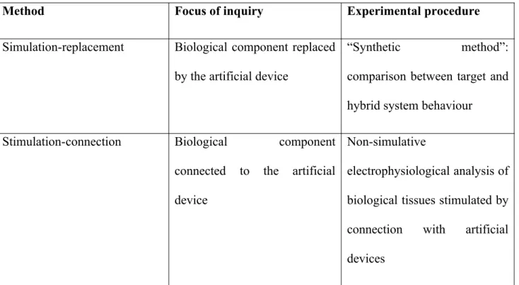
Table 1. Summary of the main differences between simulation-replacement and stimulation-connection
studies.
Method Focus of inquiry Experimental procedure
Simulation-replacement Biological component replaced
by the artificial device
“Synthetic method”:
comparison between target and
hybrid system behaviour
Stimulation-connection Biological component
connected to the artificial
device
Non-simulative
electrophysiological analysis of
biological tissues stimulated by
connection with artificial
2. Two bionics-supported studies for the discovery of brain mechanisms
2.1 The lamprey reticulo-spinal pathway
One of the goals of this article is to outline and discuss the structure of the stimulation-connection
bionics-supported methodology for the study of the brain. The key features of this methodology
may be easily identified by comparison with the simulation-replacement experimental strategy
discussed in (Datteri, 2009), which has been followed by Zelenin and colleagues (2000) to test a
mechanistic model2 of the lamprey sensory-motor system.
Lampreys are able to maintain a stable roll position by moving tail, dorsal fin, and other
body parts in response to external disturbances caused by water turbulence or other factors. A
particular portion of the lamprey nervous system – called the reticulo-spinal pathway, rs from now
on – is thought to play a crucial role in this behaviour. The goal of Zelenin and co-authors is to
discover the behaviour of rs – more precisely, to discover the relationship between the “input”
neurons of rs (the reticular neurons) and the roll angles of the animal, which vary as a function of
the activity of the “output” spinal neurons. The authors have initially formulated a relatively simple
hypothesis r(rs) about this relationship. To test it, they have built an electro-mechanical device
2 The expression “mechanistic model” is used here to refer to the description of a mechanism (Craver, 2007).
In what follows I assume that mechanistic models describe the regular behaviour of system components by means of
generalizations (Glennan, 2005; Woodward, 2002). The term “model” is used to emphasize the fact that mechanism
descriptions may be more or less abstract in the sense clarified by Suppe (1989): they characterize the behaviour of each component as depending on a restricted (though not necessarily narrow) set of factors. For example, a model might
characterize the activity of the neurons in a particular brain area as depending only on the firing rate of neurons in
another area; a less abstract model would take into account more input or boundary factors. Both models express
counterfactual generalizations stating that the behaviour of reticular neurons would be such and such, if it depended
whose input-output behaviour is r(rs). Then they have removed the reticulo-spinal component3 and
replaced it with the electro-mechanical device: the artificial component picked up the activity of the
reticular neurons and produced stabilization movements in line with the hypothesized regularity.
Finally, Zelenin and colleagues have experimentally tested whether the hybrid system exhibited
stabilization abilities comparable to those of the intact system. This has happened to be the case: the
authors have therefore concluded that the electro-mechanical device was a good substitute for the rs
component – and, as a consequence, that the rs component actually exhibited the hypothesized
input-output regularity r(rs).
2.2 Brain control of robotic prostheses in monkeys
In the study described in (Carmena et al., 2003), two monkeys chronically implanted with
micro-electrode arrays in various frontal and parietal brain areas have been trained to perform three kinds
of task. In the first one, they had to move a cursor displayed on a screen and reach a target by using
a hand-held pole. In the second one, they had to change the size of the cursor by applying a gripping
force to the pole. The third task was a combination of the first two. Neural activity was acquired,
filtered and recorded during execution of these tasks.
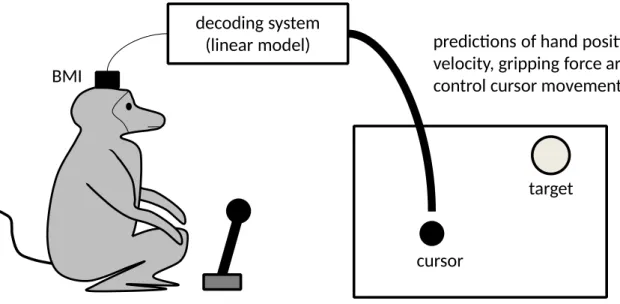
Two different uses have been made of these neural recordings in two distinct phases of the
study. During the first “pole control” phase, a reliable correlation has been identified between
neural activity and motor behaviour of the monkeys. More precisely, a linear model has been
trained to predict various motor parameters – hand position, hand velocity, and gripping force –
from brain activity (see Figure 1)
decoding system (linear model)
predictions of hand position, hand velocity, gripping force
pole control of cursor movements and size BMI
cursor
target
Figure 1 – The experimental set-up in the “pole control” phase.
After obtaining a predictively adequate model, the authors have proceeded with a so-called
“brain control” phase. During this phase, cursor position and size were totally disconnected from
pole movements: they were instead controlled by the output of the linear model receiving brain
activity as input (see Figure 2). The monkeys had to carry out the same three tasks, obtaining
rewards on successful trials.
decoding system
(linear model) predictions of hand position, hand
velocity, gripping force are used to control cursor movements and size BMI
cursor
target
Figure 2 - The experimental set-up in the brain control phase, with the decoder directly controlling the
cursor.
the movements of the robotic end-effector in space (see Figure 3), thus providing monkeys with
visual feedback on robot movements.
decoding system (linear model)
BMI
cursor
target
predictions of hand position, hand velocity, gripping force are used to control robot movements
robot control of cursor movements and size
Figure 3 - The experimental set-up in the brain control phase, with the decoder controlling the robot and
the robot controlling the cursor.
Interesting results with important engineering, therapeutic, and neuroscientific implications
have been obtained in these three experimental conditions. A first, basic result, in line with previous
studies (see for example Chapin et al., 1999), is that brain control of robotic prostheses is possible.
Indeed, after a short learning period, the monkeys became relatively proficient in brain-controlling
the cursor, both directly (Figure 2) and indirectly (Figure 3). The authors note that, at the very
beginning of the “brain control” phase, arm movements were still produced even though they were
no more needed to control the cursor. Interestingly, however, after a short period of time, the
monkeys ceased to move their limbs while continuing to brain-control the cursor.
Over and above this basic result, which paves the way to important therapeutic applications,
the authors have drawn interesting insights on the functioning of the (monkey) nervous system from
data obtained during the “pole control” (Figure 1) and “brain control” (Figures 2 and 3) stages.
Let me start from the “pole control” phase. As pointed out before, at the end of this phase a
fairly good model has been obtained, demonstrating that it is possible to predict various motor
Different brain areas have been found to contribute differently to various aspects of motor
behaviour. Moreover, by a so-called “neuron-dropping” methodology (Wessberg et al., 2000), it has
been found that the number of neurons required to make good motor predictions based on the linear
model changes from area to area (e.g., 33-56 cells in the primary motor area guaranteed accurate
predictions of all motor parameters, while 16-19 cells in the supplementary motor area were
sufficient to accurately predict hand position and velocity but not gripping force).
The achievement of good performances in the successive “brain control” phase may be
taken as indicative of decoder accuracy (even though, as often pointed out in the literature, efficient
prosthetic control can be due to the brain’s ability to compensate for decoder errors). However, it is
worth noting that the primary evidential basis on which the claims summarized in the previous
paragraph are based – stating that several motor parameters can be predicted from neural activity,
that different areas contribute differently to predicting motor behaviour, and that the quality of
prediction depends on population size – flows from data obtained during the “pole control” phase.
All these claims concern the predictive value of the model – that is to say, the relationship between
model outputs and actual pole-control movements made by the monkeys (see Figure 1). Information
on pole-control movements was clearly available during the “pole control” phase only. Only in this
phase it was therefore possible to compare model predictions with pole-control movements.4 As
pointed out before, monkeys rapidly ceased to produce limb movements in the successive “brain
control” phase – and, more generally, brain activity in the selected areas ceased to reflect
movements of the monkeys’ own limbs during the second phase of the study. For this reason, model
prediction accuracy could not be sensibly evaluated using data obtained in that phase.
According to the authors, the possibility to simultaneously extract several motor parameters
from neural ensembles in various parts of the brain suggest that “motor programming and execution
are represented in a highly distributed fashion across frontal and parietal areas and … each of these
Other insightful results obtained in the framework of this study are based on “brain control”
data. Some of them flow from control performance analysis. Performances suddenly declined after
switching from pole control (Figure 1) to brain control (Figures 2 and 3); however, they
progressively improved in successive brain control trials. According to the authors, this result could
be explained by assuming that efficient motor control requires a neural representation of the
dynamics of the controlled object. At the beginning of the “brain control” phase, the monkeys had
to control a totally novel object (a cursor on the screen and a robotic arm): no representation of it
could be available in their brains, leading to inefficient motor control. The successive improvements
in performance could be explained by hypothesizing that some adaptation process was taking place
in the brain, producing a neural representation of the new actuator.
This conjecture is further supported by other results flowing from the analysis of directional
tuning (DT) profiles of individual neurons and ensembles in the “brain control” phase. A DT profile
models the relationship between neural activity and direction of movement – for example, by
stating that a particular neuron fires maximally whenever the monkey moves her arm leftward. DT
profiles have been calculated during the “pole control” and the “brain control” phases, by modelling
the relationship between neural firing and cursor movements. Gradual changes in DT profiles have
been found during the “pole control” phase. Immediately after switching from pole to brain control,
that is to say, at the very beginning of the “brain control” phase, a general decline of DT strength
(i.e., of the strength of the correlation between firing activity and movement direction) has been
detected. A further decline has been observed when the monkeys ceased to move their limbs. Later
on, gradual increases in DT strength have been detected while the monkeys progressively improved
their brain-control proficiency, but the levels measured during pole control have been never reached
again. The emergence of clusters of neurons with similar DT profiles – that is to say, firing in
According to the authors, these results shed some light on the mechanisms of sensory-motor
control in the intact system. The sudden decrease in DT strength after switching from pole to brain
control, and especially the fact that DT strength was low even at the very beginning of the second
phase when the monkeys were still moving the pole, suggests that DT profiles do not reflect only
movement direction as signalled by proprioception (this kind of feedback was available at the very
beginning of the “brain control” phase). The successive increases in DT strength, when
proprioceptive feedback was totally uninformative of cursor direction, further support the thesis that
monkeys’ brains can progressively acquire a neural representation of the movements of the new
actuator based on visual feedback only.
Thus, we hypothesize that, as monkeys learn to formulate a much more abstract
strategy to achieve the goal of moving the cursor to a target, without moving their
own arms, the dynamics of the robot arm (reflected by the cursor movements)
become incorporated into multiple cortical representations. In other words, we
propose that the gradual increase in behavioral performance during brain control of
the BMI emerged as a consequence of a plastic reorganization whose main outcome
was the assimilation of the dynamics of an artificial actuator into the physiological
properties of frontoparietal neurons. (p. 205)
Note that this conjecture, supported by data obtained during the brain control phase, consists
in a very large-grained, tentative, and incomplete sketch of the mechanism connecting visual
feedback to changes in the behaviour of the neurons reached by the interface, that is to say, of a
neural mechanism implemented in the biological system to which the prosthesis is connected. This
observation will be more extensively justified in the ensuing contrastive methodological analysis of
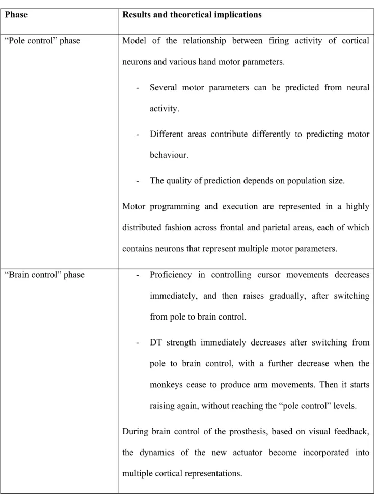
the case-studies reviewed so far. Table 2 summarizes the main results discussed in this section and
Table 2 - Summary of the main results obtained in (Carmena et al., 2003) and of their theoretical
implications
Phase Results and theoretical implications
“Pole control” phase Model of the relationship between firing activity of cortical
neurons and various hand motor parameters.
- Several motor parameters can be predicted from neural
activity.
- Different areas contribute differently to predicting motor
behaviour.
- The quality of prediction depends on population size.
Motor programming and execution are represented in a highly
distributed fashion across frontal and parietal areas, each of which
contains neurons that represent multiple motor parameters.
“Brain control” phase - Proficiency in controlling cursor movements decreases
immediately, and then raises gradually, after switching
from pole to brain control.
- DT strength immediately decreases after switching from
pole to brain control, with a further decrease when the
monkeys cease to produce arm movements. Then it starts
raising again, without reaching the “pole control” levels.
During brain control of the prosthesis, based on visual feedback,
the dynamics of the new actuator become incorporated into
3. The stimulation-connection methodology: a comparative analysis
3.1 Methodological and structural similarities
The two studies discussed in the previous section, to which I will refer as “lamprey study” and
“monkey study” respectively from now on, share some common methodological features. They
both involve structurally similar systems, that is to say, hybrid systems obtained by connecting
living systems with artificial (computer and robotic) devices. In some parts of both studies, the
artificial component of the hybrid system may functionally replace a biological component in
performing particular tasks. The robotic device used in the lamprey study functionally replaces the
reticulo-spinal pathway plus the motor organs of the animal in the posture control task. And the
device used in the “brain control” part of the monkey study replaces the animals’ arms and the
neural circuitries converting brain activity into efferent commands in the three reaching and
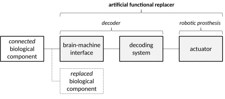
grasping tasks. To be sure, what has been called “robotic device” in the last two statements is in fact
composed of a brain-machine interface plus a computer-based decoding system generating motor
predictions based on neural signals (that is to say, outputting roll angles and hand motor parameters
in the lamprey and in the monkey study respectively), and of an electro-mechanical actuator. Let us
call “decoder” and “robotic prosthesis” these two parts of the artificial functional replacer (see
connected
biological
component
brain-machine
interface
decoding
system
actuator
decoder robotic prosthesis
artificial functional replacer
replaced
biological
component
Figure 4 – The structure of a hybrid bionic system.
It follows from this description that in the bionic preparations used in these studies one can
identify a biological component replaced by the artificial functional replacer (the reticulo-spinal
pathway plus the motor organs in the lamprey study; the arm plus a cortical decoding circuitry in
the monkey study) and a biological component connected to it (that is to say, the “rest” of the
system). Note that the replaced component is not physically removed from the intact system in
neither of the two studies: it is only made ineffective in performing the task.
The two studies are similar to each other in another methodological aspect. In both studies
the artificial device, qua functional replacer, plays a crucial experimental role in addressing
particular neuroscientific questions. Note the “qua functional replacer” remark. In principle, the
artificial device (decoder plus robotic prosthesis) or a part of it could be experimentally used in a
way that does not rests on its being a functional replacer of a biological component. For example,
one may use the brain-machine interface to pick-up neural activity while the monkey is performing
a particular task with the control system and the robotic prosthesis turned off. In this way the
artificial device would be simply used qua monitor of neural activity. Or, one may turn on both
devices without searching for correlations between (changes in) brain activity and (changes in)
As a third case, exemplified by the “pole control” part of the monkey study, one may use the
brain-machine interface and the decoding system only (with the robotic prosthesis unused or turned
off) in order to train the decoding system itself, that is to say, in order to obtain a model able to
decode particular motor parameters from brain activity. To this end, one acquires brain activity,
generates motor predictions based on the decoding system, compares them with actual movements,
and corrects the model implemented in the decoding system so as to obtain better predictions at the
next step. The formulation of a model able to predict motor behaviour from brain activity
constitutes a striking neuroscientific result, which may have interesting implications for
understanding the way movements are represented in, and decoded by, brain circuits. Moreover,
such a result would be simply unattainable without automatizing the generation of predictions and
the model correction process. It therefore flows from the deployment of innovative machine
technologies for the modelling of brain mechanisms. These technological novelties are not devalued
by noting that the machine is not used qua artificial replacer of a biological component in this case.
The fact that the machine used in the “pole control” phase of the study – namely, the interface and
the decoding system – was in principle able to drive a functional replacer of the animals’ arms has
not been crucial to obtain these results. Indeed, there was no artificial replacement at all in this
phase of the study: the model was trained with the monkeys using their own limbs to perform the
tasks.
The point is not whether the robotic part of the artificial device has been used or not. There
is a striking methodological difference between the “pole control” phase of the monkey study, on
one hand, and the lamprey study and the “brain control” part of the monkey study, on the other
hand. Functional replacement has played a crucial epistemic role in the two latter cases. It is by
measuring whether the artificial component is an efficient replacer that Zelenin and colleagues have
tested their hypothesis on the input-output behaviour of the reticulo-spinal component. And it is by
the three reaching and grasping tasks that Carmena and colleagues have discovered something
important on the mechanisms of plastic change in brain activity. The artificial devices have been
used qua functional replacers to obtain these results.
To sum up, the two studies are similar to each other not only in the fact that they both
involve a hybrid system, but also in the fact that they have made a theoretically fruitful use of the
artificial part of the hybrid system qua functional replacer of a biological component. This makes
the two studies (among other examples), and in particular the “brain control” phase of the monkey
study, not only technologically, but also methodologically novel. It is on the epistemic value of this
methodological novelty – that is to say, on the epistemic value of bionic systems used as functional
replacers of biological organs – that the ensuing analysis will be selectively focused.5
5 The distinction made here can be brought to bear on the question of what exactly is a bionics-supported
neuroscientific experiment. It may be defined as a neuroscientific experiment exploiting in vivo connections between living systems and artificial devices. This definition would be too liberal, however, as traditional neurophysiological
experiments – e.g., voltage clamp experiments – would fit it. On the contrary, it would be too restrictive to include in
the class of bionics-supported experiments only those experiments in which the target living system controls robotic
devices, as this would exclude experiments in which the subject brain-controls virtual devices (as, for example, in the
set-up illustrated in Figure 2). Then, one may include in that class all and only those neuroscientific experiments which
involve hybrid systems whose structure can be described as in Figure 4. It is worth noting, however, that the label
“hybrid bionic system” is typically used in the contemporary scientific literature to refer to systems in which the
artificial device functionally replaces a biological component. That is to say, contemporary bionics research is focused
on the realization of devices which are essential for people suffering from motor or sensory limitations to perform particular tasks, as they can replace sensory or motor organs. For this reason, here I will restrict the label
“bionics-supported neuroscientific experiment” to all and only the experiments which make an essential use of artificial devices
qua replacers of biological components. The label therefore does not apply to the experiments carried out in the “pole control” phase of the monkey study, even though they have involved a system that could be structurally described as in
Besides these fundamental analogies, the two studies differ from one another in a number of
methodological aspects. The analysis of these differences may enable one to appreciate the novelty
of the experimental approach adopted in (Carmena et al., 2003) and in similar studies with respect
to the hybrid simulative methodology discussed in (Datteri, 2009), and to critically evaluate some
arguments recently proposed by Craver (2010) and Chirimuuta (2013).
3.2 Connection vs. replacement methodology
As pointed out before, in the two studies reviewed here a biological component is connected to an
artificial device which may replace another biological component. A first methodological
difference between the two studies concerns whether the focus of inquiry is on the replaced or the
connected biological component. The lamprey study aims at modelling the behaviour of the
replaced component. On the contrary, the “brain control” part of the monkey study offers insights to
theorize on the neural mechanisms implemented in the component to which the artificial device is
connected. Let me elaborate on this point.
The question addressed in the lamprey study concerns the input-output behaviour of the
reticulo-spinal pathway controlling roll posture, which is the biological component replaced by the
artificial device. As discussed above, the authors’ strategy consists in replacing the target
component with an artificial device whose input-output behaviour is known, and checking whether
the hybrid system can produce the same behaviour as before. In that case, one may be induced to
conclude that the input-output behaviour of the replaced component matches the behaviour of the
replacer – hence, since the behaviour of the replacer is known, one would obtain a description of the
former. Bionics-supported experiments and methodologies in which functional substitution with an
artificial device enables one to inquire into the behaviour of the replaced biological component will
be called replacement experiments and methods from now on.
parameters, that is to say, a model of the behaviour of the biological component that in the “brain
control” phase has been functionally replaced by the artificial device. In the previous section I have
argued that the machine has not been used qua functional replacer in the first part of the monkey
study. On the contrary, in the “brain control” phase the authors have analysed changes in the
directional tuning of cortical neurons while the monkeys were learning to brain-control the
prosthesis, and have brought these results to bear on what happens in the brain after connection with
an artificial functional replacer. These results can be brought to bear on the mechanisms enabling
the brain to adapt to new tools and, more generally, to develop good motor control abilities (recall
the theoretical considerations summarized at the end of Section 2.2). In this part of the study, the
authors have made experimental use of the artificial device qua functional replacer to address
scientific questions on the biological component connected to the artificial device itself. Unlike the
lamprey study, this part of the monkey study can be classified as a connection bionics-supported
study.
This thesis can be further supported by reflecting on the relationship between the
input-output behaviour of the artificial device and of the replaced biological component in the lamprey
study and in the “brain control” part of the monkey study. At the beginning of the lamprey study,
the authors do not know whether the two components have the same behaviour or not: this is exactly
the question that the authors want to address. At the beginning of the “brain control” part of the
monkey study, on the contrary, the authors already know that the decoding system’s behaviour
matches to a reasonable extent the input-output behaviour of the biological component which is
now functionally replaced by it: this was exactly the goal of the previous part of the study. One
should be careful to note that other connection studies have tested the ability of the brain to adapt to
decoders which were known to be predictively inaccurate, as they had been obtained by shuffling
the weights of a previously trained model (Ganguly and Carmena, 2009, is a case in point). The
or not. Rather, it concerns whether, at the beginning of the study, one has information on the match
or mismatch between the two behaviours. Such information was unavailable at the beginning of the
lamprey study, consistently with my claim that the goal of the study was exactly to discover the
behaviour of the replaced component. On the contrary, such information was available at the
beginning of the “brain control” monkey study and of (Ganguly and Carmena, 2009), consistently
with the claim that the goal of the study was not to discover the behaviour of the biological
component replaced by the decoding system. I also take for granted that the monkey study was not
aimed at modeling the behaviour of the biological component replaced by the robotic prosthesis,
i.e., the arm. Therefore, since the artificial system used in that study was composed by a decoding
system and by a robotic prosthesis (Figure 4), and that neither system was the focus of inquiry, one
may legitimately conclude that the “brain control” monkey study was aimed at modeling the
behaviour of the biological component connected to the artificial system.
A wide variety of bionics-supported experiments of neuroscientific interest have been
performed since (Carmena et al., 2003). Indeed, there are good reasons to claim that the number of
bionics-supported connection studies that are reported in the literature is far higher than the number
of replacement studies. For example, Ganguly et al. (2011) have claimed that, although previous
studies have discovered modifications in neural activity correlated with improvements in brain
control performance, “it remains difficult to place such modifications in the context of the large
cortical network for motor control” (p. 662). To proceed towards this goal, they have studied the
relationship between changes in the activity of the neurons more directly involved in prosthetic
control and changes in the activity of what they have called “indirect” neurons, that is to say,
neurons reached by the interface but less involved in prosthetic control. Based on that, they have
speculated on the functional role that these indirect neurons might have in determining the activity
of the “direct” neurons, and on the possible layout of the cortical mechanisms involved in forming
happens upstream of the neurons more directly involved in motor control, and in particular, to
develop hypotheses on the brain mechanisms underlying plasticity (see also Koralek et al., 2012).
The claim that bionics can assist in the study of the biological components connected to the
machine is often more or less explicitly made in the scientific literature. For example, Golub et al.,
(2016) point out that bionic systems can help one to “obtain a more complete understanding of the
cognitive processes underlying sensorimotor control” (p. 53) and “to understand how different
sensory modalities contribute to sensorimotor control” (p. 55): “because of the similarity of the
cognitive processes and brain areas involved in native motor and BCI control, we view BCI as a
stepping stone toward understanding the full native sensorimotor control system. The BCI
paradigm, being a reduced system, offers vastly improved accessibility and manipulability, without
simplifying away the complexities of brain processing that we wish to understand” (p. 37, emphasis
added). It follows from this claim that the focus of inquiry in bionics-supported experiments is the
non-simplified part of the brain, that is to say, the biological components connected to the BMI.
Similarly, Orsborn and Carmena (2013) point out that “BMI also allows observation of brain areas
not directly contributing to the task. What occurs in other parts of the motor cortex as a subject
learns a neuroprosthetic skill?” (p. 4, emphasis added). According to Wander and Rao (2014),
various results flowing from motor bionics-supported experiments have suggested that, during
adaptation to a novel tool, “the brain [is] dynamically modifying internal networks to dissociate
changes in neural activity from the motor movements with which they were originally correlated”
(p. 71) and that, “even when effective control of the BCI only explicitly requires modulation of
activity in a small cortical region, frontal and parietal cortical regions are strongly task modulated
during initial performance of the task and less so after extensive training” (p. 72). All these claims
point to the role of BMIs in modelling the processes occurring in the part of the hybrid systems
connected to the machine. Even more explicitly, the same authors state that “BCIs can be a
experimentalist opportunities not only to observe sensorimotor transformations as information
travels through the brain, but also to modify the nature of these transformations in real-time. Most
importantly, BCI technology provides a scaffold for scientific experimentation that enables
investigation of the nervous system doing what it does best: incorporating new information and
rapidly adapting to new constraints” (p. 73, emphasis added).
Let me sum up by mathematical analogy. The behaviour of the replaced components is the
unknown variable in the equation the authors of the lamprey study have tried to solve. In the “brain
control” part of the monkey study, on the contrary, the behaviour of the replaced component has
been fixed by a prosthetic implant which imposes a known mapping from brain activity to
movements, in order to simplify the identification of the unknown variables representing brain
mechanisms.
In the next section I will argue that the lamprey and the “brain control” part of the monkey
study differ from each other also in the nature of the empirical basis which is brought to bear on the
theoretical hypothesis under scrutiny.
3.3 Stimulation vs. simulation methodology
Machines have been often used in cognitive science and neuroscience as simulations of theoretical
models of animal behaviour. The simulative methodology, sketched by pioneers of Cybernetics
Arturo Rosenblueth, Norbert Wiener and Julian Bigelow in (Rosenblueth and Wiener, 1945),
proceeds as follows. Let M be a model of the (cognitive or neural) mechanism hypothesized to
produce behaviour B by living system S in particular experimental conditions C. To test if this
hypothesis is true, one can build an artificial system A which implements M, and compare A’s
behaviour in conditions C with B. Behavioural similarities between A and S may induce one to
conclude that M can produce B in C. Otherwise, one may be induced to reject the conjecture.
and Simon, 1961; Simon and Newell, 1962), and contemporary biorobotics (Floreano et al., 2014;
Datteri and Tamburrini, 2007; Tamburrini and Datteri, 2005; Webb, 2001).
A notable feature of the simulative method is that theoretical conclusions on the relationship
between theoretical model M and living system S are obtained by comparing S’ behaviour with the
behaviour of the simulation. The fact that computers implementing particular models of heuristic
means-end analysis proved able to solve problems with performances comparable to those exhibited
by human beings engaged in the same tasks was the main evidential basis used by Newell and
Simon to support their theories on problem solving. The fact that the robot described in (Grasso et
al., 2000) consistently failed to reproduce lobsters’ goal-seeking behaviour was the main evidential
basis used by the authors of the study to reject their hypothesis on lobster chemiotaxis. Note that the
simulative methodology does not require one to focus on the motor or verbal output of the
simulation only. Often, internal comparisons between living and simulation systems are carried out.
For example, one may check not only if the output of an artificial neural network reproduces the
output of a biological network, but also if the patterns of spiking activity of individual artificial
neurons match those of their biological counterparts (see Chou and Hannaford, 1997, for an
example). Newell and Simon famously asked their subjects to produce verbal reports of their
reasoning processes during problem-solving tasks in order to compare their cognitive dynamics
with the representational transformations made by the simulation system.
As discussed in (Datteri, 2009), the lamprey study exemplifies a hybrid variant of the
simulative strategy. While classic, non-hybrid simulation studies typically start with a description of
M specifying the input-output behaviour of all the components of the hypothesized mechanism and
their organization, the authors of the lamprey study start with a hypothesis on the input-output
behaviour of just one component – the reticulo-spinal pathway plus the motor organs of the animal.
While in non-hybrid simulation studies one builds a fully artificial system implementing M, the
behaviour of the target biological component only. More crucially, as in classic simulation studies
of Artificial Intelligence, Cybernetics, and biorobotics, the primary evidential basis which is
brought to bear on the target hypothesis flows from the analysis of whether the hybrid system’s
posture maintenance behaviour matches the behaviour of the intact lamprey or not. For these
reasons, I take the lamprey study as a simulation bionics-supported study.
The “pole control” phase of the monkey study conforms to the simulation methodology in
some of the aspects discussed here, as the primary evidential basis to conclude that the decoding
system predicts hand motor parameters from brain activity comes from the analysis of whether the
monkey’s behaviour matches decoder outputs or not. Interestingly, the successive “brain control”
phase of the monkey study does not conform to the simulative methodology. The experimental
results obtained in this part of the study (see Table 2) flow from an analysis of the relationship
between neural and motor parameters of the system (for example, between firing rate and
movement direction), and of changes in this relationship, while the animal is learning to
brain-control the artificial replacer. In other words, even though an artificial model of the replaced
biological component is used, these results are obtained by applying relatively traditional
electrophysiological analysis techniques in a radically novel experimental setting – that is to say,
while the brain is stimulated by the presence of a new tool.6 For this reason, I consider the “brain
6 To be sure, experiments in electrophysiology often involve artificial stimulations of the target biological
tissue. For example, one may intervene on the membrane potential of particular neurons after blocking specific kinds of
ion channels in order to find the threshold above which action potentials are generated in those conditions. Note that, in
experiments of this sort, the nature and magnitude of the “input” stimulation (e.g., of changes in membrane potential)
do not systematically depend on the effects of that stimulation (e.g., on whether action potentials are generated or not).
The “input” parameters are independent of the “output” of the interventions: the experimenter explores a relevant
portion of the “input” space and measure the effects in order to find a correlation. Quite on the contrary, in
non-simulation, connection bionic experiments, the nature and magnitude of the stimulation received by the biological
control” part of the monkey brain as an example of a stimulation, non-simulative bionics-supported
methodology for the discovery of brain mechanisms.
This is not to say that comparisons between motor control proficiency in the hybrid and in
the intact monkey play no theoretical role at all. The achievement of high brain control proficiency
is crucial to interpret the detected plastic changes as related to the successful “internalization” of the
dynamics of the new tool. But the outcome of this comparison is not the main empirical basis on
which the theoretical conclusions summarized above are made to rest, as in the (hybrid) simulation
methodology: they crucially flow from a neurophysiological analysis not intended to provide data
for output or “internal” comparisons. DT analysis and the neuron-dropping procedure adopted in the
“brain control” phase are not parts of the “synthetic method”.
To sum up, the “brain control” phase of the monkey study exemplifies how machine models
of biological components can be used in a way that is interestingly different from the way machine
models have been traditionally used in cognitive science and neuroscience. The distinction sketched
here between simulation and stimulation bionics-supported experiments, as well as the distinction
between connection and replacement experiments, will be used in the next section to evaluate some
claims made in Craver (2010)’s and Chirimuuta (2013)’s analyses of the epistemic role of bionics.
4. Underdetermination, plasticity, and the methodological requirements of
“good” bionic experiments
4.1 Craver on the epistemic role of bionic experiments
In his article, Craver (2010) focuses on “what, if anything, building a prosthetic mechanistic model
adds to our confidence that we have a valid mechanistic model over and above the degree of
subject about the way her brain is moving the prosthesis. Brain activity determines prosthetic behaviour; information on
prosthetic behaviour determines changes in brain activity. Such a “circular” connection between the nature and
confidence provided by models and simulations alone” (p. 843). His answer is a qualified
“nothing”. However, at the end of the paper he draws a stronger conclusion, defending a sceptical
view as to whether “the ability to build a successful prosthesis counts as evidence that one knows
how the system works” (p. 850).
Craver’s argument runs as follows. He refers to a prosthesis as affordance valid “to the
extent that the behaviour of the simulation could replace the target in the context of a higher-level
mechanism” (p. 842). A robotic arm enabling one to perform all the movements and actions she
could perform with a biological arm – such as grasping, lifting, or pushing objects – is affordance
valid. A prosthesis is said to be phenomenally valid to the extent that “its input-output relationship
is relevantly similar to the target input-output relation” (p. 842) under standard or non-standard
conditions. A robotic arm, for example, is phenomenally (or behaviourally) valid if the relationship
between its inputs and movements is relevantly similar to the relationship between the inputs and
the movements of the biological arm it is replacing. Finally, a prosthesis is said to be
mechanistically valid to the extent that its parts, activities, and organizational features are relevantly
similar to the parts, activities, and organizational features of the target system (p. 842). A robotic
arm, for example, is mechanistically valid if its internal mechanism is relevantly similar to the
internal mechanism governing the replaced arm (mechanistic validity will be discussed more in
detail below).
Craver’s first point is that affordance valid prostheses need not be phenomenally valid. He
rightly argues that a robotic system can partially replace the functionality of a missing arm or leg
even if its input is very different from the input of the replaced biological component. The arm
prosthesis described in Carmena et al. (2003), for example, is affordance valid (the monkeys could
use a brain-controlled prosthesis to carry out tasks which they had previously carried out by using
their own hands). However, the neurons whose activity controlled the prosthesis were not those
various frontoparietal neural ensembles acquired through a multi-electrode brain-machine interface.
Even though these brain areas participate in motor control, monkey arms are not directly connected
to these areas. The input of the prosthesis was very different from the input of biological monkey
arms. For this reason, the input-output behaviour of the prostheses was radically different from the
input-output behaviour of monkey arms, and the prosthesis was therefore to be regarded as
phenomenallyinvalid.
The fact that affordance validity does not imply phenomenal validity has, according to
Craver, an important consequence as to whether bionics research can really contribute to the
discovery of brain mechanisms: being able to build an affordance valid prosthesis does not imply
having understood the input/output relationship characterizing the replaced biological component.
Another point made by Craver is that affordance and phenomenal validity do not imply
mechanistic validity: “a prosthetic model might be affordance valid and phenomenally valid yet
mechanistically invalid”; “building a functional prosthesis that simulates a mechanistic model is
insufficient to demonstrate that the model is mechanistically valid” (p. 845). Recall that a prosthesis
is mechanistically valid to the extent that its parts, activities, and organizational features are
relevantly similar to the parts, activities, and organizational features of the target. Therefore, here
Craver is arguing that a prosthesis may be affordance or phenomenally valid even though its
internal mechanism is not relevantly similar to the internal mechanism of the target. This is because
many different mechanisms, in principle, could produce the same input-output behaviour: in the
philosophical jargon, the mechanism is underdetermined by the device’s input-output behaviour, or,
equivalently, the latter is said to be multiply realizable.
Phenomena are multiply realizable in lower-level mechanisms. Multiple realizability
obstructs the inference from a model’s phenomenal validity to its mechanistic
validity. The space of phenomenally adequate simulations might well be too large
phenomenally adequate simulation are relevantly similar to the mechanistic features
of the target (p. 844).
It is important to understand what exactly Craver means with “target” in these claims. The
problem of multiple realizability obstructs inferences from the behaviour of a system to the
mechanism producing that behaviour. In a simulation study, in particular, one builds an artificial
system A simulating a theoretical model of target (living) system S (see Section 3.3). In this
context, the problem of multiple realizability can be stated as follows: the fact that simulation
system A reproduces the input-output behaviour of target system S is insufficient to conclude that
A’s internal mechanism is relevantly similar to the mechanism governing S. Now, Craver warns us
that building a prosthesis that reproduces the input-output behaviour of a biological component
(thus being phenomenally valid) is insufficient to conclude that the prosthesis reproduces the
internal mechanism of that biological component, namely of the component replaced by the
prosthesis. His point is that a phenomenally valid arm prosthesis, for example, can fail to reproduce
the mechanism governing the replaced arm. To sum up, Craver argues that being able to build a
phenomenally valid prosthesis is not sufficient to demonstrate that one has understood the
mechanism governing the biological component replaced by the prosthesis.
Taken together, the arguments discussed so far allow Craver to conclude that “affordance
valid models need not be mechanistically or phenomenally valid. This is a blessing for engineers
and a mild epistemic curse for basic researchers” (p. 850). Accurate replication of the input-output
behaviour and internal mechanism of the replaced component is not needed to build an efficient
prosthesis (this is the blessing for engineers); at the same time – Craver argues – one can build an
efficient prosthesis without having understood the behaviour of the replaced component and its
internal mechanism (this is the curse for basic researchers). The sceptical theses on the epistemic
Craver’s claims that affordance validity does not imply phenomenal validity, and that
phenomenal validity does not imply mechanistic validity of a prosthesis, are logically correct.
However, based on the distinction between simulation-replacement and stimulation-connection
methodologies made above, I will argue that they do not provide good reasons to believe that bionic
systems are of little help in discovering brain mechanisms.
Craver’s first point is that affordance valid prostheses need not be phenomenally
(behaviourally) valid. This thesis, interpreted as a general rule, is true: one can find many examples
of animals learning to control robotic prostheses whose input-output behaviour does not match the
behaviour of the replaced components (as Craver rightly points out, efficient control can be
achieved even when the input of the prosthesis is very different from the input of the replaced
component). It is worth noting, however, that in the framework of particular studies, an example
being the lamprey study, it may be safe to infer phenomenal validity from affordance validity.
Craver claims that bionic prostheses are never connected to the original brain inputs of the replaced
biological component (“no currently available [bionic] device, to my knowledge, makes use of just
those brain inputs that move limbs in typical animals”, p. 845). This is not true: the
electro-mechanical device in the lamprey study described above was driven by the same neural input (i.e.,
by the activity of the reticular neurons) that, in intact lampreys, drive the behaviour of the target
reticulo-spinal component. And the fact that the prosthesis proved able to functionally replace the
target component (thus being affordance valid) was taken as a basis to conclude that its input-output
behaviour matched the target component’s input-output behaviour (i.e., that it was phenomenally
valid too). Another study in which the prosthesis is driven by the same inputs that drive the
behaviour of the replaced component is reported in (Le Masson et al, 2002).
The fact that, in the framework of particular studies, it is safe to infer phenomenal validity
from affordance validity does not invalidate Craver’s thesis that affordance valid prostheses need
not be improperly taken as supporting a too sceptical thesis on the epistemic value of prosthetic
models. In some cases, having built an affordance valid prosthesis implies having understood the
input-output behaviour of the replaced component.
Another point made by Craver is that affordance and phenomenal validity do not imply
mechanistic validity: being able to build an artificial device which reproduces the input-output
behaviour of a biological component does not imply that one has understood the mechanism
governing it. It is important to clarify what exactly we can learn from this claim. Evidently, as
argued before, Craver is warning us that building a working (affordance or phenomenally valid)
prosthesis does not imply that we have understood the mechanism governing the behaviour of the
replaced biological component (for example, of the replaced arm, in the case of an arm prosthesis).
His argument, therefore, specifically targets the epistemic value of bionic experiments in which the
target component (i.e., the component whose behaviour or internal mechanism is to be discovered)
is replaced by the prosthesis (i.e., those experiments referred to in this paper as replacement
experiments). Moreover, it is intended to support a sceptical view as to whether such experiments
can assist in discovering the internal mechanism of the replaced component.
I will reply to Craver’s argument in two steps. First, as borne out by many successful
model-oriented simulation studies, the fact that the input-output behaviour underdetermines the internal
mechanism does not imply that building a phenomenally valid prosthesis provides no evidence at
all regarding the structure of the replaced component’s internal mechanism. Second, as suggested
earlier, bionic technologies enable experimental strategies that are significantly different from the
simulative replacement methodology described in section 2.1, and which contribute to the discovery
of brain mechanisms by shedding light on the input-output behaviour of the replaced component
rather than on the replaced component’s internal mechanism. Craver’s multiple realizability
argument does not apply to these strategies, and therefore does not support a generalized sceptical
Let me begin from the first step. Craver is right in pointing out that, strictly speaking,
phenomenal validity does not imply mechanistic validity. However, this argument does not exclude
that building a phenomenally valid prosthesis can provide evidence contributing to the discovery of
the internal mechanism of the replaced component. Craver construes evidence as “a finding that
shapes (or constrains) the space of possible mechanisms for a given phenomenon” (Craver, 2010, p.
843). Now, the fact that the output behaviour of the prosthetic component matches the
input-output behaviour of the replaced component – thus, that the prosthesis is phenomenally valid – is
not a conclusive reason to claim that the prosthesis’ internal mechanism is relevantly similar to the
replaced component’s internal mechanism. Nevertheless, it is a good reason to include the
prosthesis’ internal mechanism within the space of the possible mechanisms governing the replaced
component. Similarly, realizing that the prosthesis is not phenomenally valid may be legitimately
taken to be a good reason to exclude its internal mechanism from the space of how-possibly models
of the replaced component. Purely behavioural tests often lead Artificial Intelligence and
biorobotics researchers to expand or restrict the space of the possible models of the target living
system. An example is the model-oriented biorobotic study reported in (Grasso et al., 2000), in
which mismatches between the overt behaviours of the simulation and of the target system – with
no “internal” comparison between the two systems – were taken as reasons to conclude that the
mechanism implemented in the robot could not be a good model of the animal. To sum up, Craver
is correct in claiming that phenomenal validity does not imply mechanistic validity. Nevertheless,
his underdetermination argument does not exclude that phenomenal validity can provide evidence
constraining the space of the possible models of the target system – thus, that it can contribute to
model discovery.
The second step is more easily taken. Craver’s underdetermination argument focuses on the
fact that it is mistaken to infer a prosthesis’ mechanistic validity from its phenomenal validity.
governing the replaced component. Therefore, Craver’s underdetermination argument supports a
sceptical view as to whether building a phenomenally valid prosthesis can contribute to discovery of
the mechanism governing the replaced limb. However, as discussed before, in bionic connection
studies (for example, in the “brain control” phase of the monkey study) one theorizes on the
behaviour of a biological component by connecting it to, rather than by replacing it with, artificial
devices. The underdetermination argument has no force against these kinds of studies. Consider
also that the argument applies only when the mechanism governing the behaviour of the replaced
component is inferred from the behaviour of the prosthesis or the behaviour of the hybrid system
only. Inferences of this kind are made in what have been called here simulation bionic experiments.
As argued in section 3.3, in stimulation bionic studies neural mechanisms are inferred not only from
the analysis of overt hybrid system behaviours, but also from the analysis of neural activity while
the subject is learning to control the prosthesis. The underdetermination argument, as proposed by
Craver, has therefore nothing to say on experiments of the latter kind – thus, among other examples,
on (Carmena et al., 2003).
One should also be careful to note that Craver’s underdetermination argument concerns
inferences from the replaced component’s behaviour to its internal mechanism. That is to say, his
argument is intended to support a sceptical view as to whether bionic technologies can assist in
discovering the internal mechanism of a component of a sensory-motor mechanism. But it does not
rule out the possibility that bionic technologies can assist in discovering the input-outputbehaviour
of a component of a sensory-motor mechanism – a discovery that may be of theoretical interest with
respect to the broader goal of discovering the structure of the mechanism governing the containing
system. For example, the goal of (Zelenin et al., 2000) was to identify the behaviour of the
reticulo-spinal component, and not its internal mechanism. Nevertheless, by achieving this result, they have
contributed to the modelling of the whole lamprey sensory-motor system. Craver’s
In sum, Craver (2010) does not offer strong arguments for excluding that bionic
technologies can provide evidence for the discovery of brain mechanisms. In particular, his
arguments – though logically correct – apply neither to stimulation-connection experiments, nor to
simulation-replacement experiments aimed at identifying the behaviour of the target component and
not its internal mechanism (as in the lamprey study).
To conclude, another claim made by Craver in his article is that bionic methodologies do not
offer significant epistemic advantages over other kinds of methodologies currently adopted in
neuroscience: “the effort to build a prosthetic model allows a decisive test of affordance validity but
offers no distinct advantages for assessing the model’s phenomenal and mechanistic validity” (p.
841). However, as acknowledged by Craver himself, simulations in general can significantly speed
up the process of evaluating the behavioural implications of the mechanistic model at issue. In some
cases, this is likely to be also true for bionic devices. Consider the lamprey study. To assess whether
the reticulo-spinal pathway performed the hypothesized regularity, the authors could well have
recorded reticular activity and measured roll movements in an intact, swimming lamprey, in search
of a correlation between the two. Even though it is difficult to say a priori whether building such a
recording apparatus would have been more difficult than setting-up the bionic preparation described
in the article, the bionic solution offered a relatively direct means of assessing whether the authors’
hypothesis was correct: instead of searching for a correlation, they just checked if the hybrid animal
was able to stabilize itself. And consider the stimulation-connection experiments described in
Section 2.2. It is reasonable to believe that connection with an actual prosthetic device played a
crucial role in the development of the various theoretical hypotheses on the neural assimilation of
external tools reported in (Carmena et al., 2003; Lebedev et al., 2005; Nicolelis, 2011) – and
therefore, that the deployment of bionic technologies has made a decisive contribution, vis-à-vis
other experimental methodologies, to achieving these results. There are no sufficiently general