REVIEW
Review on the current status of polymer
degradation: a microbial approach
Vinay Mohan Pathak
*and Navneet
Abstract
Inertness and the indiscriminate use of synthetic polymers leading to increased land and water pollution are of great concern. Plastic is the most useful synthetic polymer, employed in wide range of applications viz. the packag-ing industries, agriculture, household practices, etc. Unpredicted use of synthetic polymers is leadpackag-ing towards the accumulation of increased solid waste in the natural environment. This affects the natural system and creates vari-ous environmental hazards. Plastics are seen as an environmental threat because they are difficult to degrade. This review describes the occurrence and distribution of microbes that are involved in the degradation of both natural and synthetic polymers. Much interest is generated by the degradation of existing plastics using microorganisms. It seems that biological agents and their metabolic enzymes can be exploited as a potent tool for polymer degradation. Bacte-rial and fungal species are the most abundant biological agents found in nature and have distinct degradation abili-ties for natural and synthetic polymers. Among the huge microbial population associated with polymer degradation, Pseudomonas aeruginosa, Pseudomonas stutzeri, Streptomyces badius, Streptomyces setonii, Rhodococcus ruber, Coma-monas acidovorans, Clostridium thermocellum and Butyrivibrio fibrisolvens are the dominant bacterial species. Similarly, Aspergillus niger, Aspergillus flavus, Fusarium lini, Pycnoporus cinnabarinus and Mucor rouxii are prevalent fungal species. Keywords: Polymer, Microbial degradation, Bacteria, Fungi, Natural polymers, Synthetic polymers, Polysaccharide, Hydrolytic enzyme, Pollution, Organic pollutants, Waste management, Biofilm, Surfactants, LDPE, Aerobic degradation, Anaerobic degradation, UV irradiation, Manmade compound, Plastic waste, SEM, Sturm test, FT-IR
© The Author(s) 2017. This article is distributed under the terms of the Creative Commons Attribution 4.0 International License (http://creativecommons.org/licenses/by/4.0/), which permits unrestricted use, distribution, and reproduction in any medium, provided you give appropriate credit to the original author(s) and the source, provide a link to the Creative Commons license, and indicate if changes were made.
Background
Developments in science and technology, especially over the last 2 decades, have led to the production of a num-ber of synthetic polymers worldwide. The polymers are chains of monomers linked together by chemical bonds. Polymers such as lignin, starch, chitin, etc., are present in the environment naturally. Nowadays, synthetic polymers are used in several industries, of which packaging appli-cation covers 30% of plastic use throughout the world (Shah et al. 2008b; Dey et al. 2012; Kumar et al. 2011). In the nineteenth and twentieth centuries plastic played a revolutionary role in the packaging industries. There-after, approaches to transportation were changed with the introduction of carrying bags made of polyethylene (Nerland et al. 2014). Synthetic polymers are widely used
because of their durability and low cost, but disposal of packaging material has emerged as a challenge for solid waste management, and it is a major source of pollution (Song et al. 2009; Dey et al. 2012). Now such types of synthetic compounds have become a nuisance affecting natural resources like water quality and soil fertility by contaminating them (Bhatnagar and Kumari 2013; Ojo 2007; Arutchelvi et al. 2008). In the 1990s, plastic waste was found to have tripled and is continuously increas-ing in the marine environment (Moore 2008). The level of debris materials increased markedly from 1990 to 1995 on Bird Island of South Georgia; similarly, the garbage amount doubled in the coastline area of the UK during 1994–1998 (Walker et al. 1997; Barnes 2002). It was esti-mated that neuston plastic increased ten-fold between 1970 and 1980 in Japan (Moore 2008). The total demand for plastic was 107 million tons in 1993, which increased to 146 million tons in 2000. The growth rate of the plas-tic industry in Pakistan is 15% per annum (Shah et al.
Open Access
*Correspondence: [email protected]
Page 2 of 31 Pathak and Navneet Bioresour. Bioprocess. (2017) 4:15
2008b). Plastic waste is being generated rapidly
world-wide. The UK, China and India contribute 1 million tons, 4.5 million tons and 16 million tons, respectively (Kumar et al. 2011). India generates around 10 thousand tons of plastic waste (Puri et al. 2013). The annual production of plastic was estimated as 57 million tons in Europe in 2012. Polyethylene is one of the common forms of plas-tic compared to others (polyvinyl chloride, polypropyl-ene, etc.) (Nerland et al. 2014). Plastic materials have become versatile, competitive and reliable substitutes for traditionally used metal, leather and wood materials in the past 5 decades because of their toughness, flexibility and physical properties (Sivan 2011; Singh and Sharma 2008). Durability and undesirable accumulation of syn-thetic polymers are major threats to the environment. Plastic waste recycling has largly unsuccessful outcomes; of the over 1 trillion plastic bags dumped per annum in the US, only 5% are recycled. Apparently, waste manage-ment (bioremediation) is one of the ways to reduce the adverse effects and can serve as a potential tool (Shah et al. 2008b; Ojo 2007; Ali et al. 2014).
In-vitro degradation of synthetic polymers is a time-consuming process (Schink et al. 1992; Bhatnagar and Kumari 2013). Production of synthetic polymers, espe-cially polyethylene (140 million tons per annum), is caus-ing problems with the waste management, and their consumption is increasing day by day at a rate of 12% per annum (Kumar et al. 2011; Sivan 2011; Shah et al. 2008b; Koutny et al. 2006).
Plastic waste in the form of litter enters running water in different ways according to nature and ultimately contaminates the marine environment (Obradors and Aguilar 1991). The proliferation rate of plastic materials is very fast, and the marine environment is affected by such wastes throughout the world. Plastic waste causes eight intricate problems in the marine environment: (1) plastic trash pollutes, (2) plastic entangles marine life, (3) ingestion of plastic items, (4) biodegradation of petroleum-based plastic polymers is time-consuming, (5) broken plastic and its pellets disturb the food web, (6) interference with sediment inhabitants, (7) marine litter destroying the primary habitat of new emerging life and (8) marine plastic litter causes major damage to vessels. In a 1970s study on 247 plankton samples in the Atlantic Ocean, 62% of the samples found plastic matter. Similarly, in the North Atlantic during the 1960s–1990s sampling of plankton showed a considerable increase of microscopic plastics in the marine environment (Moore 2008).
Distribution of different types of polymers
Polymers are made up from non-renewable as well as renewable feedstock. These polymers are well known for their diverse applications in industries, domestic
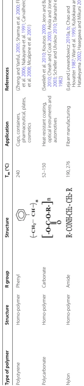
appliances, transportation, construction, shelters, storage and packaging practices. Such polymers are differentiated according to their chemical nature, structural arrange-ment, physical properties and applications as shown in Table 1 (Shah et al. 2008b; Dey et al. 2012; Kumar et al. 2011; Smith 2005).
Natural polymers
Natural polymers are found abundantly in nature in the forms of biopolymers and dry material of plants as shown in Table 2 (Leschine 1995). The constitution of the plant cell wall differs with the composition of the lignocellu-losic biomass (cellulose, hemicellulose and lignin), which provides strength (Premraj and Doble 2005). Lignocellu-loses play a critical role in developing plant biomass, in which cellulose, hemicellulose and lignin are the major building blocks of the natural polymer (Perez et al. 2002).
Synthetic polymers
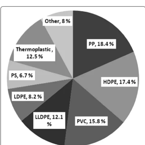
Plastics are manmade compounds that consist of a long chain of polymeric molecules and unusual bonds, with excessive molecular mass and halogen substitutions. Nowadays plastic manufacturing involves different inor-ganic and orinor-ganic materials, including carbon, hydrogen, chloride, oxygen, nitrogen, coal and natural gases (Shah et al. 2008b). The most widely used polymers contribut-ing to plastic waste are low-density polyethylene (LDPE), high-density polyethylene (HDPE), polyvinyl chloride, polystyrene and polypropylene with 23, 17.3, 10.7, 12.3 and 18.5%, respectively, and the remaining 9.7% of other types of polymer (Puri et al. 2013). The polymer produc-tion in 2012 was estimated as polyethylene 30% (LLDPE and LDPE 18%, HDPE 12%), polypropylene 19%, polyvi-nyl chloride 11%, polystyrene 7%, polyethylene tereph-thalate 7% and polyurethanes 7% worldwide (Nerland et al. 2014). The sales distribution and amount in per-centage of synthetic polymer consumed in North Amer-ica during 1995 and 2004 are shown in Table 3 and Fig. 1, respectively (Summers 1996; Zheng and Yanful 2005).
Standards for polymer degradation
Table 1 T yp es of p olymer ( A ver ous and P ollet 2012 ; B abul et al . 2013 ) Type of polymer Struc tur e R g roup Struc tur e Tm (°C ) A pplica tion Ref er enc es Bio -based polymers Poly (3-h ydr ox yvalerat e) (PHV ) Homo -polymer Eth yl 118 Industr ial
, drug deliv
er
y
(A
ver
ous and P
ollet
2012
; Liu et
al . 2014 ; O jumu et al . 2004 ; T ur esin et al . 2000 ; Bonar tse v et al . 2007 ) Poly (3-h ydr ox ybut yrat e) (PHB) Homo -polymer M eth yl 168–182 Phar
maceutical and drug
deliv
er
y
(A
ver
ous and P
ollet
2012
; Liu et
al . 2014 ; V elde and Kiek ens 2002 ; Nur
bas and Kutsal
2004 ; O jumu et al . 2004
; Jirage et
al . 2011 ; T ur esin et al . 2000 ) Poly (3-h ydr ox yoc tade -canoat e) (PHO d) Homo -polymer Penta decano yl 54–55 M edicine ar ea (A ver
ous and P
ollet
2012
; Guo et
al . 2013 ; Dhar et al . 2008
; Giudicianni et
al
.
2013
; Bonilla and
Per illa 2011 ) Poly (3-h ydr ox yoc tanoat e) (PHO) Homo -polymer Pent yl 40–60 M edical applications (A ver
ous and P
ollet 2012 ; S ouza 2013 ; Basnett et al . 2012
; Liu et
al
.
2011
; Basnett et
al . 2013 ) Poly (3-h ydr ox ydecanoat e) (PHD) Homo -polymer Hept yl 54 Fiber industr y (A ver
ous and P
ollet 2012 ; W er ner et al . 2014 ; S ong et al . 1998 ; R amesh war
i and M
eenakshisunda -ram 2014 ) Poly (3-h ydr ox ybut yrat e-co -3-h ydr ox yhexanoat e) (PHBH x ) Co -polymer M eth yl
, with pr
op yl 10%HH × 120 M edical applications (Babul et al . 2013 ; A ver
ous and P
ollet 2012 ; Chang et al . 2014 ; C
oen and D
ehor
ity
1970
; Xie et
al . 2009 ) Poly (h ydr ox ybut yrat e-co -hy dr ox yvalerat e) (PHB V ) Co -polymer M eth
yl with eth
yl 165–175 Phar maceutical ( drug deliv er y) (Ner land et al . 2014 ; G erar d et al . 2014 ; N wachk wu et al . 2010 ; Hatak ka 2005
; Danis et
al . 2015 ; Zembouai et al . 2014 )
Synthetic polymers Poly
eth ylene Homo -polymer H ydr ogen 140–143 In wir
es (as insulating
matt er), bags (T obin 2010 ; Nak ayama et al . 1991 ; M enon et al . 2010 ; P etr e et al . 1999 ) Poly vin yl chlor ide Homo -polymer Chor ine [– CH 2 –CHCl–] n 115–245 Leather , pipe , bottles (W ilk es et al . 2005 ; Summers 2008 ; Summers 1996 ; M enon et al . 2010 ) Polypr op ylene Homo -polymer M eth yl 165 Fabr ic mat er ial , car pets (Ruiyun et al . 1994 ; T ripathi 2002 ; M ccallum et al . 2007 ; Y am 2009 ; P er ez et al . 2014 ; M enon et al . 2010 ; P etr e et al . 1999 ) Poly eth ylene t er ephthalat e Homo -polymer Car bo
xyl and h
ydr ox yl 280 Pack ag ing applications , bottles , f ood wrappers , pipes (Zheng and Y anful 2005 ; P er ez et al . 2002
; Liu et
al . 2011 ; J effr ie 1994 ; Kw on et al . 2009 ) Polyur ethane Het er o-polymer Isoc yanat
e and poly
ol 400 Fibers , f oams , paints , coat -ing , pack ag ing (Zheng and Y anful 2005
; Slade et
Page 4 of 31 Pathak and Navneet Bioresour. Bioprocess. (2017) 4:15
Table
1
c
on
tinued
Type of polymer
Struc
tur
e
R g
roup
Struc
tur
e
Tm
(°C
)
A
pplica
tion
Ref
er
enc
es
Polyst
yr
ene
Homo
-polymer
Phen
yl
240
Cups
, containers
,
phar
maceutical
, plat
es
,
cosmetics
(Zheng
and Y
anful
,
2005
; Shar
ma et
al
.
2000
; F
la
vel
et
al
.
2006
; Nak
ayama et
al
.
1991
; C
ar
valheir
o
et
al
.
2008
; M
calpine et
al
.
2001
)
Poly
car
bonat
e
Homo
-polymer
Car
bonat
e
52–150
Heat
-r
esistant coating
,
optical instruments and aut
omotiv
es
(Sw
eileh et
al
.
2010
; K
outsos
2009
; J
eon and Baek
2010
; Cheah and C
ook
2003
; Ak
ola and J
ones
2003
; S
cheller and Ulvsk
ov
2010
; T
ak
anashi et
al
.
1982
)
N
ylon
Homo
-polymer
Amide
190, 276
Fiber manufac
tur
ing
(L
eja and L
ewando
wicz
2010
a,
b
; Chao and
Ho
vatt
er
1987
; W
an et
al
.
1995
; Kubok
awa and
Hatak
eyama
2002
; Hasega
wa and M
ik
uni
2014
Composting Council) and BPI, shown in Fig. 2 (Kolybaba et al. 2003; http://www2.congreso.gob.pe/sicr/cendocbib/ con2_uibd.nsf/4EF8A31F2BF5D3480525772A0053CD80/$
FILE/Ensayo_biodegradables_pl%C3%A1sticos_by.pdf).
Diversity of polymer degradation
Living organisms are involved in the breakdown of plas-tic material, and consequently the recycled form reverses back to the environment. Anaerobic microbial degra-dation releases greenhouse gas (methane) in landfills, which increases global warming. Aerobic conditions are essential for fungal degradation while bacterial degrada-tion proceeds in aerobic as well as anaerobic condidegrada-tions (Kumar et al. 2011; Chandra and Rustgi 1998). Plastic can be reduced in an eco-friendly manner with the help of soil bacteria and proper water availability. Decomposition of the polymer depends on its chemical composition, which supports the growth of microorganisms in the form of nutrient sources. The starch-based polymer is favorable for microbial attack, and hydrolytic enzymes act on the polymer matrix to reduce their weight. Polymer made from starch or flax fiber shows greater biodegradability as compared to other synthetic polymers. Microorgan-isms also play an important role in the degradation of petroleum-based polymers. Petroleum-based polymers such as polyolefins are degraded through photo-degrada-tion (Kumar et al. 2011; Sen and Raut 2015).
Emerging technology is continuously involved in improving the processing of biopolymers by using an additive (benzophenone) during their construction. Additives play a significant role in the chemical process during photo-degradation. Such amendments affect their thermal sensitivity and UV-absorbing capacities. Chemi-cally sensitive polymers have a better biodegradabil-ity rate compared to other polymers. Similarly, thermal Table 2 Types of bio-based polymers (Babul et al. 2013; Averous and Pollet 2012)
Microorganism based Biotechnology based Agro-based
Polyhydroxyalkanoates (mcl-PHA,
PHB, PHB-co-V) Polylactides, PBS, PE, PTT, PPP Polysaccharides and lipids (starch, cellulose, alginates) Proteins–animal proteins (casein, whey, colagen/gelatin), plant protein (zein, soya, gluten)
Table 3 Plastic sales in North America, 1995 (Summers
1996)
Type of polymer Billions of pounds
LDPE/LLDPE 14–16
PVC 12–14
HDPE 12–14
PP 10–12
PS 6–7
Polyester 4
PC <2
Fig. 1 Percentage distribution of synthetic polymer (PP polypropyl-ene, HDPE high-density polyethylene, PVC polyvinyl chloride, LLDPE linear low-density polyethylene, LDPE low-density polyethylene, PS polystyrene, thermoplastics and others) sales in North America in 2004 (Zheng and Yanful 2005)
Page 6 of 31 Pathak and Navneet Bioresour. Bioprocess. (2017) 4:15
exposure is also involved in the breakdown of a polymer into simpler forms that increase the availability of micro-organisms. Nodax is alkaline in nature and generally involves in the structural change of the polymer (Kumar et al. 2011; Augusta et al. 1993).
Renewable resources are also used in the formation of biodegradable materials. Animal and plant originated compounds are susceptible to microbial degradation (Schink et al. 1992). Development of bio-based mate-rials is beneficial for our environment’s sustainability, maintenance of greenhouse gas emissions, etc. (Song et al. 2009). This type of material’s manufacturing prac-tice plays a significant role in the environment by reduc-ing the amount of dumped polymer waste (Leja and Lewandowicz 2010a, b). Many synthetic polymers are degraded under exposure to solar ultraviolet (UV) radiation, photo-oxidative, thermo-oxidative and pho-tolytic reactions (Singh and Sharma 2008). In the natu-ral environment, hydrolytic properties of seawater, oxidative properties of the atmosphere and sunlight radiation (UVB) make the polymers fragile and eventu-ally break them into smaller pieces (Moore 2008). The American Society for Testing and Materials (ASTM) and International Standards Organization (ISO) pro-vided the analytical protocol for plastic degradation on the basis of alteration in chemical structure and loss of physical properties of plastic (Kumar et al. 2011; http:// www2.congreso.gob.pe/sicr/cendocbib/con2_uibd. nsf/4EF8A31F2BF5D3480525772A0053CD80/$FILE/
Ensayo_biodegradables_pl%C3%A1sticos_by.pdf).
Biodegradation of polymers
Degradation of polymers is a process that alters the strength and color of polymeric material under con-trolled conditions. Disruption of the chain length initi-ates the primary breakdown (aging), and several external factors such as temperature and chemicals also enhance the rate of degradation. The term “aging” is used for the change in properties. It is utilized as polymer recycling, which reduces the outcome of pollution load (Kumar et al. 2011; Bhardwaj et al. 2012a). Anaerobic degradation is another way to dispose of plastic materials through landfilling (Schink et al. 1992; Shah et al. 2008b). Cur-rently, the recycling process is increasing but the recy-cling rate is very low for most plastic materials because of the use of more additives in their manufacturing (Song et al. 1998). The recycling rate of thermosets is very low but thermoplastics can be easily recycled (Moore 2008). Plastic comprises 60–80% of litter, and its persistence and discharge to the surroundings create harmful effects on wildlife as well as agriculture and forest land. Furans and dioxins are persistent organic pollutants (POPs) and are formed through the burning of polyvinylchloride (PVC).
Plastic waste may come from post consumption and dif-ferent stages of production (Nerland et al. 2014; Shah et al. 2008b).
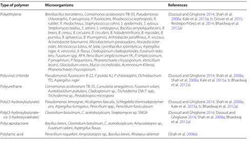
Worldwide research for the last 3 decades has focused on the biodegradation of plastic (Shimao 2001). Biodeg-radation is compatible (microbial mineralization) com-pared to other waste management techniques (Schink et al. 1992). Bioremediation serves as the best way to manage waste material in an eco-friendly manner. Pol-luted sites are increasing constantly because of improper waste management strategies; such waste comes from industrial areas and community activities. Biologi-cal agents, both prokaryotic (bacteria) and eukaryotic (fungi, algae and plant), are involved in the bioremedia-tion process. Pseudomonas, Streptomyces, Corynebacte-rium, Arthrobacter, Micrococcus and Rhodococcus are the prominent microbial agents being used for bioremedia-tion as illustrated in Table 4 (Bhatnagar and Kumari 2013; Kathiresan 2003; Dussud and Ghiglione 2014; Shah et al.
2008a; Kale et al. 2015a, b; Grover et al. 2015;
Restrepo-Flórez et al. 2014; Bhardwaj et al. 2012a).
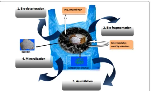
Different steps in the polymer biodegradation mechanism Microorganisms break down the compounds into a simpler form through biochemical transformation. Bio-degradation of polymer is described as any alteration of the polymer properties such as digestion by micro-bial enzymes, reduction in molecular weight, and loss of mechanical strength and surface properties, in other words, the breakdown of material into fragments via microbial digestion. Degraded particles are redistributed and probably non-toxic to the environment. In nature, microorganisms form catalytic enzymes for biodegrada-tion (Hadad et al. 2005). This approach is proficient for environmental waste management, and microorganisms involved in this process for oxidation serve as a tangible alternative mode to maintain the healthy environment (Singh and Sharma 2008). The degradation process is accomplished by microorganisms via different enzy-matic activities and bond cleavage. This degradation occurs in sequential steps, bio-deterioration (altering the chemical and physical properties of the polymer), bio-fragmentation (polymer breakdown in a simpler form via enzymatic cleavage) and assimilation (uptake of molecules by microorganisms) and mineralization (pro-duction of oxidized metabolites (CO2, CH4, H2O) after
degradation), which are shown in Fig. 3. Mineralization of polymers takes place in both aerobic and anaerobic conditions. In the aerobic condition, CO2 and H2O are
formed, while under anaerobic conditions, CH4, CO2
and H2O are produced (Singh and Sharma 2008). The
able to utilize polyester and polyurethane at a slower rate (Dey et al. 2012; Schink et al. 1992). Starch- or cellulose-based plastics are biodegradable; they degrade easily through composting, which can reduce landfilling and solve the waste management problem. Biodegradation with the help of microorganisms is an approachable way to clean up such plastic waste. Microorganisms are able to utilize synthetic polymers, but the composition of the polymer and manufacturing process need to be defined for the biological activity on the polymer material (Sivan 2011; Song et al. 2009; Leja and Lewandowicz 2010a, b; Kumar et al. 2011). Biodegradability of synthetic poly-mers with chemical groups that are susceptible to micro-bial attack can be carried out with polycaprolactone, poly-β-hydroxyalkanoates and oil-based polymers (Leja and Lewandowicz 2010a, b; Song et al. 2009). Enzymes of microbial origin are employed to control pollution and contribute to developing an eco-friendly environment. Diverse forms of microflora are known to utilize them through the mineralization process.
Microbial metabolism and physiological processing of polymer degradation
Bacteria and fungi are a widely distributed group of microorganisms that play a significant role in the process-ing of polymer compounds in the natural environment (Upreti and Srivastava 2003). These microorganisms are
used to convert the insoluble biopolymer into a solu-ble biopolymer. Naturally occurring polymers consist of lipids, carbohydrates and proteins. Microbial enzymes are the ultimate source to hydrolyze only low molecu-lar weight and soluble macromolecules. These soluble compounds are exploited by microorganisms for energy production (Gallert and Winter 2005). Microbial deg-radation of polymers leads to alteration of the physico-chemical properties of materials. The bioconversion or degradation of biomaterials is well understood by study-ing the mechanical properties, degradation kinetics and recognition of the degraded products. The bioconversion process also alters the efficiency of the host response, cel-lular growth, material function, etc. (Azevedo and Reis
2005).
The tricarboxylic acid (TCA) cycle serves as one of the main metabolic pathways for energy generation from most of the organic compounds. In the TCA cycle, acetyl-CoA acts as the key intermediate and is exploited in cellular activities like CO2 formation by oxidation,
acetate formation, biosynthesis, etc. The major contri-bution of the TCA cycle is to generate ATP and provide energy to the cell. Two molecules of ATP are synthesized by oxidation of 2 mol of acetate, while 34 mol of ATP is synthesized by the electron transport chain (ETC) through phosphorylation. The metabolism and efficiency of energy production vary according to the microbial Table 4 List of microorganisms associated with polymer degradation
Type of polymer Microorganisms References
Polyethylene Brevibacillus borstelensis, Comamonas acidovorans TB-35, Pseudomonas chlororaphis, P. aeruginosa, P. fluorescens, Rhodococcus erythropolis, R.
rubber, R. rhodochrous, Staphylococcus cohnii, S. epidermidis, S. xylosus,
Streptomyces badius, S. setonii, S. viridosporus, Bacillus amyloliquefaciens, B. brevis, B. cereus, B. circulans, B. circulans, B. halodenitrificans, B. mycoides, B. pumilus, B. sphaericus, B. thuringiensis, Arthrobacter paraffineus, A. viscosus, Acinetobacter baumannii, Microbacterium paraoxydans, Nocardia aster-oides, Micrococcus luteus, M. lylae, Lysinibacillus xylanilyticus, Aspergillus niger, A. versicolor, A. flavus, Cladosporium cladosporioides, Fusarium
redo-lens, Fusarium spp. AF4, Penicillium simplicissimum YK, P. simplicissimum,
P. pinophilum, P. frequentans, Phanerochaete chrysosporium, Verticillium lecanii, Glioclodium virens, Mucor circinelloides, Acremonium Kiliense, Phanerochaete chrysosporium
(Dussud and Ghiglione 2014; Shah et al.
2008a; Kale et al. 2015a, b; Grover et al. 2015; Restrepo-Flórez et al. 2014; Bhardwaj et al.
2012a)
Polyvinyl chloride Pseudomonas fluorescens B-22, P. putida AJ, P. chlororaphis, Ochrobactrum
TD, Aspergillus niger (Dussud and Ghiglione Shah et al. 2008a; Kale et al. 2014; Shah et al. 2015a, b; Bhardwaj 2008a;
et al. 2012a) Polyurethane Comamonas acidovorans TB-35, Curvularia senegalensis, Fusarium solani,
Aureobasidium pullulans, Cladosporium sp., Trichoderma DIA-T spp.,
Trichoderma sp., Pestalotiopsis microspora
Poly(3-hydroxybutyrate) Pseudomonas lemoignei, Alcaligenes faecalis, Schlegelella
thermodepolymer-ans, Aspergillus fumigatus, Penicillium spp., Penicillium funiculosum (Dussud and Ghiglione Kale et al. 2015a, b; Bhardwaj et al. 2014; Shah et al. 2012a2008a) ;
Poly(3-hydroxybutyrate-co-3-hydroxyvalerate)
Clostridium botulinum, C. acetobutylicum, Streptomyces sp. SNG9 (Dussud and Ghiglione 2014; Dussud and
Ghiglione 2014; Shah et al. 2008a; Bhardwaj et al. 2012a)
Polycaprolactone Bacillus brevis, Clostridium botulinum, C. acetobutylicum, Amycolatopsis sp., Fusarium solani, Aspergillus flavus
Page 8 of 31 Pathak and Navneet Bioresour. Bioprocess. (2017) 4:15
growth conditions. Aerobic bacteria are able to respire carbohydrates, but one-third of the starting energy is not utilized by the cell and is lost in the form of heat; the remaining energy is conserved biochemically. During the processing of wastewater, activated sludge reactors lose much of their energy as heat. Under growth-limiting con-ditions, the ATP consumption rate is increased and less energy is available for cellular growth and metabolism (Gallert and Winter 2005).
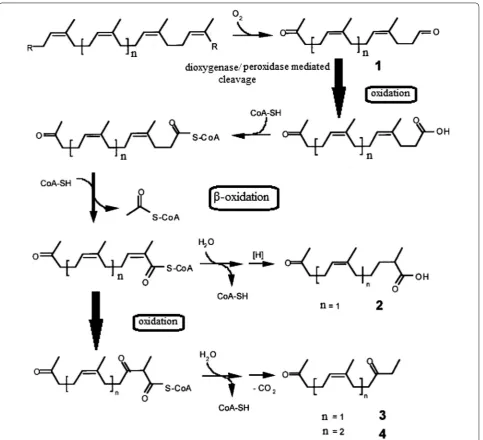
Bode et al. (2000) investigated the physiological and chemical process of biodegradation of synthetic poly (cis-1,4-isoprene) polymer and found that two bacterial strains, i.e., Streptomyces coelicolor 1A and Pseudomonas citronellolis, were able to utilize degraded vulcanized natural rubber and synthetic poly(cis-1,4-isoprene). They observed the growth of these bacteria on polymer was better as compared to Streptomyces lividans 1326, and they were exploited under controlled conditions. Three degraded products have been identified from the culture suspension of S. coelicolor 1A, and vulcanized rubber was used as substrate. These degraded products were deter-mined as (5Z,9Z)-6,10-dimethyl-penta-dec-5,9-diene-2,13-dione, (5Z)-6-methyl-undec-5-ene-2,9-dione and (6Z)-2,6-dimethyl-10-oxo-undec-6-enoic acid. They also
proposed the oxidative pathway for conversion of poly(cis-1,4-isoprene) into methyl-branched diketones by follow-ing different steps, i.e., aldehyde intermediate to carboxylic acid oxidation, β-oxidation, oxidation of the conjugated bond (double bond) to β-keto acid and the decarboxyla-tion process. The authors proposed a hypothetical model for poly (cis-1, 4-isoprene) degradation as shown in Fig. 4.
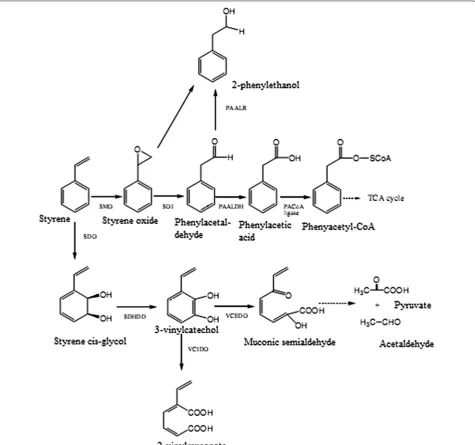
Similarly, Mooney et al. (2006) studied the microbial degradation of styrene. Styrene is one of the identical xenobiotic compounds and serves as a potent toxic pol-lutant to the environment as well as human health. It comes from industrial practices that involve polymer and petrochemical processing. Mooney et al. (2006) reported bacterial enzymes involved in styrene biodeg-radation. The detailed process of styrene bioconver-sion to its metabolites and degraded products is shown in Fig. 5. Pyruvate, acetaldehyde 2-phenylethanol and 2-vinylmuconate are some of the degradative metabolites obtained during styrene biodegradation. Similarly, phenyl acetyl-CoA obtained via styrene degradation enters into the tricarboxylic acid (TCA) cycle. The TCA cycle thus plays a vital role in energy production, essential for cellu-lar and metabolic events, and also produces CO2 during
Aerobic and anaerobic biodegradation of polymers
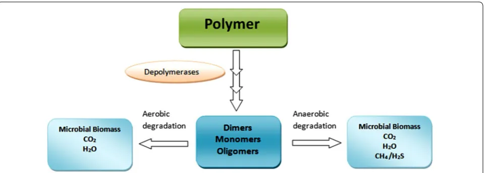
Polymer biodegradation depends on the physical and chemical properties of the polymer. Molecular weight and crystallinity are key properties of polymers that affect the biodegradation efficiency of microorganisms. The enzymes responsible for polymer degradation are cat-egorized into two groups, i.e., extracellular depolymerase and intracellular depolymerase (Gu 2003). Exoenzymes are generally involved in the degradation of complex pol-ymers to simple units like monomers and dimers. These are further exploited by microorganisms as energy and carbon sources as shown in Fig. 6.
Polymer degradation (mineralization) forms new prod-ucts during or at the end of processes, e.g., CO2, H2O
or CH4. Natural polymers like cellulose, PHB and chitin
are susceptible to microbial degradation and serve as biodegradable polymers (Gu 2003; Chahal et al. 1992; Brune et al. 2000). The degradation process depends on the availability of O2. Polymer degradation
accom-plished under anaerobic conditions produces organic acids, H2O and gases (CO2 and CH4) (Gu 2003). Under
aerobic conditions, the biodegradation of the polymer forms CO2 and H2O in addition to the cellular biomass of
Page 10 of 31 Pathak and Navneet Bioresour. Bioprocess. (2017) 4:15
polymer degradation forms H2S, CO2 and H2O (Gu and
Mitchell 2006; Barlaz et al. 1989; Gu 2003; Merrettig-Bruns and Jelen 2009). The aerobic process is more effi-cient than the anaerobic process by means of energy production as less energy is produced in anaerobic pro-cesses because of the lack of O2, which serves as an
elec-tron acceptor, and this is more efficient in comparison to CO2 and SO42− (Gu 2003).
In solid waste treatments, denitrifiers are categorized as aerobic organisms and used for nitrate or nitrite exploitation. Under anoxic conditions, nitrate or nitrite serves as a terminal electron acceptor in the metabolic respiration process. Extracellular solubilized biopoly-mers (natural polybiopoly-mers like carbohydrates, lipids and proteins) from CO2 and H2O form during the respiration
process, while in anaerobic conditions CO2, CH4, NH3,
and H2S are produced from the degradation of soluble
carbon compounds via methanogens or sulfate reducers. In anaerobic conditions, if SO42− is present, then SO32−
and CO2 will form. On the other hand, if SO42− is absent,
then CH4 and CO2 will form under anaerobic conditions
(Gallert and Winter 2005; Mohan and Srivastava 2011; Leja and Lewandowicz 2010a, b). A larger amount of CO2
is produced at the lowest pH with lignolytic fungi (Kale et al. 2015a, b).
Huang et al. (2005) investigated sludge microorganisms for degradation of polyethylene glycols (PEGs) under aerobic and anaerobic conditions. They studied the effect of molecular weight (MW) on the efficiency of aerobic as well as anaerobic biodegradation processes of poly-ethylene glycols (MW 600, 6000 and 20,000) and found that aerobic degradation is more efficient than anaerobic degradation by means of PEG degradation abilities; 80% of biodegradability was reported in 5 days under aerobic conditions with diverse molecular weight PEGs. While the PEG biodegradation under the anaerobic condi-tion showed 50% biodegradability of PEG 600 in 9 days, 40% biodegradability of PEG 6000 in 10 days and 80% biodegradability of PEG 20,000 obtained after 6 days of incubation. Huang et al. (2005) further investigated the nutrition effect on the degradation process under anaero-bic conditions. The enriched organic media showed its positive effect on PEG 6000 biodegradation by increasing the biodegradability. Similarly, Sridevi et al. (2012) stud-ied the metabolic pathways for phenol biodegradation and found that under aerobic conditions phenol biodeg-radation can form catechols and serves as intermediate products by ring cleavages at the ortho or meta position. In aerobic conditions, initially phenol is converted into catechol and then intermediates of the central metabo-lism by ortho or meta fission and catalyzed by catechol 1,
2 dioxygenases or catechol 2,3 dioxygenase, respectively. They found that anaerobic biodegradation of phenol is slower as compared to aerobic biodegradation. In anaero-bic biodegradation the end product is CH4 and CO2, and
under aerobic biodegradation the end product is CO2.
Microbial development as biofilms on polymer
The growth of microorganisms is observed under differ-ent environmdiffer-ental conditions. These microorganisms are able to adapt to conditions as experience reflects changes, and they undergo a transition. In normal con-ditions, plankton is found in free swimming form, and under transition state these assemblages of microorgan-isms attach irreversibly to the surface and form biofilms.
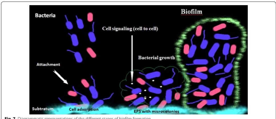
Escherichia coli O517:H7 and Myxococcus xanthus attach to the surface under nutrient-rich environments (O’Toole et al. 2000; Sharma et al. 2015; Simoes et al. 2010). Micro-organisms in biofilms show novel phenotypic charac-teristics, specific mechanisms to attach to surfaces and respond to the external signals. Biofilm formation is a highly synchronized and intricate process, as shown in Fig. 7. Some microorganisms are reported to change their morphology (e.g., Bacillus subtilis, Caulobacter crescen-tus) under harsh conditions, which allows them to exist in a hostile environment (O’Toole et al. 2000). In the ini-tial stage, microorganisms produce some proteins such as collagen, fibrin, fibronectin and laminin as a coating material, which help in cell-to-cell adhesion in biofilm (Bryers et al. 2006; Simoes et al. 2010). Biofilms exist as a matrix of extracellular polymeric substance (EPS) that depends on the surrounding conditions; it consists of a series of biological and physical changes during matrix formation and from the three-dimensional structure (Prakash et al. 2003; Bryers et al. 2006; Simoes et al.
2010).
Page 12 of 31 Pathak and Navneet Bioresour. Bioprocess. (2017) 4:15
Microbial species grow on a wide variety of surfaces, i.e., inert or living. Inert surfaces include piping systems (biofilms on noncellular materials), medical devices (bio-films composed of microorganisms with blood compo-nents) and living tissues, which have served as surfaces for the development of biofilms and consist of single or mixed populations of microorganisms. Single species of gram-positive (Staphylococcus aureus, Staphylococ-cus epidermides and enterococci) and gram-negative (E. coli, Pseudomonas aeruginosa, Pseudomonas fluorescens
and Vibrio cholerae) bacteria were widely reported for biofilm formation. However, multiple species popula-tions of microorganisms have been reported prepon-derantly in biofilms (O’Toole et al. 2000; Prakash et al. 2003). Microorganisms that form biofilms show elaborate growth by increasing their antibiotic resistance through gene regulation. Microorganisms reflect unique inhab-itance in biofilms with enhanced defense mechanisms against therapeutic agents and drugs, and they relieve their survival under sessile conditions (osmotic stress, desiccation, UV radiation and pH changes) (Prakash et al. 2003; Sharma et al. 2015; Bogino et al. 2013; Simoes et al. 2010). Some dyes are used to visualize biofilm formation, i.e., safarinin and crystal violet. The term surface attach-ment defective (sad) is used for the group of mutant microorganisms unable to form biofilms (O’Toole et al. 2000). Heterogeneity of the biofilm matrix is one of the distinctive features that maintain the nutritional require-ment of biofilms. The matrix is composed of polysac-charides, macromolecules and water, which provide heterogeneity and form enclosed exopolysaccharides in cells; these structures protect the biofilms from the external environment (O’Toole et al. 2000; Prakash et al.
2003). In biofilms, microorganisms are hugely differen-tiated compared to free-swimming organisms and form multicellular (microcolony) complex structures with interstitial voids (Sivan et al. 2006). Microorganisms are present on the upper layer of biofilms and actively divid-ing compared to microbes in proximity to the surface and also differ in metabolism. Interstitial voids (water channels) in the matrix help to separate the microbial colonies and facilitate diffusion of nutrients, water, gases, enzymes, waste, signals, etc., throughout the biofilms. The heterogeneity in a matrix is visible in both kinds of biofilms, i.e., single species populations of microbial bio-films and mixed species populations of microbial biobio-films (Prakash et al. 2003; Bogino et al. 2013). The structural components, i.e., curli fimbriae, extracellular polymeric substances, outer membrane proteins, flagella and pili, hugely participate and are important for biofilm forma-tion. The relieve form biofilms of flagellated motile bacte-ria compared to non-motile bactebacte-ria and other structural components such as curli and mannose sensitive type I, IV pili (TfP) encoding genes also play an important role in cell-cell or cell-surface attachments. Type IV pili play a significant role in bacterial motility on the surface and are important for quorum sensing. OmpR is reported to be a functional gene that promotes biofilm formation (Prakash et al. 2003; O’Toole et al. 2000; Bogino et al.
2013).
Biofilm formation is a highly regulated event, and
communication, e.g., AI 1, AI 2 and N-acetyl homoserine lactones (Vibrio fisheri or Aliivibrio fisheri). Such cellu-lar communication accomplished in addition to quorum sensing signaling systems, i.e., the Rh1I/Rh1R system observed in P. aeruginosa and the AfeI/AfeR system in
Acidithiobacillus ferroxidans (Vu et al. 2009).
In addition to extracellular appendages, cellular hydro-phobicity plays a key role in surface attachment and bio-film formation (Simoes et al. 2010). Synthetic polymers are resistant to water because of their hydrophobic prop-erties and non-polar nature that prevents water absorp-tion (Khoramnejadian 2013). Usually non-polar polymers show limitations in biodegradation because of their hydrophobic surface properties, and the formation of biofilms overcomes this barrier. Microorganisms are eas-ily attached to non-polar surfaces (plastics and Teflon), which support microbial attachment compared to hydro-philic materials. Non-polar polymer shows an enhanced surface hydrophobicity. The fimbrial structure contains hydrophobic amino acid and mycolic acid of gram-pos-itive bacteria, and the fimbriae are important in estab-lishing the cell attachment to the hydrophobic surface (Prakash et al. 2003; Sarjit et al. 2015). The hydrophobic nature of the surface is one of the major concerns during synthetic polymer degradation and microbial attachment to the surface.
In gram-negative bacteria O-antigen has a significant role in governing the hydrophilic properties that hinder the cell attachment to the hydrophobic surface. Inversely, O-antigen of lipopolysaccharides and extracellular poly-meric substance (EPS) promotes the cell attachment to the hydrophilic surface (Prakash et al. 2003). Mutants for O-antigen also showed biofilm attachment on the plastic surface, e.g., Bradyrhizobium japonicum mutant (Bogino et al. 2013). In addition to fimbriae, other structures such as the EPS serve as the primary material in the biofilm matrix; it is a hydrated structure and chemically found as colonic acid and alginate in E. coli and P. aeruginosa, respectively (Prakash et al. 2003). Biofilm formation is important for degradation in the natural environment. Biofilm formation on the surface of synthetic polymers facilitates biodegradation (Sivan et al. 2006). Low-den-sity polyethylene (LDPE) serves as the most widely used synthetic polymer. Still, many microorganisms (bacte-ria, fungi) are known for polymer degradation. Tribedi and Sil (2013a) reported that the degradation of LDPE is done by the Pseudomonas sp. AKS2 strain; it forms a biofilm on the surface of LDPE. Microbial adapta-tion is a key factor for bioremediaadapta-tion. Biofilm forma-tion depends on the adaptable nature of Pseudomonas
sp. AKS2 and shows positive results for LDPE degrada-tion by means of increased hydrolytic activity up to 31%, and the cellular surface hydrophobicity increases by
about 26% (Tribedi et al. 2015). Tribedi and Sil (2013a) reported 5 ± 1% LDPE degradation with Pseudomonas
sp. AKS2 and enhanced LDPE biodegradation were achieved with biofilm formation. Tribedi and Sil (2013a) conducted an experiment on hydrophobicity and found that Tween 80 had negative effects on hydrophobicity. Sivan et al. (2006) isolated the biofilm-producing bacte-rial strain Rhodococcus ruber C208 and reported a 0.86% per week biodegradation rate. Orr et al. (2004) reported earlier that mineral oil accelerated the degradation rate up to 50% with R. ruber C208 and enhanced the biofilm potential by increasing microbial colonization on poly-ethylene surface. Similarly, Penicillium frequentans and
Bacillus mycoides exhibited biofilm formation and poly-ethylene degradation. B. mycoides colonized in a myce-lial network formed by P. frequentans on a polyethylene surface. Degradation was determined by percent weight loss, CO2 evolution and gas chromatography (Sangale
et al. 2012; Arutchelvi et al. 2008). Metabolic activities of cells in biofilms were studied with the help of fluorescein diacetate (FDA) analysis and ATP assay (Arutchelvi et al. 2008). The development of biofilms was observed and its three-dimensional hydrated microstructure and metabo-lism examined under confocal scanning laser microscopy with fluorescent in situ hybridization and 16-23S rRNA hybridization (Kokare et al. 2009). Biofilm formation also played an important role in biocontrol. Gene transfer was seen within the biofilms of Streptococcus sp. as conjuga-tive transposons and confers the antibiotic (tetracycline) resistance.
Cellulose
The terrestrial system is known for the plentiful produc-tion of cellulose biopolymers. The occurrence of cellulose in decaying microorganisms takes place in sundry envi-ronments (soil, aquatic, compost, anaerobic digestors and also the carbon cycle) (Leschine 1995). Several microorganisms (bacteria and fungi) were reported to have cellulose-degrading activity, and such cellulose degrading microbes exist in nature with non-cellulolytic microorganisms. Biodegradation of cellulose proceeds under aerobic (form CO2 and H2O) as well as
anaero-bic (form CO2, CH4 and H2O) conditions (Perez et al.
Page 14 of 31 Pathak and Navneet Bioresour. Bioprocess. (2017) 4:15
the end of the chains, respectively. Cellobiose molecules generated from cellulose by the endoglucanases and cel-lobiohydrolases are hydrolyzed by β-glucosidases and releases two units of glucose. CBHs act on crystalline cel-lulose effectively. Breakdown of celcel-lulose makes carbon available to microorganisms. Pseudomonas, Streptomy-ces and Cellulomonas are reported for cellulase activity (Perez et al. 2002; Leschine 1995; Souza 2013; Kamesh-war and Qin 2016). Fungal degradation of cellulose was reported with members of Basidiomycetes and Chytridi-omycetes (Souza 2013). Fungi are also reported to have cellulase activity, including Phanerochaete chrysosporium
and Trichoderma reesei. Anaerobic biodegradation of cellulose is very complex compared to aerobic degrada-tion (Leschine 1995; Souza 2013; Kameshwar and Qin 2016). Larroque et al. (2012) studied CBM1 (carbohy-drate-binding module) containing proteins from fungi and Oomycetes (Phytophthora parasitica). In the case of Oomycetes, CBM1 is involved in plant immunity by adhesion to polysaccharide (cellulose) substrates, while CBM1 of fungi is reported for cellulose degradation.
Anaerobic degradation contributes 5–10% of the total cellulose-degradation. In the case of anaerobic degrada-tion of gram-positive bacteria, Clostridium thermocellum
was reported to have cellulolytic activity, and cellulosome is a functional hydrolytic enzyme unit. Clostridium ther-mocellum has an excellent cellulose degradation ability in cellulosomal and free-enzyme systems. Carbohydrate-active enzymes (CAZymes) play an important role in their activity and form large protein complexes (primary and secondary scaffoldings) that bond with the cell wall of bacteria. Xu et al. (2016) studied the “cell-free” cellu-losomal system and characterized two types of cellulo-somal systems by using mutants with scaffolding gene deletions. CipA (primary scaffolding) has a significant role in cellulose degradation compared to other scaf-foldings. Xu et al. (2016) also reported that primary scaf-folding (CipA) plays a key role in C. thermocellum for cellulose degradation.
Pretreatment of cellulose material also makes it suscep-tible to hydrolysis by reducing the barrier [chemical and physical (UV)] effects (Jonsson and Martín 2016; Arutch-elvi et al. 2008). Pretreatments were employed in milling, acid, alkali and other treatments (Karimi and Taherza-deh 2016). Some cellulase genes (9, 45, GH5 family gene) were reported in insects belonging to the taxa Crustacea, Gastropoda and Annelida (Cragg et al. 2015). Blouzard et al. (2007) reported a hydrolytic system for cellulose degradation in Clostridium cellulolyticum and found that hydrolase belongs to the GH2, GH9, GH10, GH26, GH27, and GH59 gene families and cellulases encoding the gene from the GH9 family. GH9 enzymes were purified from E. coli and characterized from one of the enzymes,
Cel9 V, belonging to this family, which showed similar-ity with cellulosomal cellulase, i.e., Cel9E (Ravachol et al. 2014). Berlemont and Martiny (2013) examined 21,985 genes in 5123 bacterial genomic sequences that encode for cellulases and found 24% potential opportunistic strains with 56% β-glucosidases activity throughout all sequences.
Maamar et al. (2004) studied the mutant strain C. cel-lulolyticum cip CMut1 and concluded that the cellulose (crystalline) hydrolyzing genes were located on the ‘cel’ cluster that encodes for cellulases. Lopez-Mondejar et al. (2016) isolated Paenibacillus O199 reported for cellulose deconstruction and studied the O199 enzyme system. The researcher suggested the exploitation of Paeniba-cillus O199 as a second-generation technology for agri-cultural waste (lignocellulosic material) management strategies and biofuel production.
Dimarogona et al. (2012) reported polysaccha-ride monooxygenase (PMO) (or GH61) enzymes that enhanced the degradation capacity of cellulases; external electrons are important to accelerate the PMO activity. Some hydrolytic enzymes also play a key role in cellulose degradation, e.g., lytic polysaccharide monooxygenases (LPMOs), CBM33 (carbohydrate-binding module fam-ily 33) and cellobiose dehydrogenases (CDHs). Similarly, Zhang et al. (2016) reported carbohydrate-binding mod-ule 1 (CBM1) for cellulose degradation; it contains cel-lulases and is found in coprophilic Podospora anserine
(coprophilous fungus) and P. chrysosporium (typical rot fungus).
Hemicellulose
1994; Menon et al. 2010; Werner et al. 2014; Carrier et al. 2012). Higher xylan degradation was found with carbonic acid and was easier to hydrolyze enzymatically compared to cellulose (Carvalheiro et al. 2008; Perez et al. 2002; Horn et al. 2012). Xylanases remove the side chain wall by attacking the backbone (Jeffrie 1994). Xylan fermenta-tion is accomplished by strains of Ruminococcus flavefa-ciens, Ruminococcus albus and Clostridium sp. (Coen and Dehority 1970). Xylanases exist in two forms: endo 1,4 β-xylanase and xylan 1,4 β-xylosidase. P. chrysosporium
is a white-rot fungus known to produce hemicelluloses that degrade endo-xylanases, acetyl xylan esterases and mannosidases. Heat-stable endo-xylanases are drived from thermophilic fungi, e.g., Thermoascus aurantiacus
and Talaromyces emersonii, while Butyrivibrio fibrisol-vens (ruminal bacterium) and Bacillus sterothermophilus
possess β-xylosidase. Bacillus spp. produces two types of alkaline xylanases, i.e., xylanase N and xylanase A, with an active pH range of 5, 7 and 11, respectively (Perez et al. 2002; Jeffrie 1994; Kameshwar and Qin 2016). Some helper enzymes like xylan esterases, α-1 arabinofuranosi-dases and α-4 O-methyl glucuronosiarabinofuranosi-dases are required for complete degradation (Perez et al. 2002). Endo-xyla-nase works in combination with acetyl xylan esterase for efficient xylan degradation; these acetyl xylan esterases are derived from Aspergillus niger, Schizophylum com-mune, Rhodotorula mucilaginosa, Trichoderma reesei
and Fibrobacter succinogenes. Similarly, Streptomyces spp. are also reported for α-O-methylglucuronidases produc-tion. Phytophthora also produces hydrolytic enzymes that target the hemicellulose (Jeffrie 1994; Souza 2013; Cragg et al. 2015). Mastotermes darwiniensis gut con-tains approximately 106–107 xylan degrading bacteria (Cazemier 1969). Glycoside hydrolases (GHs) are the key enzymes for hemicellulose degradation by the break-down of the glycosidic linkages and remove the phenolic, methyl and acetyl esters. Lytic polysaccharide monooxy-genases also reported this on hemicelluloses (Cragg et al. 2015). In T. reesei, 200 genes encode for glycosyl hydro-lases and are involved in the degradation of hemicel-lulose and celhemicel-lulose (Saloheimo and Pakula 2012). Sun et al. (2012) identified 353 genes and 34 proteins (sec-retary) from Neurospora crassa, which were involved in the breakdown of hemicellulose in the plant cell wall by hydrolytic enzymes. They reported TF and XLR-1 (xylan degradation regulator 1) played a central role in lulose biodegradation by regulating some of the hemicel-lulase-encoding genes in N. crassa and orthologs to genes found in Trichoderma and Aspergillus, i.e., XYR1 and XlnR, respectively. In addition to the five hemicellulases reported in C. thermocellum, C. fimi and Nasutitermes species, they also have genes involved in the hydrolysis of hemicellulose (French 2009; Kameshwar and Qin 2016).
Gene prediction analysis showed 587 gene-encoded enzymes involved in hemicellulose and pectin hydrolysis and 259 ORFs reported for gene (endo-xylanases gene) expression for hemicellulose degradation (Kameshwar and Qin 2016).
Lignin
Lignin is a relatively water-insoluble aromatic heter-opolymer complex. It is a phenolic polymer that provides strength to the plant cell wall via increased internal bond-ing. With its distinct characteristics, it confers resistance against biological stresses (Perez et al. 2002; Leschine 1995; Souza 2013; Petre et al. 1999; Hatakka 2005; Wer-ner et al. 2014). It consists of three monolignol phenyl propionic alcohols, i.e., coniferyl, sinapyl and p-coumaryl. These alcohols present in lignin in the form of guaia-cyl, syringyl and p-hydroxyphenyl propanol (Perez et al. 2002; Horn et al. 2012; Giudicianni et al. 2013; Car-rier et al. 2012). Softwood lignin has coniferyl alcohol or guaiacyl propanol as the principal component, while lignin of hardwoods consists of guaiacyl and syringyl propanol as the dominant constituents (Perez et al. 2002; Horn et al. 2012; Hatakka 2005). Lignin present in grass contains all three types of propanol units. Polymerization of lignin consists of carbon-carbon linkages, aryl ether linkages and an aryl glycerol β aryl ether structure. At the stage of cell wall maturation, lignin polymers accumulate with a carbohydrate cross-linked complex (Perez et al. 2002; Jeffrie 1994). Degradation of lignin polymer is car-ried out by specific extracellular enzymatic systems, such as oxidoreductases (Perez et al. 2002; Horn et al. 2012). Alkaline-based solubilization is also effective in lignin degradation (Carvalheiro et al. 2008). P. chrysosporium,
Page 16 of 31 Pathak and Navneet Bioresour. Bioprocess. (2017) 4:15
Jeffrie 1994; Souza 2013). Pyrococcus (archaeon) hav-ing GH12 endoglucanase is reported for lignocellulose degradation at elevated temperature (Cragg et al. 2015). Basidiomycetes (white-rot fungi) and actinomycetes are known for lignin biodegradation by forming peroxidases; these hydrolytic enzymes are accountable for lignin deg-radation (Souza 2013). Junnarkar et al. (2016) studied the lignin peroxidase (LiP) production at 39 °C by using
P. chrysosporium MTCC 787. Enterobacter lignolyticus
SCF1 was reported as a novel strain for anaerobic lignin degradation, and lignin was used as the carbon source (Fisher and Fong 2014). In biotechnological studies C. subvermispora (Polyporales) is widely used for selec-tive lignin biodegradation. Agaricales species also serve as a prominent lignin biodegrader. Fernandez-Fueyo et al. (2016) reported lignin degradation with Pleurotus ostreatus. According to Ma et al. (2016), P. ananatis Sd-1 produces ligninolytic relevant enzymes in addition to cel-lulases and hemicelcel-lulases. Pereira et al. (2016) studied the catabolic pathway for β-aryl ether subunits of lignin. This catabolic pathway identified in Sphingobium sp. SYK-6 NAD-dependent dehydrogenases and glutathione-dependent lyase is important for the β-ether degradation pathway by approaching the early and late hydrolytic enzymes. Lignin peroxidases (LiPs) and versatile peroxi-dases (VPs) are able to hydrolyze the non-phenolic con-figuration of lignin and laccases and affect the linkage of β-aryl ether bonds (Álvarez et al. 2016). Kato et al. (2015) conducted the anaerobic degradation of lignin-derived aromatics (syringate and vanillate) by methanogenic microorganisms.
Polyethylene succinate
Polyethylene succinate (PESu) is an aliphatic polyester polymer and is synthesized by the polycondensation of succinic acid and ethylene glycol. In the polycondensa-tion method, initially succinic acid and ethylene gly-col undergo esterification in the presence of catalyst (tetrabutoxytitanium) and then polycondensation with a heat stabilizer (polyphosphoric acid) (Chrissafis et al. 2006; Seretoudi et al. 2002). Such polyethylene succinate polymers are hydroxyl terminated elastomers (Shanks and Kong 2012). Wu and Qiu (2012) reported that poly-ethylene succinate is miscible with other polymers, e.g., polyvinyl phenol, polyethylene oxide and polyhydroxy-butyrate. Polyethylene succinate has a melting point of 103–106 °C, glass transition at −11.5 °C and cold crystal-lization at 55 °C, showing similarity with non-biodegrad-able polymers like polypropylene (Chrissafis et al. 2005; Tribedi and Sil 2013c). The crystallization nature of poly-ethylene altered the biodegradation rates of PESu (Sere-toudi et al. 2002; Qiu et al. 2003).
Polyethylene succinate serves as a biodegradable poly-ester; it contains ester bonds, which are hydrolyzable in nature and susceptible to microbial attack. The Pseu-domonas sp. AKS2 strain was reported for polyethylene succinate degradation due to its enhanced cell surface hydrophobicity, which is serving as a determining fac-tor for surface attachment. Pseudomonas sp. AKS2 cell surface hydrophobicity is important to the interaction between the cell and hydrophobic surface of polyethylene succinate (Liu et al. 2012; Qiu et al. 2003; Tribedi and Sil
2013b). Biostimulation (in situ bioremediation) has
con-structive effects on PESu degradation by establishing a new community and shows a founder effect (Tribedi and
Sil 2013c).
Thermophilic actinomycetes were used for polyethyl-ene succinate degradation, and Microbispora were able to degrade polyethylene succinate film in 6 days at 50 °C (Duddu et al. 2015; Seretoudi et al. 2002; Hoang et al. 2007). Tseng et al. (2007) reported thermophilic actino-mycetes from Taiwan with 29.0% polyethylene succi-nate degradation efficiency. Tseng et al. (2007) reported 31 isolates belonging to the genus Saccharomonospora,
Streptomyces, Microbispora, Thermoactinomyces and
Actinomadura for polymer degradation. Biodegrada-tion of plastic employs different ways, and thermophilic composting serves as a promising method for removal of degradable plastic from the natural environment. Cala-bia and Tokiwa (2004) isolated a thermophilic microor-ganism (Streptomyces sp.) from soil and reported poly (D-3-hydroxybutyrate) degradation at 50 °C; Streptomy-ces sp. also showed a capacity for polyethylene succinate degradation. The strain Bacillus sp. TT96 also serves as a thermophilic polyethylene succinate degrader (Tokiwa et al. 2009).
Polyhydroxyalkanoates
Polyhydroxyalkanoates (PHAs) are well-known bac-terial-originated polymers and serve as storage poly-esters. In abnormal growth conditions, bacterial cells are committed to PHA [3-hydroxybutyrate (PHB) and 3-hydroxyvalerate (PHBV)] production. The 3-hydroxy-butyrate (PHB) used in various medical devices was found to accumulate in several bacteria, e.g., Azotobacter vinelandii and Alcaligenes eutrophus. The use of poly-hydroxybutyrate in pharmaceutical practices is due to their bio-acceptance in patients (Bonartsev et al. 2007; Leja and Lewandowicz 2010a, b). Polyhydroxybutyrate (PHB) is a polyester and extremely crystalline (>50%) in nature. The melting point of polyhydroxybutyrate is high in contrast to polyesters (Averous and Pollet 2012). Poly-hydroxybutyrate serves as an energy storage source for microorganisms. Polyhydroxybutyrate and its copolymer (3-hydroxyvalerate, etc.) are utilized in the production of degradable plastics (Ohura et al. 1999; Shimao 2001; Premraj and Doble 2005). Kadouri et al. (2002) reported three genes in Azospirillum brasilense strain Sp7 that are involved in PHB synthesis, i.e., phbA, phbB and phbC genes for β-ketothiolase, acetoacetyl coenzyme A reduc-tase and PHB synthase, respectively. Biodegradation of PHA is accomplished by microbiological mineraliza-tion and generates CO2 and H2O (Bonartsev et al. 2007;
Leja and Lewandowicz 2010a, b). Bacterial-originated PHA depolymerases are known, which may help in the breakdown of polymer (polyhydroxyalkanoate). PHA depolymerases are multi-subunit enzymes, and each sub-unit has a specific property including a signal sequence, substrate-binding domain, catalytic domain and inter-connecting domain (fibronectin type III or threonine similar sequence). Instead, the interconnecting domain of PHB depolymerase contains a cadherin-like sequence found in Pseudomonas stutzeri. PHA depolymerases and PHB depolymerases also differ in substrate-binding domains; PHA depolymerases have one domain, while PHB depolymerases have two domains (for a giant sub-strate). Polymers that contain cyclic or chain (R) oli-gomers are sensitive to enzymatic (PHB depolymerases) degradation. Alcaligenes faecalis T1 utilizes the oligomer of 3-hydroxybutanoate, and their depolymerase activity was also studied (Ohura et al. 1999; Shimao 2001; Prem-raj and Doble 2005). Degradation of PHB was also done by Ilyobacter delafieldii in the absence of oxygen (Schink et al. 1992; Jirage et al. 2011). During the cell lysis pro-cess, PHB granules are released outside and hydrolyzed by extracellular PHB depolymerase. PHB depolymer-ases are enzymes that initiate the intracellular degrada-tion of PHBs. In addidegrada-tion to other genes (phaY, phaX,
phaW), phaZ is known as the first structural gene for PHB degradation and releases the D-3-hydroxybutyrate
monomer, which can be further oxidized by 3-hydroxy-butyrate dehydrogenase and the final product assimilated by the TCA cycle (Sharma and Dhingra 2016). Korotkova and Lidstrom (2001) reported the depA and depB genes, which encode PHB depolymerases for PHB degradation. Volova et al. (2010) reported an enhanced biodegrada-tion rate of polymer films under the marine environment (seawater) instead of compact pellets, and Gracilibacil-lus sp., Enterobacter sp. and Bacillus sp. were identified. Similarly, Mabrouk and Sabryb (2001) found that the marine bacterium Streptomyces sp. SNG9 is involved in PHB utilization. Weng et al. (2011) investigated the bio-degradability of PHAs (PHB, PHBV) under controlled conditions (composting) and found that the degradation rate decreases with decreasing percentages of HV (40, 20 and 3%). Oda et al. (1995) reported a Paecilomyces lilaci-nus fungal isolate and observed the degradation of PHB by secreting PHB depolymerases and glucose/lactose. Aburas (2016) isolated 20 fungal isolates to examine their PHB degradation potential; out of 20 isolates 11 (belong-ing to Aspergillus, Penicillium, Fusarium, Trichoderma
and Alterneria genera) showed the PHB hydrolysis, with
Aspergillus oryzae having an optimal PHB degradation potential at 30 °C and pH 6.5 in 1% PHB containing mini-mal medium. Foster and Tighe (2005) introduced a new hydrolytic system for PHB degradation that served as an accelerated model (70 °C and pH 10.6) and modified the degradation characteristics.
Shah et al. (2007) purified the 37-kDa enzyme (PHBV depolymerase) from Bacillus sp. AF3 and found a posi-tive impact on PHBV degradation. Shah et al. (2010) con-ducted similar studies with Actinomadura sp. AF-555 for PHBV biodegradation and achieved reproducible results for degradation, determined by SEM analysis (roughness and pits on the surface) and FTIR analysis [(cleavage of –C–H bonds and O–R groups or ester (>C=O)].
Polyesters
Page 18 of 31 Pathak and Navneet Bioresour. Bioprocess. (2017) 4:15
Pseudomonas sp. (Chandra and Rustgi 1998; Flavel et al. 2006). Microorganisms are capable of breaking the ure-thane or ester bonds because of the capability of micro-organisms to attack the thermoset polymer and utilize it as a carbon source as well as nitrogen source (Zheng and Yanful 2005). The reliability of the polymer degradation rate is greatly impacted by the environmental conditions. The marine environment consists of different trophic lev-els; it is observed that the polyester (PCL, PHB/V) sam-ple was ruined after 12 months incubation in the deep sea (Sekiguchi et al. 2011). Hazen et al. (2010) reported that the plume of hydrocarbon in sea depths stimulated the growth of ϒ-proteobacteria. ϒ-Proteobacteria was reported as an indigenous petroleum degrading bacte-ria from the depth of the sea, and such oil contaminated places are known sources for hydrocarbon degrading genes.
Sekiguchi et al. (2011) reported five bacterial species from the Tenacibaculum, Alcanivorax and Pseudomonas
genus for polyester (PCL) degradation. Yagi et al. (2014) reported eubacteria and archaea are involved in anaero-bic degradation of polyesters (PCL, PLA). Mesorhizobium
sp. and Xanthomonadaceae bacteria were identified as PLA degrading eubacteria, and A. thereius was involved in PCL degradation. Archaea Methanosaeta concilii and
Methanobacterium petrolearium are involved in anaero-bic biodegradation of PLA and PCL, respectively. Zafar et al. (2014) studied commercial composting for polyes-ter (polyurethane) degradation at maturation phase and found that low temperature supports the fungal growth on the surface of the polymer and also in compost. Poly-ester (polyurethane foam) also is a huge environmental problem and one of the major challenges to waste man-agement. Pseudomonas chlororaphis ATCC 55729 effec-tively degraded polyester polyurethane foam in in vitro conditions (Gautam et al. 2007).
Nakajima-Kambe et al. (1999) reported using strain
Comamonas acidovorans TB-35 for PUR esterase pro-duction, which is involved in polyester-polyurethane degradation. Degradation was accomplished via hydro-phobic substrate binding and the catalytic domain of PUR esterase. Both of the domains play key roles in poly-ester-polyurethane hydrolysis. Gene pudA from C. acido-vorans TB-35 encoded the enzyme PUR esterase.
Polylactic acid (PLA)
Polylactic acid (PLA) is a polymer used in medicine and also biodegradable plastics. Bacillus brevis, Amycolatop-sis sp. Fusarium moniliforme, Penicillium roquefort and
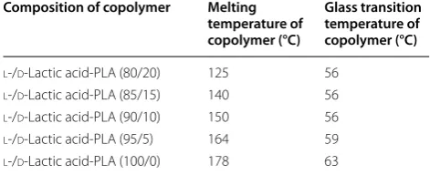
B. brevis participate in polylactic acid biodegrading (Ikada and Tsuji 2000; Tomita et al. 1999; Shah et al. 2008b). Pro-duction of polylactic acid through the polymerization of the monomer of lactic acid is shown in Table 5.
Lactic acid (hydroxyl carboxylic acid) is a basic com-ponent of aliphatic polyester polymer (polylactic acid). It is a bio-based bacterial fermented product. Fermenta-tion dependent l or d forms of lactic acid contribute to the formation of several forms of polylactic acid and its derivative. T16-1 strain of Actinomadura keratinilytica
NBRC 104111 reported for production of PLA-degrading enzyme Amycolatopsis sp. also shows ~60% degradation of PLA film degradation within 14 days (Babul et al. 2013; Garlotta 2002; Sukhumaporn et al. 2012; Pranamuda et al. 1997). Similarly, Prema and Uma (2013) reported mesophilic bacteria Bacillus amyloliliquefaciens for the degradation of polylactic acid. Iovino et al. (2008) inves-tigated aerobic biodegradation of polylactic acid under a composting environment and checked the effect of the presence of thermoplastic starch (TPS) and short natu-ral fibers (coir). In the initial stage, TPS degrades faster than PLA, and TPS is most susceptible to degradation as compared to PLA. Particle size is also an important factor for biodegradation; fine particles (nano-particles) involve a complex mechanism as compared to micro-particles (Kunioka et al. 2006). Pranamuda et al. (1997) isolated Amycolatopsis and studied it for PLA degrada-tion, finding 60% degradation of PLA film within 14 days. PLA depolymerase of Amycolatopsis sp. serves as a pro-tease instead lipase and degrades casein silk fibroin and Suc-(Ala) 3-pNA. In addition to Amycolatopsis, Saccha-rotrix was also reported for PLA degradation (Tokiwa et al. 2009). According to Hidayat and Tachibana (2012) 12, 21, 30 and 48% polylactic acid degradations in 1, 2, 3 and 6 months, respectively, were achieved with P. ostreatus. Biodegradation of lactic acid polymers and its copolymers was widely affected by the amount of lactic acid content in polymers. Different fungal species were reported for lactic acid and its copolymer degradation, e.g., Rhizopus sp., Mucor sp., Aspergillus sp. and Alter-naria sp. Eubeler et al. (2009) found the effect of UV irradiation on enzymatic hydrolysis of polylactic acid. The researchers expected that UV irradiation caused a chain alteration by C=C bonds formation. Proteinase K Table 5 Effect of l-lactic acid or d-lactic acid copolymer
composition of polylactic acid on melting temperature and glass transition (Babul et al. 2013)
Composition of copolymer Melting temperature of copolymer (°C)
Glass transition temperature of copolymer (°C)
l-/d-Lactic acid-PLA (80/20) 125 56
l-/d-Lactic acid-PLA (85/15) 140 56
l-/d-Lactic acid-PLA (90/10) 150 56
l-/d-Lactic acid-PLA (95/5) 164 59