This is an open access journal, and articles are distributed under the terms of the Creative Commons Attribution-Non Commercial-ShareAlike 4.0 License, which allows others to remix, tweak, and build upon the work non-commercially, as long as appropriate credit is given and the new creations are licensed under the identical terms.
© 2018 Journal of Advanced Pharmacy Education & Research | Published by SPER Publication
73
Falciform Ligament plug in Laparoscopic Hiatus Hernia repair
Mohamed El-Kordy, Sameh Gabr Attia, Ibrahim Aboulfotoh Mohamed, Hesham Wefky Anwar,
EidRezk Elgamal, Mohamed Ibrahim Shalamesh*, Alsayed Basiony Moghazy
General Surgery Department, Faculty of Medicine (Boys), Al-Azhar University, Cairo, Egypt.
Correspondence: Mohamed Ibrahim Shalamesh, General Surgery Department, Faculty of Medicine (Boys), Al-Azhar University, Cairo, Egypt. e-mail:
ABSTRACT
Objectives: Laparoscopic repair of a para-esophageal hiatal hernia is a challenging procedure. Several methods have been developed in efforts to achieve tension-free reconstruction of the esophageal hiatus. The falciform ligament is used as an autologous onlay flap to achieve tension-free closure of the crural defect of a para-esophageal hernia (PEH). Aim of the work: To assess the use of falciform ligament for hiatus hernia repair and to evaluate the results. Methods: Hernias were diagnosed with esophago-gastroscopy and esophageal manometry and barium contrast X-ray study. Results: Fifty patients underwent falciform ligament plug repair while eight patients underwent crural approximation and fundoplication repairs. The classical traditional practice period for repairs was significantly (p=0.004) longer in duration. Period of stays in the hospital, average contraction amplitudes, and lower esophageal sphincter pressure during the rest were non-significantly different among the groups. Discussion: The well-established benefits of laparoscopic hiatal hernia repair include shorter period of hospitalization, less pain; and rapid recovery rate, but with high failure rates than in open repairs of patients. Conclusion: Different data were recorded about demographic distribution, steps of operation and symptoms pre-and post-operation. Application of the falciform ligament for induction of vascularization on lay flap is effective and safe. The technique was used for adequate closing of the crural anomaly of para-esophageal hiatus hernia. The recommended instructions for the position of the falciform ligament is that, it must be mobilized freely in the region of anterior abdominal wall to avoid lateral pressure on the flap, in the same time avoiding induction of devascularization. For fixing the falciform ligament to the left and right hiatal crura, interrupted vertical mattress sutures should be done.
Keywords: Hiatal hernia, Gastro-esophageal reflux, Laparoscopy, Falciform ligament plug.
Introduction
The general termination of para-esophageal hernia (PEH) is a projection of the gastric fundus within the diaphragmatic space whereas the lower esophageal sphincter is still in its usual anatomic location (hiatal hernia, type II). Commonly, mutually the lower esophageal sphincter and the fundus are herniated into the chest (hiatal hernia, type III) [1].
Since late 1980's laparoscopy was familiarized and was rapidly
approved by GIT surgeons as chance to afford effective repair to avoid the side effects of the open operations. Subsequently, we and other investigators have recognized the safety and practicability of a laparoscopic method to massive para-esophageal hernia repair [2].
Currently, laparoscopy interference is used as the ordinary methodology for treatment of gastro-esophageal reflux disease surgically, in addition to its wide use in PEH repair. Although this technique requires special skillfulness, this method offers good coverage of the site of operation than open trans-abdominal approaches; in addition to frequency of morbidity, period of stay in the hospital is very short, pain drugs requests are reduced, and healing time is quick. These benefits may be valuable particularly in cases of PEH people as most of patients are of advanced years and have several comorbid disorders. PEHs is characterized by a high recurrence rate and are primarily non-symptomatic, therefore, contrast imaging is required to diagnose the relapses precisely [3-5].
Access this article online
Website: www.japer.in E-ISSN: 2249-3379
How to cite this article: Mohamed El-Kordy, Sameh Gabr Attia, Ibrahim
74 Journal of Advanced Pharmacy Education & Research | Oct-Dec 2018 | Vol 8 | Issue4 Restoration of the esophageal hiatus by re-approximation of the
right and left hiatal crura with intermittent closures (posterior hiatoplasty) frequently outcomes in marked pressure on the restoration. The main reason for hernia relapse is the disappointment of the posterior hiatoplasty [6, 7].
Many operational methods have been proposed to reduce the rate of repetition, such as trials at producing a tension-free healing by the application of synthetic and biological lattice [8].
Synthetic lattice or meshes, still, have been accompanied with severe complications at site of the hiatus, like, esophageal structuring, fibrosis, gastric fibrosis and erosion [9].
To avoid the side effects of plastic meshes, the biologic mesh substances were introduced recently to replace the plastic one, in expectations of escaping these possibly sever complications even though attaining a healthy stable healing. Theoretically, these substances lead to little reactions to these foreign bodies and as an alternative doing as a momentary matrix to innate tissue development and restoration. Many researchers have found that hiatal hernia strengthening with biologic mesh is accompanied with low independent repetition rates which ranged from 0.0 to 11% [10, 11].
When peri-operative outcomes are compared between laparoscopy and open surgical method regarding length of stay in hospital, blood loss, postoperative morbidity and mortality, they are shift significantly for laparoscopy with great reduction in the mentioned parameters than open method [12].
The application of the falciform ligament by means of a vascularized autologous flap for induction of healing without pressure in case of a large hiatal weakness is considered as a smart decision for getting healed without stress of a PEH deprived from the risks linked with synthetic substances or the substantial charge of biologic mesh. There are some differences in anatomy features; where the blood source for the falciform ligament is mainly via branches of the left inferior phrenic and left hepatic arteries which union to constitute an arcade, besides from smaller perforators originated from the parietal peritoneum. It is essential to sustain the reliability of the arterial arcade throughout mobilization of the ligament and this is preeminent confirmed by protection of the separation line as adjacent to its connection to the anterior abdominal wall as probable [13].
The main cause of repair failure is attributed to the low value of the crus and the diaphragm, in which the tissue does not adequately grasp suture well and can easily slit. Correspondingly, the faults are usually long-lasting and bulky, and the initial closure of the hernia is continuously under pressure. These issues create the repetition of the hernia further expected and may be accompanied with signs of relapse such as dysphagia [14].
Patients and Methods
From January 2015 to august 2017, a total of 24 patients were included in the study, with para-esophageal hiatus hernia underwent falciform ligament onlay rape and hiatus plug at
General Surgery Department, Al-Azhar University Hospitals. Pre-operative assessment was performed, including history, examination, Gastro-esophageal reflux evaluation with esophago-gastroscopy and esophageal manometry and barium contrast X-ray study, full laboratory study, and abdominal ultrasonography. Patients were divided randomly into: group I (Falciform ligament repair), and group II (crural approximation with or without fundoplication). Patients were scheduled for 3, 6, and 12 months visits for assessment.
All patients were subjected to follow-up barium contrast study, esophago-gastroscopy and abdominal ultrasonography (US) at 3, 6, and 12 months post-operative.
Eligible patients were between 20 and 46 years of age and all patients provided consent to participate in the retrospective review of the prospectively collected data.
Patients who are candidates for PEH repair undergo a complete pre-operative workup for both the surgical pathology and any medical co-morbidity relevant to undergo a general anesthetic and hiatus hernia repair.
Informed consent was obtained from each patient before initiation of the study, and hospital ethical committee approval was obtained for the protocol of the study, as Photo 1.
Photo 1
Para esophageal Hiatus Hernia
Pre-operative Assessment
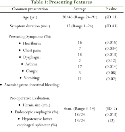
Laparoscopic repair of para-esophageal hernia begins with a careful preoperative evaluation. We obtain a careful history, including assessment of typical symptoms of gastro-esophageal reflux (heartburn and regurgitation) and dysphagia. Additional symptoms that are common include chest or epigastric pain, recurrent aspiration with or without associated pneumonia, cough, shortness of breath, and dyspnea on exertion. Signs of compromised blood flow to the herniated stomach may be subtle, such as iron-deficiency anemia. Patients often report knowledge of a “hiatal hernia” for many years.
Next, we obtain a radiographic evaluation by barium swallow. Abnormal esophageal motility is often evident on barium swallow in patients with para-esophageal hernia; barium swallow is useful for identifying the location of the gastro-esophageal junction (GEJ), assessing the degree to which the stomach is herniated into the chest. Because the esophagus is often tortuous and placement of a manometry catheter across the lower esophageal sphincter into the stomach can be difficult, we rarely perform a complete motility study.
Journal of Advanced Pharmacy Education & Research | Oct-Dec 2018 | Vol 8 | Issue4 75
albumin levels for evaluation of nutritional status. We obtain pulmonary function testing in patients with a complaint of shortness of breath to understand whether the breathing difficulties are because of restriction of lung function due to compression of adjacent lung by herniated stomach or to co-existing intrinsic lung disease, which may in fact be related to long-standing reflux, aspiration, and lung injury.
Surgical Procedures
Patient Preparation, Positioning, and Port
Placement:
After the pre-operative evaluation is complete, the patient is advised of the risks and benefits of the operation, and informed consent is obtained. Once this is accomplished, the patient is taken to the operating room. Our preferred position of the patient is supine, in reverse Trendelenburg position, with the surgeon on the patient’s right side and the assistant on the left. Sequential compression devices are placed on the legs bilaterally. Patients should also receive 5,000 IU of heparin subcutaneously prior to induction of anesthesia [15].
The patient is placed in the low lithotomy position. The operating surgeon now stands between the patient’s legs with the first and second assistants on the patient’s left and right, respectively. Pneumo-peritoneum is established via insertion of a Veress needle to insufflate the abdomen to a pressure of 12– 15 mmHg. A 10 mm 30° laparoscopic telescope is then introduced under direct visualization via a vise port, approximately one third to one half of the distance between the umbilicus and xiphoid process. All further ports are placed under direct visualization. A 5 mm port is placed at the xiphoid process to facilitate retraction of the left lobe of the liver, 5 mm working ports are placed in the left and right hypochondriac, and a 5 mm assistant’s port is placed in the left flank just inferior to the left costal margin.
Care is taken to avoid dissection into the falciform ligament. Because of the extensive dissection within the mediastinum, this port must be placed in the upper third of the abdomen [16].
Surgical Technique
The hernia contents are reduced by gentle traction. The gastro-splenic ligament and short gastric vessels are divided with harmonic scalpel or the ultrasonic shears; this allows access to the areolar attachments of the hernia sac to the mediastinal structures. Care is taken to identify both the anterior and posterior vagus, and re-establishing adequate intra-abdominal esophageal length. Great care must be taken not to cause an iatrogenic esophageal injury at this point, as Photo 2.
Photo 2
Division of vascularized falciform ligament
After mobilization of the anterior and left sides of the esophagus, attention is turned to the lesser omentum. The pars flaccida is divided, and the base of the right crus is identified. Strict attention to maintaining the integrity of the peritoneal lining over the crura is essential for the success of primary closure. Without this lining, the crural musculature has no intrinsic strength and, thus, will not hold suture sufficiently to prevent dehiscence of the crural repair. We assess the location of the GEJ for adequate intra-abdominal length in a neutral resting position in the abdomen [17] ,as Photo 3.Photo 3
Approximation of two crura
A Penrose drain is passed around the esophagus and is used for gentle retraction to expose the base of the suture approximation of the crura, a falciform flap is developed and used as an onlay mesh to achieve a tension-free closure of the defect, and the tip of the flap is rolled upon itself to plug the rest of esophageal hiatus.
We repair the hiatus in all patients regardless of the decision, and an attempt is made to re-approximate the crura by one or two stitches of 2-0 prolin, anterior to the aorta, as Photo 4.
76 Journal of Advanced Pharmacy Education & Research | Oct-Dec 2018 | Vol 8 | Issue4
Falciform ligament plug fixation
The falciform ligament is mobilized at the level of its attachment to the anterior abdominal wall starting just superior to the umbilicus and continuing superiorly, and anterior of the liver with harmonic scalpel dissection through the left flank port, and retracted with a traumatic grasper via the port in the left hypochondrium.
The falciform flap is guided under the lateral segment of the left lobe of the liver and maneuvered to cover the hiatal defect. A 54-Fr bougie is introduced trans-orally and advanced into the stomach under laparoscopic visualization. The bougie is used as a guide to prevent excessive narrowing of the hiatus and plug the flap on the posterior wall of the esophagus.
Post-operative Care
Following completion of the operation, the patient is typically extubated and transferred to the recovery room and admitted to the intensive care unit (ICU) for post-operative observation. We routinely perform barium swallow prior to discharge to document sub-diaphragmatic positioning of the fundoplication wrap and look for unrecognized esophageal or gastric injury. The median post-operative length of hospital stay was 3 days. Routine follow-up, including barium oesophagram and symptom assessment with validated measures for gastro-esophageal reflux disease health-related quality of life [18].
All patients are seen 2 weeks after surgery and again 3 and 6 months and 1 year after surgery. Barium oesophagram is performed 1 year after surgery and then at 2-year intervals.
Symptoms of Hernia Recurrence
Common post-operative complaints include dysphagia, heartburn, gas bloat, and diarrhea.
Results
All cases in the study were scheduled a selective procedure. Nissen fundoplication was performed on 10 patients, two patients had no fundoplication.
An esophageal lengthening procedure (Collis gastroplasty) was not performed in study groups.
Mean operative time was 100 minutes in group I (falciform ligament repair), with a median of 120 minutes (range 100 to 180 minutes), while in group II (crural approximation with or without fundoplication) it was 140 minutes, median of 140 minutes (range 120 to 220 minutes).
Post-operative stay in the hospital was 3 days as median stay, ranged from 1 to 8 days (Table 1).
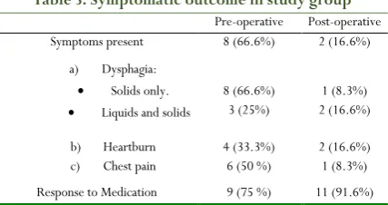
Post-operative follow-up exceeded 6 months, in group I (falciform ligament repair) of these 10 patients, 7 (75%) had no esophageal signs. Heartburn was the more general grumble and was found in 2 individuals (20%). Also, dysphagia was recorded in 2 subjects (20%); one of them found with ingestion of solids only, whereas, other patient was fed on liquids too. One patient (10%) complained from chest pain.
Table 1: Presenting Features
P value Average
Common presentation
(SD 13) 20/46 (Range 24–91)
Age (yr.)
(SD 43) 12 (Range 1–24)
Symptom duration (mo.)
(0.015) (0.034) (0.013) (0.12) (0.014) (0.08) (0.02) 16
7 18 2 17 3 11 Presenting Symptoms (%):
•Heartburn:
•Chest pain:
• Dysphagia:
•Asthma:
•Cough:
•Vomiting:
• Anemia/gastro-intestinal bleeding:
(SD 2) (0.013) (12) 6cm. (Range 3–14)
18/24 13/24 Pre-operative Evaluation:
•Hernia size (cm.):
• Endoscopic esophagitis (%):
• Hypotensive lower esophageal sphincter (%)
Post-operative physiologic valuation (manometry, 24-hour pH tests) and endoscopy were not implemented regularly. Contrast barium meal esophago-image were achieved regularly at 6 to 12 months post-operation, and/or signs were measured during follow-up period (Table 2).
Pre- and post-operative signs were matched; noticeable progress was observed in all measured parameters (P<0.001). A remarkable reduction in the administered antacids therapy from pre-operative (77%) to post-operative (11%) assessment was also noted (P< 0.001).
Statistical analysis of the obtained data using, chi-square/Fisher exact tests were used for comparison of disconnected variables and the two-tailed t-test was performed for continuous data. Significance was defined as P<0.05. Summary of the data are presented as mean± SD.
Table 2: Characteristics of the study groups
Point of difference Group I (falciform plug) Group II (classical repair) P value
Number 12 12
Gender M: 9 (75%); F: 3 (25%) M: 11 (91.6%); F: 1 (8.3%)
Approach Laparoscopic: 12 (100%) Laparoscopic: 12 (100%)
Conversions to Open
Approach 0 0
Operative time (minutes)
120 (132.9±40.4
min) 140 (190±84.7) P=0.004 Estimated bleeding (mL) 14.7 (0 - 100) 14.7 (0 - 100) P=0.1
Hospital stay (days) 3 (1 - 8) 3 (3 - 10) P=0.08
Incidence of recurrence 0 (0%) 2 (16.6%)
Journal of Advanced Pharmacy Education & Research | Oct-Dec 2018 | Vol 8 | Issue4 77
longer than falciform ligament cases group (132.9±40.4 min, p=0.004) (Table 2).
Regardless of the higher difficulty of cases and the operation continue for a long of time, individuals in the falciform ligament group had simple primary post-operative sequences and were cleared from the hospital at parallel times to their non-falciform ligament ones (day 2.26±0.94 vs. 2.24±0.90, p=0.94). Falciform ligament cases were realized on post-operative day 24.9±10.7 and the non-falciform ligament patients followed up on day 23.0±17.0 (p=0.57).
Generally, during the follow-up for the 2nd time for
non-falciform ligament patients, it is observed that there were more symptom complaints such as abdominal pain, severe chest pain, and incapability to eruct to release embarrassment.
Non-falciform ligament cases had developed heartburn scores than cases who had bio-mesh repair of the hiatal hernias. Falciform ligament patients tend to complain from dysphagia and bloating during eating solid foods, but the differences were non-significant.
A routine examination must be performed for esophageal manometric at the 6-month visit. Whereas, an inactive LES pressure was similar for both hernia repair groups; the outstanding pressure in a mesh repair patient group was found to be significantly higher (13.7±6.6 vs. 1.7±5.8 mmHg, p=0.0001).
Barium esophagograms were happening in 6/24 (37.5%) patients during the period of more than 6 months of follow-up. The remainders of patients have either declined to participate in the investigation or have not been appraised in the place of work for more than 6 months post-operatively.
The obtained data revealed the incidence of repeated hiatal hernias in 2 patients (8.3%) by using radiographic imaging or 16.6% of those undertaking contrast x-ray evaluation as seen in table (3).
Table 3: Symptomatic outcome in study group
Post-operative Pre-operative
2 (16.6%) 8 (66.6%)
Symptoms present
1 (8.3%) 2 (16.6%) 8 (66.6%)
3 (25%) a) Dysphagia:
• Solids only.
• Liquids and solids
2 (16.6%) 1 (8.3%) 4 (33.3%)
6 (50 %) b) Heartburn
c) Chest pain
11 (91.6%) 9 (75 %)
Response to Medication
Discussion
With regards to the esophageal hiatus dynamic nature, where the continuous movement of the esophagus, diaphragm, pericardium and stomach occurs, in addition to ascent pressure from the chest to abdomen, which is highlighted by Valsalva movements (e.g., sneezing, laughing), are all donating causes to failure of the hiatal healing [19].
Impediments accompanied with the application of synthetic mesh in the healing of a PEH can be overcome by the
application of the falciform ligament as an onlay flap and plug. This results in buttressed or tension-free repair with fundamentally an autologous well-vascularized pedicle flap, the ultimate “biologic” substance.
Many investigators applied different methods in order to achieve a tension-free healing of the esophageal hiatus. These methods comprise the using of many variant biologic meshes and non-absorbable synthetic meshes [20].
One of the most advanced techniques in the repair is the using of biological mesh which is used for re-enforcement of hiatal hernia repair. These meshes are composed of collagenous matrix originated from porcine intestinal sub-mucosa, porcine or human dermis or bovine pericardium. Theoretically, after inserted, they are ultimately substituted by blood vessels and host collagen. While displaying pronounced capacity in a rare select presentation, their protagonist in assistant long-term robust hiatal repairs is far from recognized [21].
The model of ligamentumteres strengthening of a hiatal repair has been formerly described by Varga et al. In their studies which included 26 patients, subsequent efficacious posterior hiatoplasty accomplished by two methods laparoscopically and open techniques, the ligamentumteres was used to support the hiatal cessation. Their data confirmed the method to convey minimal morbidity (11 %) and mortality (0 %) with a radiologic repetition of 15 % at an average of 3 years follow-up [22].
The radiological recurrence rate, following laparoscopic suture repair of a PEH, has been reported as shig has 42%. It is assumed that radiologic indication of disappointment of a HH healing does not essentially associate with clinical indication of failure. Subsequently, subjects who have undertaken healing of GPEH can be establish to have anatomic confirmation (to varying degrees) of hernia repetition without clinical sign or return of symptoms [23].
This results of a vascularized falciform ligament flap technique that can be applied to both support a finalized hiatoplasty and, more uniquely, bridge a fault when the crura cannot be re-approximated. This offers a block to outlet of bowel or other abdominal contents into the mediastinum and was, in fact, the motivation for developing the method when no other incomes of hiatal healing were accessible. Biologic meshes cannot be used to length a weakness, and non-absorbable meshes should not be applied to span a hiatal fault.
Conclusion
78 Journal of Advanced Pharmacy Education & Research | Oct-Dec 2018 | Vol 8 | Issue4
References
1. Hashemi M, Sillin LF, Peters JH. Current concepts in the management of paraesophageal hiatal hernia. J Clin Gastro-enterol 1999; 29:8–13.
2. Luketich JD, Nason KS, Christie NA, Pennathur A, Jobe BA, Landreneau RJ, Schuchert MJ. Outcomes after a decade of laparoscopic giant paraesophageal hernia repair. J Thorac Car-diovasc Surg. 2010; 139(2):395–404, 404 e391.
3. Wu JS, Dunnegan DL, Soper NJ. Clinical and
radiologic assessment of laparoscopic paraesophageal hernia repair. SurgEndosc 1998; 13:497–502.
4. Laine S, Rantala A, Gullichsen R, Ovaska J.
Laparoscopic vs. conventional Nissen fundoplication: A prospective ran-domized study. SurgEndosc 1997;11:441–444.
5. Schauer PR, Ikramuddin S, McLaughlin RH, Graham TO, Slivka A, Lee KK, Schraut WH, Luketich JD. Comparison of laparoscopic versus open repair of paraesophageal hernia. Am J Surg 1998;176:659–665. 6. Ally A, Munt J, Jamieson GG, et al. Laparoscopic repair
of large hiatal hernias. Br J Surg. 2005; 92(5): 648– 653.
7. Smith GS, Isaacson JR, Draganic BD, et al.
Symptomatic and radiological follow-up after para-esophageal hernia repair. Dis Esophagus. 2004; 17(4): 279–284.
8. Varga G, Cseke L, Kalmár K, et al. Prevention of recurrence by re-inforcement of hiatal closure using ligamentumteres in laparoscopic repair of large hiatal hernias. SurgEndosc. 2004; 18: 1051– 1053.
9. Griffith PS, Valenti V, Qurashi K, Martinez-Isla A. Rejection of goretex mesh used in prosthetic cruroplasty: a case series. Int J Surg. 2008; 6: 106–9. 10. Oelschlager BK, Barreca M, Chang L, Pellegrini CA.
The use of small intestine sub-mucosa in the repair of paraesophageal hernias: initial observations of a new technique. Am J Surg. 2003; 186: 4–8.
11. Lee E, Frisella MM, Matthews BD, Brunt LM.
Evaluation of a cellular human dermis reinforcement of the crural closure in patients with difficult hiatal hernias. Surg Endosc. 2007; 21: 641–5.
12. Hakanson BS, Thor KB, Thorell A, Ljungqvist O. Open vs laparoscopic partial posterior fundoplication. A prospective randomized trial. Surg Endosc. 2007;21(2):289–298.
13. Li XP, Xu DD, Tan HY, et al. Anatomical study of the morphology and blood supply of the falciform ligament and its clinical signif-icance. Surg Radiol Anat. 2004; 26: 106–109.
14. Oelschlager BK, Pellegrini CA, Hunter J, Soper N, Brunt M, Sheppard B, Jobe B, Polissar N, Mitsumori L, Nelson J, Swanstrom L. Biologic prosthesis reduces recurrence afterlaparoscopicparaesophageal hernia repair: a multicenter, prospec-tive, randomized trial. Ann Surg. 2006; 244: 481–90.
15. Zurawska U, Parasuraman S, Goldhaber SZ. Prevention of pulmonary embolism in general surgery patients. Circulation. Mar 6 2007; 115(9):e302-307.
16. Zollinger RJ, Zollinger RS. Cholesystectomy, Hasson Open Technique, Laparoscopic. In: Jr ZR, Sr ZR, eds. Zollinger's Atlas of Surgical Operations, 8th edn. New York: The McGraw-Hill Companies, Inc; 2003. 17. Whitson BA, Hoang CD, Boettcher AK, Dahlberg PS,
Andrade RS, Maddaus MA. Wedge gastroplasty and reinforced crural repair: important components of laparoscopic giant or recurrent hiatal hernia repair. J Thorac Cardiovasc Surg. 2006; 132(5):1196– 1202 e1193.
18. Velanovich V, Vallance SR, Gusz JR, Tapia FV,
Harkabus MA. Quality of life scale for gastroesophageal reflux disease. J Am Coll Surg. Sep 1996; 183(3):217– 224.
19. Varga G, Cseke L, Kalmár K, et al. Prevention of recurrence by reinforcement of hiatal closure using ligamentumteres in laparo-scopic repair of large hiatal hernias. SurgEndosc. 2004; 18: 1051– 1053.
20. Champion JK, Rock D. Laparoscopic mesh cruroplasty for large paraesophageal hernias. SurgEndosc. 2003; 17: 551–553.
21. Oelschlager BK, Pellegrini CA, Hunter JG, Brunt ML, Soper NJ, Sheppard BC, Polissar NL, Neradilek MB, Mitsumori LM, Rohr-mann CA, Swanstrom LL. J Am Coll Surg. 2011; 4:461–468.
22. Varga G, Cseke L, Kalmar K, et al. Prevention of recurrence by reinforcement of hiatal closure using ligamentumteres in laparoscopic repair of large hiatal hernias. SurgEndosc. 2004; 18: 1051– 1053.
23. Carlson MA, Condon RE, Ludwig KA, et al.