Alicja Kędzia
1, A–F, Małgorzata Janeczko
1, B, D, Katarzyna Miśkiewicz
1, B, D,
Krzysztof Dudek
2, B–DMorphometry of Human Musculus Gluteus Maximus
in Foetal Period
1 Department of Normal Anatomy, Wroclaw Medical University, Poland
2 Institute of Machines Design and Operation, Wroclaw University of Technology, Poland
A – research concept and design; B – collection and/or assembly of data; C – data analysis and interpretation;
D – writing the article; E – critical revision of the article; F – final approval of article; G – other
Abstract
Background. Magnus gluteal muscle (musculus gluteus maximus) belongs to the group of lower limb girdle mus-cles. It is one of the biggest muscles in human organism and is located mostly superficially in gluteal region. Literature provides discussion concerning its role in movement such as walking, running and climbing as well as plastic surgery in reconstructive operations of trochanter. Magnus gluteal muscle plays an important role in orthopaedic surgery.
Objectives. The goal of the study was to analyse the human magnus gluteal muscle in the foetal period.
Material and Methods. The analysis was carried out on 154 muscles originating from human foetuses (including 30 females – 39%) belonging to the collection of Normal Anatomy Dept. of Wroclaw Medical University. The body length was assessed with the use of vertex-tuberal (v-tub) length and it was included in the range 107–205 mm, which corresponds with the period 17–30 weeks of foetal life. The survey incorporated the following methods: anthropological, preparational and image acquisition which was acquired with the use of high-resolution digital camera. In order to take computer measurements, the following systems were exploited: Image J and Scion for Windows. Statistical analysis was carried out with the use of STATISTICA package v. 9 (t-Student test).
Results. The magnus gluteal muscle was analysed in respect to sexual dimorphism and symmetry. On the basis of elicited parameters, the model of muscle increase in foetal period was defined. The following measurements were taken: v-tub, vertex-plantare (v-pl), body mass, muscle particular sides lengths and distance between corresponding measurement points. In every muscle, the lengths of four sections forming the circumference as well as the area were measured.
Conclusions. No difference was observed in foetal magnus gluteal muscle sexual dimorphism or symmetry (p > 0.05). The correlation diagram was used to calculate the muscle weekly increase in foetal period. The results suggest that lesions and pathologies in the region of magnus gluteal muscle are acquired in post foetal period (Adv Clin Exp Med 2014, 23, 1, 9–16).
Key words: humanmusculus gluteus maximus, prenatal period, morphometry.
Adv Clin Exp Med 2014, 23, 1, 9–16 ISSN 1899–5276
ORIGINAL PAPERS
© Copyright by Wroclaw Medical University
Magnus gluteal muscle (musculus gluteus
maximus) belongs to the group of lower limb
gir-dle muscles. It is one of the biggest muscles in hu-man organism and is located mostly superficially in gluteal region [1]. This muscle’s characteristic size, anatomy and function makes distinguishes it from the analogous muscles of monkeys and oth-er primates [2]. The litoth-erature presents discussions concerning its role in movement such as walking, running and climbing [3] as well as plastic surgery in reconstructive operations of trochanter [4, 5]
far less accurate and reliable than those applied in these surveys.
Material and Methods
Examination material consisted of 154 mag-nus gluteal muscles originating from 77 human foetuses (30 females – 39%) belonging to Nor-mal Anatomy Dept., Wroclaw Medical Univer-sity. Body length assessed on the basis of vertex-tuberale (v-tub) length was included in the range 107–205 mm, which corresponds with the period 17–30 weeks of foetal life. The survey incorporated anthropological and preparational methods as well as image acquisition achieved with high resolution digital camera. These methods have already been utilized by other authors using the above material from the collection mentioned above [11, 12].
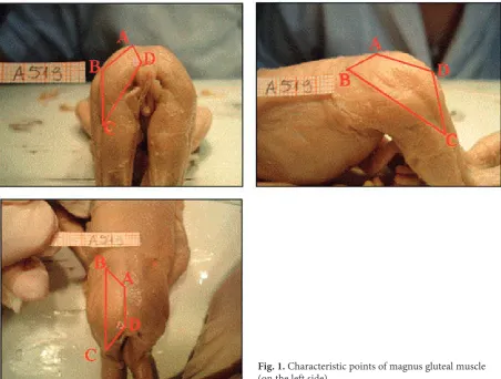
Magnus gluteal muscles were taken from three sides (Fig. 1) with the use of long focal length lens. On the basis of these photos, the following mea-surements were made: vertex tuberale (v-tub), ver-tex plantare (v-pl), body mass, each muscle four sides lengths (the total composed the circumfer-ence), angles included between the muscle sides and the distances between the measurement cor-responding points.
On total, 1386 measurements were made with the use of free and available online computer sys-tems Image J and Scion Image for Windows[13, 14]. The two advantages of these systems were the possibility of multiple repetition of demanded measurement, accuracy and the option of using the muscle photo for conducting measurements (fig-ure). Other papers exploring the problem of foetal muscles metrology also incorporated the two sys-tems [11, 12 – sartorius and scapula]. The quanti-tative assessment was made as well. Statistical anal-ysis was carried on with the use of STATISTICA v.9 package (t-Student test).
The muscles were analysed in respect to sexual dimorphism and symmetry and their somatic fea-tures were defined.
Results
Table 1 presents basic somatic features (v-tub, v-pl, body mass) of the examined material. Foetus-es of both sexFoetus-es were compared in rFoetus-espect of thFoetus-ese features and in the analysed material, male and fe-male foetuses did not differ significantly (Fig. 2 – p > 0.05). In the analysed material female and
male foetuses did not differ significantly in respect of somatic features (Fig. 2 – p > 0.05)
Symmetry Analysis
Significant differences at p < 0.05 level were marked.
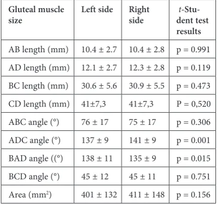
No statistically significant difference was found between left and right side sizes (p > 0.05) (Fig. 3, Table 2). Only ADC angle and BAD angle sizes re-veal asymmetry. However, this may have resulted from the foetus’ uneven position during the pho-tos being taken.
Table 1. Characteristics of somatic features of 77 foetuses
Age group N Sex Vertex tuberale (mm) Vertex plantare (mm) Body mass (g)
M SD M SD M SD
I
(17. and 18. week) 0 8 8 F M FM – 118.2 118.2 – 9.4 9.4 – 165.6 165.6 – 15.7 15.7 – 94 94 – 22 22 II
(19. and 20. week) 8 7 15 F M FM 148.4 148.4 148.4 5.2 5.7 5.2 211.4 215.0 213.1 7.9 9.3 8.4 208 230 218 57 34 48 III
(21. and 22. week) 1420 34 F M FM 166.2 168.5 167.6 7.0 5.5 6.2 241.6 237.4 239.2 19.9 26.5 23.7 273 290 283 62 36 48 IV
(23, 24 and 22. week) 812 20 F M FM 188.8 185.1 186.6 8.3 7.8 8.0 277.2 260.2 267.1 10.3 25.0 21.8 431 348 381 70 92 92 M – mean; SD – standard deviation.
Table 2. Basic statistics (mean value ± standard deviation) of magnus gluteal muscle of 77 foetuses and t-Student test results for related variables
Gluteal muscle
size Left side Right side dent test t -Stu-results
AB length (mm) 10.4 ± 2.7 10.4 ± 2.8 p = 0.991 AD length (mm) 12.1 ± 2.7 12.3 ± 2.8 p = 0.119 BC length (mm) 30.6 ± 5.6 30.9 ± 5.5 p = 0.473 CD length (mm) 41±7,3 41±7,3 P = 0,520 ABC angle (°) 76 ± 17 75 ± 17 p = 0.306 ADC angle (°) 137 ± 9 141 ± 9 p = 0.001 BAD angle ((°) 138 ± 11 135 ± 9 p = 0.015 BCD angle (°) 45 ± 12 45 ± 11 p = 0.751 Area (mm2) 401 ± 132 411 ± 148 p = 0.156
Significant differences at p < 0.05 level were marked.
Fig. 2. Comparison of somatic features in female and male foetuses, and t-Student test results for unrelated variables.
t = -0.689; p = 0.493
F M 160 162 164 166 168 170 172 174 176 V-tub (mm)
t = 0.382; p = 0.704
F M 230 235 240 245 250 255 V-pl (mm)
t = 0.041; p = 0.967
F M 250 260 270 280 290 300 310 320 330 340
Body mass (g
Sexual Dimorphism Analysis
As the analysed material in age group I (17–18 weeks) did not contain female foetuses, sexual di-morphism analysis was carried on in groups II, III and IV (Fig. 4).
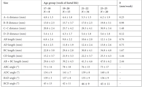
Due to no statistically significant symmetry (Table 3), magnus gluteal muscle size was taken on the left and right sides conjointly and for this group, mean values as well as standard deviations were defined (Table 4).
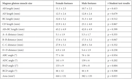
No statistically significant sexual dimorphism was observed. Length and area sizes of both sexes do not differ significantly (p > 0.05).
AB and AD distances were taken in YZ plane, BC distance was measured in XZ plane and CD dis-tance was taken in XY plane (Fig. 6). Magnus glu-teal muscle area was defined as the total of ABD, BDE and CDE triangles areas.
Magnus gluteal muscle area increases irregular-ly. From 17th till 20th, area accession amounts to 59 mm2, from 21th to 25th week to 70 mm2 on
av-erage (Fig. 7).
Mathematical model describing this process (R2 = 0.798) is an exponential model:
F = 18.06 × exp(0.144 × week).
Fig. 3. AB lengths comparison on the left and right
sides and t-Student test results for related variables Fig. 4. AD lengths comparison in female (F) and male (M) foetuses and t-Student test result for unrelated variables
Table 3. Basic statistics (mean value ± standard deviation) of magnus gluteal muscle sizes in 69 foetuses and t-Student test results for unrelated variables
Magnus gluteus muscle size Female foetuses Male foetuses t-Student test results
AB length (mm) 11.1 ± 2.5 10.7 ± 2.2 p = 0.413 AD length (mm) 12.5 ± 2.4 12.8 ± 2.4 p = 0.679 BC length (mm) 32.0 ± 5.2 31.3 ± 4.0 p = 0.512 CD length (mm) 22.9 ± 4.1 23.1 ± 4.0 p = 0.867 AB+BC length (mm) 43.2 ± 6.9 42.0 ± 4.9 p = 0.399 A–A distance (mm) 5.1 ± 1.9 5.5 ± 1.7 p = 0.355 B–B distance (mm) 17.6 ± 3.4 17.5 ± 3.2 p = 0.847 C–C distance (mm) 27.9 ± 5.1 28.9 ± 3.6 p = 0.352 D–D distance (mm) 4.9 ± 1.8 5.4 ± 1.9 p = 0.230 ABC angle (°) 77 ± 16 76 ± 16 p = 0.934 ADC angle (°) 141 ± 9 139 ± 6 p = 0.202 BAD angle ((°) 133 ± 9 139 ± 8 p = 0.004 BCD angle (°) 46 ± 12 44 ± 8 p = 0.308 Area (mm2) 444 ± 131 382 ± 135 p = 0.053
t = -0.415; p = 0.679
F M
11,0 11,5 12,0 12,5 13,0 13,5 14,0 14,5 15,0
AD
(mm)
t = -0.004; p = 0.997
L R
Side 9,6
9,8 10,0 10,2 10,4 10,6 10,8 11,0 11,2
AB
Discussion
Muscular system development with special re-gard to particular muscles has been repeatedly ex-amined by many authors. In one of the first avail-able publications, Bardeen and Lewis [15] followed the first stages of human embryo formation and observed lower limb structures differentiation till the end of 7th week of foetal period [15]. Other papers present early embryonal stages of foetal de-velopment and discuss mainly its histological as-pect [16, 17].
The latest techniques enabling image ac-quisition as well computer programmes for
Fig. 5. Correlation diagram of AB and BC sec-tions total length and foetal age
Table 4. Basic statistics (mean value ± standard deviation) of linear and angular sizes of magnus gluteal muscle as well as linear regression index of b size and age of 77 foetuses
Size Age group (week of foetal life) b
(mm/week) 17–18
N = 8 19–20N = 15 21–22N = 34 23–25N = 20
A–A distance (mm) 4.8 ± 1.3 4.4 ± 1.8 5.3 ± 1.5 6.2 ± 1.9 0.23 B–B distance (mm) 13.0 ± 2.5 15.7 ± 3.7 17.0 ± 2.5 19.8 ± 3.1 0.98 C–C distance (mm) 20.8 ± 2.4 25.7 ± 4.2 28.3 ± 4.1 30.9 ± 3.4 1.48 D–D distance (mm) 5.4 ± 1.1 4.3 ± 1.7 5.4 ± 1.8 5.6 ± 1.8 0.12 AB length (mm) 6.8 ± 2.4 9.8 ± 2.2 10.6 ± 2.0 12.1 ± 2.6 0.76 AD length (mm) 8.4 ± 2.5 11.8 ± 1.9 12.4 ± 2.4 13.8 ± 2.6 0.75 BC length (mm) 22.8 ± 3.8 29.4 ± 2.8 30.8 ± 4.1 34.8 ± 4.8 1.67 CD length (mm) 15.2 ± 3.7 21.9 ± 3.3 22.2 ± 3.7 25.3 ± 4.3 1.44 AB + BC length (mm) 29.6 ± 4.5 39.2 ± 4.5 41.3 ± 4.6 47.0 ± 6.2 2.44 ABC angle (°) 73 ± 14 78 ± 18 76 ± 15 73 ± 17 – ADC angle (°) 134 ± 9 141 ± 7 139 ± 8 140 ± 8 – BAD angle (°) 139 ± 3 137 ± 8 135 ± 9 136 ± 9 – BCD angle (°) 45 ± 13 42 ± 11 46 ± 9 45 ± 11 –
measurements and statistical analyses allow high-ly accurate morphometric observation of foetal muscles [11, 12]. Available literature provides on-ly one publication presenting magnus gluteal mus-cle morphometry analysis in human foetuses. The paper by Kołaczkowski and Taboła [10] based on smaller material (25 foetuses) with the use of prim-itive (available at those times) instruments such as a magnifying glass and a nonius scale scroll bar. The authors presumed that magnus gluteal mus-cle was diamond-shaped. In this shape the follow-ing measurements were made: proximal, medi-al and distmedi-al widths, superior and inferior lengths and attachments lengths. The results were not
AB + BC = -10.6 + 2.44 * Age
r = 0.678
16 17 18 19 20 21 22 23 24 25 26
Age (week) 20
25 30 35 40 45 50 55 60
Fig. 6. Example photographs of A519 foetus (F, 20th week, v-tub = 154 mm) magnus gluteal muscle coordinate sys-tem and three-dimensional model of magnus gluteal muscle
Fig. 7. Correlation diagram of magnus gluteal muscle area and foetal age
Area = 18.06 * exp(0.144 * Week)
16 17 18 19 20 21 22 23 24 25 26 Week
0 100 200 300 400 500 600 700 800
Area (mm)
statistically analysed. However, the abovemen-tioned authors’ results correspond with this pa-per’s conclusion. They pointed to the good for-mation of the muscle and its similarity to the ones found in adult individuals. In the presented study, no asymmetry, sexual dimorphism, somatic vari-ability or developmental anomalies were found in the examined muscle. However, magnus gluteal muscle dissimilarities, such as its duplication, have already been observed [1]. It suggests that the ma-jority of magnus gluteal muscle abnormalities and deformations get acquired during individual life. Magnus gluteal muscle morphology stability is im-portant in the aspect of its use in plastic and re-constructive surgery procedures. The available lit-erature provides a number of papers discussing
muscular or dermatomuscular flaps application as autologous grafts in genitourinary interseptum re-construction [8], breast rere-construction after neo-plastic disease [18, 19] or gluteal area defects due to bedsores [7]. Magnus gluteal muscle plays an important role in orthopaedic surgery. In his pa-per, Whiteside presents operative procedure de-scription of stitching down the belly of the gluteal muscle into magnus trochanter. The operation was performed in 11 patients after complete hip arthro-plasty in whom Trendelburg’s positive symptom had been observed. The procedure aimed at hip abduction restoration and walking stability resto-ration. In 9 patients, the goal was achieved [3, 4] which confirms magnus gluteal muscle impor-tance in human organism.
References
[1] Kirici Y, Ozan H: Double gluteus maximus muscle with associated variations in the gluteal region. Surg Radiol Anat 1999, 21, 397–400.
[2] Lieberman DE, Raichlen DA, Pontzer H, Bramble DM: The human gluteus maximus and its role in running. J Exp Biol 2006, 209, 2143–2155.
[3] Reiman MP, Bolgla LA, Loudon JK: A literature review of studies evaluating gluteus maximus and gluteus medius activation during rehabilitation exercises. Physiother Theory Pract 2012, 28, 257–68. Epub 2011 Oct 18.
[4] Whiteside LA: Treating abductor deficiency: a transference technique. Orthopedics 2011, 34, e470-472. doi: 10.3928/01477447-20110714-34.
[5] Whiteside LA: Surgical technique: Transfer of the anterior portion of the gluteus maximus muscle for abductor deficiency of the hip. Clin Orthop Relat Res 2012, 70, 503–510.
[6] Ikenaga M, Miyazaki M, Yasui M, Mishima H: A case of total pelvic exenteration and reconstruction of perianal skin defect using a VY advancement of bilateral gluteus maximus musculocutaneous flaps for anal canal cancer associated with anal fistula. Gan To Kagaku Ryoho 2010, 37, 2650–2652.
[7] Kim JT, Kim YH, Naidu S: Perfecting the design of the gluteus maximus perforator-based island flap for coverage of buttock defects. Plast Reconstr Surg 2010, 125, 1744–1751.
[8] Anderin C, Martling A, Lagergren J, Ljung A: Short term outcome after gluteus maximus myocutaneous flap reconstruction of the pelvic floor following extra-levator abdominoperineal excision of the rectum. Colorectal Dis 2011, 8, doi: 10.1111/j.1463-1318.2011.02848.x. [Epub ahead of print)
[9] Carnevale A, del Castillo V, Sotillo AG, Larrondo J: Congenital absence of gluteal muscles. Report of two sibs. Clin Genet 1976, 10, 135–138.
[10] Kolaczkowski Z, Tobola S: The gluteus maximus muscle in the human fetus. Folia Morphol (Warsz) 1970, 29, 135–140.
[11] Kędzia A, Ziajkiewicz M, Seredyn A, Dudek K: Computer morphometric analysis of the Palmaris longus muscle in fetal period. Adv Clin Exp Med 2009, 18, 5, 437–447.
[12] Kędzia A, Wałek E, Podleśny K, Dudek K: Musculus sartorius metrology in the fetal period. Adv Clin Exp Med 2011, 20, 5, 567–574.
[13] Image J: Image Processing and Analysis in Java, http://rsbweb.nih.gov/ij/.
[14] [Scion) – http://www.scioncorp.com
[15] Bardeen CR, Lewis WH: Development of the limbs, body-wall and back in man. Amer J Anat 1901, 1, 1–37.
[16] Stickland NC: Muscle development in the human fetus as exemplified by m. sartorius: a quantitative study. J Anat 1981, 132, 557–579.
[17] Hewer EE. The development of muscle in the human foetus. J Anat 1927, 62, 72–78.
[18] Matar N, Quilichini J, Bosc R, Benjoar MD: Breast reconstruction with superior gluteal artery perforator (SGAP) flap without intraoperative setup change. About eight cases. Ann Chir Plast Esthet 2010, 55, 539–546. Epub 2010 Oct 16.
Address for correspondence:
Alicja KędziaDepartment of Anatomy Wroclaw Medical University Chałubińskiego 6a
50-368 Wrocław Poland
E-mail: [email protected]
Conflict of interest: None declared