Address for correspondence
Guo Fu Wang
E-mail: 1090983005@qq.com
Funding sources
none declared
Acknowledgements
The study was supported by the open funds of the Zhejiang Provincial Key Lab of Geriatrics, research funds of the Zhejiang Hospital (2013YJ006), major national science and technology projects (2013ZX09303005), the National Natural Science Foundation of China (31201040, 31301139), the Science Technology Department of Zhejiang Province (2012C24005), the Health Bureau of Zhejiang Province (2013KYA003 & 2013ZDA002), and the Zhejiang Provincial Administration of Traditional Chinese Medicine (2012-XK-A04).
Conflict of interest
none declared
Received on August 07, 2015 Revised on October 09, 2015 Accepted on January 19, 2016
Abstract
Background. Mesenchymal stem cells (MSC) are considered promising in tissue repair and regeneration medicine due to their proliferation and differentiation ability. Many properties of MSC are affected by cy-tokines, and IFN-γ has been shown to regulate MSC in many aspects. Senescence affects the proliferation, differentiation and cytokine secretion of MSC.
Objectives. To investigate the effects of IFN-γ on the senescence-associated properties of MSC.
Material and methods. The MSC used in our study were isolated from the bone marrow (BM) of mice. Cell vitalities were measured by CCK8. The phenotypes and ROS of mBM-MSC were analyzed by flow cy-tometry. Cellular senescence was detected using SA-β-gal stains. IL-6 and CXCL1 secretions were measured by ELISA.
Results. mBM-MSC can differentiated into osteocytes and adipocytes. They expressed CD29, CD106, and Sca-1, and did not express CD31, CD45 or FLK1. Our study showed that the cell vitalities of mBM-MSC were significantly reduced after IFN-γ treatment for 5 days, and the cell numbers were obviously lower after IFN-γ treatment for 5, 10 or 15 days. The IFN-γ group increased SA-β-gal-positive cells and reactive oxygen species (ROS) significantly after 15 days of IFN-γ treatment. Moreover, IL-6 and CXCL1 secretions were up-regulated by IFN-γ.
Conclusions. Our study shows IFN-γ can induce senescence-like characteristics in mBM-MSC, suggesting a novel target for anti-aging therapy.
Key words: IFN-γ, senescence, mesenchymal stem cells
DOI
10.17219/acem/61431
Copyright
© 2017 by Wroclaw Medical University This is an article distributed under the terms of the Creative Commons Attribution Non-Commercial License (http://creativecommons.org/licenses/by-nc-nd/4.0/)
IFN-γ induces senescence-like characteristics
in mouse bone marrow mesenchymal stem cells
Zhou Xin Yang
A–D, F, Gen Xiang Mao
A–C, F, Jing Zhang
B, E, F, Xiao Lin Wen
B, C, Bing Bing Jia
B, C,
Yi Zhong Bao
B, Xiao Ling Lv
C, Ya Zhen Wang
B, Guo Fu Wang
A, C, E, FZhejiang Provincial Key Lab of Geriatrics, Zhejiang Hospital, Hangzhou, China
A – research concept and design; B – collection and/or assembly of data; C – data analysis and interpretation; D – writing the article; E – critical revision of the article; F – final approval of article
Mesenchymal stem cells (MSC) have been isolated from many kinds of tissues, and MSC derived from bone marrow have been studied the most. MSC proliferate in culture flasks quickly in vitro and differentiate into cells of the mesoderm, such as osteoblasts, adipocytes, chondro-cytes, tendon and muscle cells under certain conditions.1
Further research has also shown that MSC can differenti-ate into cells of the ectoderm and endoderm.2 Thus, MSC
is supposed to be promising in tissue repair and regenera-tion medicine.
MSC undergoes genomic mutation after long term in vitro culture, and becomes senescent.3 The senescence
of MSC in vitro has been shown to be a continuous pro-cess, and senescence progress starts in the early passages.4
Additionally, MSC can be induced to premature senes-cence. Oxidative stress was one of the major inducers of premature senescence of MSC.5 Like other cells, the
pro-liferations of MSC were reduced by cellular senescence, and senescent MSC were stained by SA-β-gal.6
The oste-ogenic and adipThe oste-ogenic differentiation potential of MSC were reduced by cellular senescence.7 Moreover, the
cy-tokine secretion of MSC was affected.8 Thus, senescence
affects many characteristics of MSC, and influences their in vivo function.
MSC are able to regulate the immune response and change the cytokine secretion of immune cells.9 IFN-γ is
one of the major cytokines for Th1 response, which is crit-ical for cellular immunity. MSC can suppress IFN-γ pro-duction of CD4+, CD8+ and NK cells, and IFN-γ changes
many properties of MSC.9 IFN-γ induces MSC-expressed
IDO1, which inhibits the proliferation of immune cells, and MSC itself.10,11 IFN-γ also inhibits the osteogenic and
adipogenic differentiation potential of human and mouse MSC.11 CD106 and CD54 expressions on MSC could be
up-regulated by IFN-γ, and they are important for the hematopoietic support and immune modulation abili-ties of MSC.12 Moreover, IFN-γ is also an inducer of the
chemokine secretion of MSC.13 IFN-γ has been shown to
induce senescence of many kinds of cells, including can-cer cells, melanocytes and endothelial cells.14–16 IFN-γ
re-duces proliferation of these cells while inducing ROS and DNA damage signaling. However, research on the effects of IFN-γ on stem cell senescence is limited.
In our study, we studied the effect of IFN-γ on mBM-MSC senescence-associated properties. We discovered that IFN-γ could suppress the proliferations of mBM-MSC and induce senescence-like characteristics of mBM-MSC. Additionally, IFN-γ regulated the senescence-associated cytokine IL-6 and CXCL1 production of mBM-MSC.
Material and methods
Generation of mouse bone marrow
mesenchymal stem cells
Bone marrow cells were collected from 6–10 week old C57BL/6 mice by flushing femurs and tibias with 2 mL needles. The cells were seeded in a flask at a density of 106/cm2. The basic culture medium for the isolation
of MSC was the MesenCult™ Proliferation Kit
(Stem-cell Technologies, Vancouver, Canada) or Dulbecco’s modified Eagle’s medium containing 10% FBS. Three days later, non-adherent cells were removed from the culture. The cells were cultured at 37°C in an atmosphere main-taining 5% CO2 and passed at 80% confluence. Cells of
passage 7 to passage 12 were used. The experimental pro-cedures used in this study had been approved by the eth-ics committee within Zhejiang Hospital.
Flow cytometric analysis
The phenotypes of mBM-MSC were analyzed using the following antibodies: Cy7-conjugated-CD31; PE-conjugated-CD29, CD45, CD106; APC-conjugated-FLK1 and Sca-1. Non-specific isotype-matched antibodies ser-ved as controls. All of the antibodies were purchased from eBioscience (San Diego, USA). Cells were analyzed using flow cytometry in a Beckman Coulter FC 500, and the data was analyzed using the FlowJo software (FlowJo LLC, Ashland, USA).
Osteogenic and adipogenic differentiation
mBM-MSC were plated in 24-well plates at a density of 3000 cells/cm2. The medium was changed with
spe-cific induction medium 24 h later. For osteogenic and adipogenic differentiation induction, kits were purchased from Cyagen Biosciences Inc. (Santa Clara, USA). After 3 weeks of induction, the cells were stained using Alizarin red S or oil red O solution.
Proliferation assay
For the cell vitality assay, mBM-MSC were seeded 103
per well in 0.1 mL of DMEM with 10% FBS in 6-well plates. In the IFN-γ stimulated group, IFN-γ (1 ng/mL or 10 ng/mL) was added. The cells were analyzed using a Cell Counting Kit-8 (Beyotime Biotechnology, Shang-hai, China). For cumulative population doublings (CPD) assay, mBM-MSC were seeded 5 × 10⁴ per well in 2 mL of
DMEM with 10% FBS in 6-well plates, and the medium changed after adherence to the flask. In the IFN-γ stimu-lated group, IFN-γ (10 ng/mL) was added. Cells were de-tached in the 5th, 10th and 15th days. The cells were
Senescence-associated beta-galactosidase
(SA-β-gal) staining
mBM-MSC stimulated by IFN-γ (10 ng/mL) for 15 days were studied. The control group was mBM-MSC cultured without IFN-γ for 15 days. The culture mediums were discarded, and cells were washed with phosphate buffered saline. SA-β-gal staining was performed using a SA-β-gal staining kit (Genmed Scientifics Inc., Plymouth, USA) fol-lowing the supplier’s instructions. The percentages of SA-β-gal positive cells out of the total number of cells were counted.
Reactive oxygen species detection
mBM-MSC stimulated by IFN-γ (10 ng/mL) for 15 days were studied. Culture mediums were discarded, and the cells were washed with phosphate-buffered saline. The cells were incubated with 10 μM H2DCFDA (Sigma-Aldrich, St. Louis, USA) for 30 min and ROS were de-tected by flow cytometry.
ELISA
mBM-MSC stimulated by IFN-γ (10 ng/mL) for 15 days were isolated and seeded 3 × 10⁴ per well in 0.5 mL of
DMEM with 10% FBS in 24-well plates. The IFN-γ group
was treated with 10 ng/mL. Cell-free supernatants were collected 24 h later and kept in a refrigerator at –80°C. IL-6 and CXCL1 ELISA kits were purchased from eBio-science (San Diego, USA), which were used following the supplier’s instructions.
Statistical analysis
The data was analyzed for statistical significance using GraphPad Prism software (San Diego, USA). The data was presented as mean ± SEM. Student’s unpaired t-test and ANOVA with Bonferroni post-hoc test were used to determine significance. P < 0.05 was considered to be sta-tistically significant.
Results
Isolation of mouse bone marrow
mesenchymal stem cells
After seeding, the bone marrow cells adhered to the flask, and proliferated quickly in the culture. Unlike in humans, several kinds of cells with different appearances could proliferate in the first few passages. Flow cytom-etry analysis showed that there were large amounts of CD45+ cells.The percentage of CD45+ cells became very
Fig. 1. Isolation of mBM-MSC. Cells of passage 7 to passage 12 were used. mBM-MSC were tested from 3 C57BL/6 mice and shown. Images for morphology and differentiation of mBM-MSC were taken by LEICA DMIL LED microscope Fig. 1a. Morphology of mBM-MSC Fig. 1b. Adipogenic differentiation potential of mBM-MSC. Cells were stained using oil red O
low after passage 6 and the cells become uniform (Fig. 1a). Thus, the cells of passage 7 to passage 12 were used for the experiments. We tested their differentiation abilities and phenotypes. They differentiated into osteocytes and adipocytes under certain differentiation-inducing condi-tions (Figs. 1b, c). They expressed CD29, CD106, and Sca-1, while not expressing CD3Sca-1, CD45 and FLK1 (Fig. 1d).
IFN-γ inhibit proliferation of mBM-MSC
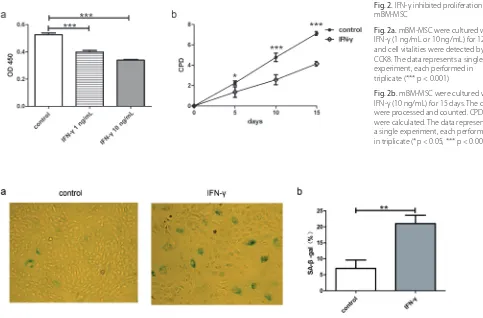
We tested the effects of IFN-γ on mBM-MSC prolifera-tion. One ng/mL and 10 ng/mL IFN-γ were used to stim-ulate mBM-MSC. After 120 h, CCK8 were used to test cell vitalities. The cell vitalities were significantly reduced by either concentration of IFN-γ (Fig. 2a, p < 0.001), and the OD in the 10 ng/mL IFN-γ group were lower than the OD in the 1 ng/mL IFN-γ group. Because of that, we used 10 ng/mL IFN-γ to treat mBM-MSC for 15 days and tested its effect on mBM-MSC proliferation. In the IFN-γ group, proliferation of mBM-MSC was reduced signifi-cantly after 5, 10 or 15 days’ treatment (Fig. 2b, p < 0.05 after 5 days, p < 0.001 after 10 and 15 days).
IFN-γ induce mBM-MSC SA-β-gal and ROS
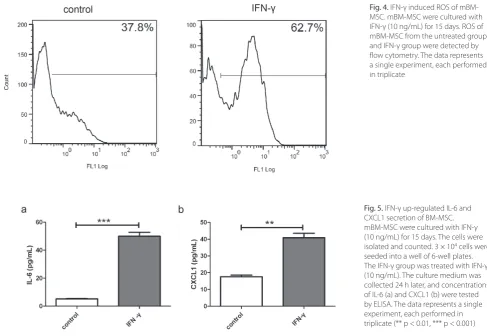
As the proliferation potentials of mBM-MSC were re-duced by IFN-γ, we supposed that IFN-γ might be an in-ducer of mBM-MSC senescence. Therefore, we stained the β-gal on the mBM-MSC. The percentages of SA-β-gal positive cells in the IFN-γ group were almost 3 times that of the control group (Figs. 3a and b, p < 0.01). ROS were suggested to be important for premature senescence. As expected, ROS were up-regulated in mBM-MSC after 15 days of IFN-γ treatment (Fig. 4).
IFN-γ up-regulated IL-6 and CXCL1
production of mBM-MSC
We tested the IL-6 and CXCL1 production after 15 days of IFN-γ treatment. IL-6 secretions were sig-nificantly up-regulated after 15 days of IFN-γ treat-ment (Fig. 5a, 5.21 ± 0.27 pg/mL in the control group vs 49.97 ± 2.86 pg/mL in the IFN-γ group, p < 0.001). Addi-tionally, the CXCL1 secretions were up-regulated signifi-cantly (Fig. 5b, 17.61 ± 1.00 pg/mL in the control group vs 40.92 ± 2.64 pg/mL in the IFN-γ group, p < 0.01).
Fig. 2. IFN-γ inhibited proliferation of mBM-MSC
Fig. 2a. mBM-MSC were cultured with IFN-γ (1 ng/mL or 10 ng/mL) for 120 h, and cell vitalities were detected by CCK8. The data represents a single experiment, each performed in triplicate (*** p < 0.001)
Fig. 2b. mBM-MSC were cultured with IFN-γ (10 ng/mL) for 15 days. The cells were processedand counted. CPD were calculated. The data represents a single experiment, each performed in triplicate (*p < 0.05, *** p < 0.001)
Fig. 3. IFN-γ induced SA-β-gal of mBM-MSC. mBM-MSC were cultured with IFN-γ (10 ng/mL) for 15 days
Fig. 2a. Cells of none-confluent state were washed with PBS, fixed with 4% formaldehyde and stained in a staining solution containing 1 mg/mL 5-bromo-4-chloro-3-indolyl-β-D-galactoside for 16 h. Images were taken by LEICA DMIL LED microscope
Discussion
Mesenchymal stem cells are populations of stem cells possessing great proliferation potential, while senescence limits its proliferate and function. Many studies have been done on replicative senescence of MSC before our research, and our data shows that the Th1 cytokine IFN-γ accelerates senescence-like characteristics in mBM-MSC.
Consistent with other research, IFN-γ reduced the pro-liferations of mBM-MSC in our study. A long-term in vitro culture with IFN-γ induced SA-β-gal expression of MSC, suggesting the role of IFN-γ on the dysfunctions of many organs in aging. Studies have shown that MSC could regulate the immune response, especially inhibit-ing the Th1 response.10,17,18 It has been suggested that the
IFN-γ produced by T/B/NK cells could activate the im-mune modulation effects of MSC, and MSC inhibits the proliferation and IFN-γ production of these cells.10,18 Our
data suggests high concentrations of IFN-γ could be an inducer of MSC senescence, and inflammatory cytokines could cause damage to MSC. It has been suggested that IFN-γ is an inducer of reactive oxygen species in endo-thelial cells, hepatocytes and melanocytes.15,16,19 In our
study, reactive oxygen species rose after 15 days of IFN-γ treatment. ROS are thought to be one of the major factors that cause cellular DNA damage, and subsequent senes-cence.20 Thus, we suppose that IFN-γ induced ROS could
be the cause of senescence of mBM-MSC.
Cellular senescence induces many cytokines, which was suggested to be the senescence-associated secretory phe-notype (SASP).21 SASP is controlled by NF-κB, and could
exacerbate cellular senescence.22 IL-6 and CXCL1 were
important cytokines in the SASP of mice.23
The up-regu-lation of IL-6 and CXCL1 by IFN-γ were consistent with the inducing senescence of MSC by IFN-γ. IL-6 is an im-portant cytokine that regulates immune cells like B cells, Th17 cells and monocytes, so the effects of increasing IL-6 production of MSC might affect the immune responses.24
Additionally, CXCL1 is related to immune response and angiogenesis.25,26 Thus, 15 days of IFN-γ treatment might
change the immune modulation and supportive effect of mBM-MSC. Other cytokines might also be influenced, and further studies are needed.
In summary, we have discovered that IFN-γ could in-duce senescence-like characteristics of mBM-MSC, sug-gesting a novel target for anti-aging therapy. Further stud-ies will focus on the change of differentiation, immune modulation and supportive potential of MSC, and the mechanisms of IFN-γ induced MSC senescence.
References
1. Caplan AI. Mesenchymal stem cells. J Orthop Res. 1991;9:641–650. 2. Jiang Y, Jahagirdar BN, Reinhardt RL, et al. Pluripotency of
mesenchy-mal stem cells derived from adult marrow. Nature. 2002;418:41–49. Fig. 4. IFN-γ induced ROS of mBM-MSC. mBM-MSC were cultured with IFN-γ (10 ng/mL) for 15 days. ROS of mBM-MSC from the untreated group and IFN-γ group were detected by flow cytometry. The data represents a single experiment, each performed in triplicate
Fig. 5. IFN-γ up-regulated IL-6 and CXCL1 secretion of BM-MSC. mBM-MSC were cultured with IFN-γ (10 ng/mL) for 15 days. The cells were isolated and counted. 3 × 10⁴ cells were
3. Wang Y, Zhang Z, Chi Y, et al. Long-term cultured mesenchymal stem cells frequently develop genomic mutations but do not undergo malignant transformation. Cell Death Dis. 2013;4:e950. 4. Wagner W, Horn P, Castoldi M, et al. Replicative senescence of
mes-enchymal stem cells: A continuous and organized process. PLoS One. 2008;3:e2213.
5. Choo KB, Tai L, Hymavathee KS, et al. Oxidative stress-induced pre-mature senescence in Wharton’s jelly-derived mesenchymal stem cells. Int J Med Sci. 2014;11:1201–1207.
6. Estrada JC, Torres Y, Benguria A, et al. Human mesenchymal stem cell-replicative senescence and oxidative stress are closely linked to aneuploidy. Cell Death Dis. 2013;4:e691.
7. Bonab MM, Alimoghaddam K, Talebian F, Ghaffari SH, Ghavamza-deh A, Nikbin B. Aging of mesenchymal stem cell in vitro. BMC Cell Biol. 2006;7:14.
8. Kasper G, Mao L, Geissler S, et al. Insights into mesenchymal stem cell aging: Involvement of antioxidant defense and actin cytoskel-eton. Stem Cells. 2009;27:1288–1297.
9. Nauta AJ, Fibbe WE: Immunomodulatory properties of mesenchy-mal stromesenchy-mal cells. Blood. 2007;110:3499–3506.
10. Krampera M, Cosmi L, Angeli R, et al. Role for interferon-gamma in the immunomodulatory activity of human bone marrow mesen-chymal stem cells. Stem Cells. 2006;24:386–398.
11. Croitoru-Lamoury J, Lamoury FM, Caristo M, et al. Interferon-gam-ma regulates the proliferation and differentiation of mesenchyInterferon-gam-mal stem cells via activation of indoleamine 2,3 dioxygenase (ido). PLoS One. 2011;6:e14698.
12. Ren G, Zhao X, Zhang L, et al. Inflammatory cytokine-induced intercellular adhesion molecule-1 and vascular cell adhesion mol-ecule-1 in mesenchymal stem cells are critical for immunosuppres-sion. J Immunol. 2010;184:2321–2328.
13. Ren G, Zhang L, Zhao X, et al. Mesenchymal stem cell-mediated immunosuppression occurs via concerted action of chemokines and nitric oxide. Cell Stem Cell. 2008;2:141–150.
14. Braumuller H, Wieder T, Brenner E, et al. T-helper-1-cell cytokines drive cancer into senescence. Nature. 2013;494:361–365.
15. Wang S, Zhou M, Lin F, et al. Interferon-gamma induces senescence in normal human melanocytes. PLoS One. 2014;9:e93232.
16. Kim KS, Kang KW, Seu YB, Baek SH, Kim JR. Interferon-gamma induc-es cellular seninduc-escence through p53-dependent DNA damage signal-ing in human endothelial cells. Mech Ageing Dev. 2009;130:179–188. 17. Chen K, Wang D, Du WT, et al. Human umbilical cord mesenchymal
stem cells huc-mscs exert immunosuppressive activities through a pge2-dependent mechanism. Clin Immunol. 2010;135:448–458. 18. Aggarwal S, Pittenger MF. Human mesenchymal stem cells
modu-late allogeneic immune cell responses. Blood. 2005;105:1815–1822. 19. Watanabe Y, Suzuki O, Haruyama T, Akaike T. Interferon-gamma
induces reactive oxygen species and endoplasmic reticulum stress at the hepatic apoptosis. J Cell Biochem. 2003;89:244–253. 20. Weyemi U, Lagente-Chevallier O, Boufraqech M, et al.
Ros-gener-ating nadph oxidase nox4 is a critical mediator in oncogenic h-ras-induced DNA damage and subsequent senescence. Oncogene. 2012;31:1117–1129.
21. Coppe JP, Patil CK, Rodier F, et al. Senescence-associated secreto-ry phenotypes reveal cell-nonautonomous functions of oncogenic ras and the p53 tumor suppressor. PLoS Biol. 2008;6:2853–2868. 22. Chien Y, Scuoppo C, Wang X, et al. Control of the
senescence-asso-ciated secretory phenotype by nf-kappab promotes senescence and enhances chemosensitivity. Genes Dev. 2011;25:2125–2136. 23. Coppe JP, Patil CK, Rodier F, et al. A human-like
senescence-associ-ated secretory phenotype is conserved in mouse cells dependent on physiological oxygen. PLoS One. 2010;5:e9188.
24. Chomarat P, Banchereau J, Davoust J, Palucka AK. IL-6 switches the differentiation of monocytes from dendritic cells to macrophages.
Nat Immunol. 2000;1:510–514.
25. Ritzman AM, Hughes-Hanks JM, Blaho VA, Wax LE, Mitchell WJ, Brown CR. The chemokine receptor cxcr2 ligand kc (cxcl1) mediates neutrophil recruitment and is critical for development of experi-mental lyme arthritis and carditis. Infect Immun. 2010;78:4593–4600. 26. Dhawan P, Richmond A. Role of cxcl1 in tumorigenesis of