Alicja E. Grzegorzewska
1, Aniela Ratajewska
2, Agnieszka Wiesiołowska
3Echocardiographic Indices in Hemodialysis Patients
in a Six-Month Observation
Wskaźniki echokardiograficzne u hemodializowanych chorych
w obserwacji 6-miesięcznej
1 Department and Clinic of Nephrology, Transplantology and Internal Diseases, Poznan University of Medical
Sciences, Poznań, Poland
2 International Dialysis Center, Rawicz Branch, Cardiology Outpatient Clinic of the Rawicz District Hospital,
Rawicz, Poland
3 Department of Computer Science and Statistics, Poznan University of Medical Sciences, Poland
Abstract
Background. Cardiovascular diseases are the leading cause of premature mortality and disability of dialyzed patients. Generally, inthe course of hemodialysis (HD) treatment, the prevalence and severity of left ventricular dysfunction (LVD) increase.
Objectives. To investigate changes in echocardiographic parameters that could be early signs of the development or further deterioration of LVD in HD patients.
Material and Methods. Echocardiography (two-dimensional, pulsed wave, continuous wave and tissue Doppler) was performed on 48 patients before and after HD sessions at the beginning of the study and after 6 months of HD treatment using Pro-Sound 4000 (Aloka, Japan). HD adequacy and laboratory parameters were evaluated. Unfavorable differences in the echocardiographic parameters of patients without changes in classification of left ventricular function over the study period were assumed to be early predictors for the development or deteriora-tion of LVD.
Results. In 31 patients, left ventricular function remained stable, in 12 it deteriorated and in 5it improved. In the patients with stable function, left atrial (LA) diameter and area were greater before HD sessions, and the differences induced by HD sessions in left atrial and right atrial (RA) diameters were significantly greater at the end of the study than at the beginning (p < 0.05). The ultrafiltration volume (2155 ± 926 mL vs. 2177 ± 952 mL, p = 0.666) and inferior vena cava diameter (22.8 ± 2.6 mm vs. 22.7 ± 2.9 mm, p = 0.764) were not different at the beginning and at the end of the study. Differences in LA and RA diameters (among others) were also noted in 12 patientswho showed a worsening in their left ventricular function classification.
Conclusions. Increasing atrial diameters under comparable hypervolemic conditions before HD sessions may be considered an early predictive sign of deterioration of left ventricular function in HD patients (Adv Clin Exp Med 2011, 20, 4, 431–440).
Key words: echocardiography,hemodialysis adequacy, left ventricular dysfunction, residual urine volume, ultra-filtration.
Streszczenie
Wprowadzenie. Choroby sercowo-naczyniowe są główną przyczyną przedwczesnej śmiertelności i inwalidztwa dializowanych chorych. W czasie leczenia hemodializą (HD) zwiększa się rozpowszechnienie i ciężkość zaburzeń czynności mięśnia sercowego lewej komory (LVD).
Cel pracy. Pokazanie zmian w parametrach echokardiograficznych, które mogą być wczesnymi oznakami rozwoju lub progresji istniejącej LVD u chorych leczonych powtarzaną HD.
Materiał i metody. Badanie echokardiograficzne (dwuwymiarowe, fali pulsacyjnej, fali ciągłej, Dopplera tkanko-wego) wykonano u 48 chorych przed zabiegiem HD i po nim na początku i końcu (po 6 miesiącach leczenia HD) okresu badawczego, używając aparatu Pro-Sound 4000 (Aloka, Japonia). Oceniano adekwatność HD i wskaźniki laboratoryjne. Niekorzystne zmiany w parametrach echokardiograficznych, występujące u chorych bez towarzy-szących zmian w klasyfikacji czynności lewej komory w ciągu okresu badawczego, uznano za wczesne predyktory rozwoju lub pogorszenia LVD.
Adv Clin Exp Med 2011, 20, 4, 431–440 ISSN 1230-025X
ORIGINAL PAPERS
Cardiovascular diseases are the leading cause of premature mortality and disability among dia-lyzed patients. Generally, in the course of hemo-dialysis (HD) treatment, both the prevalence and the severity of left ventricular dysfunction (LVD) increase. This progression results from the coex-istence of multiple metabolic disorders, cardio-vascular risk factors and the HD treatment itself [1]. Non-invasive methods for assessing the risk of LVD progression in HD patients are still being sought. Since it was first developed, echocardiog-raphy has been used to evaluate left ventricular systolic function [2]. The introduction of tissue Doppler echocardiography (TDE), which as-sesses intracardiac blood flow and tissue motion, made echocardiography useful for evaluating left ventricular diastolic function as well. Periodic as-sessment of both these cardiac functions provides valuable prognostic information [3–6].
Because cardiovascular events remain the pri-mary cause of mortality in HD patients, the cur-rent study was undertaken to investigate changes in echocardiographic parameters for left ventric-ular systolic and diastolic function that could be early signs of the development or further deterio-ration of LVD during HD treatment.
Material and Methods
The Patients
and the Study Protocol
After having obtained written informed con-sent, 56 stable patients with stage 5 chronic kid-ney disease being treated with intermittent HD at a single dialysis center (in Rawicz, Poland) were enrolled as participants in a prospective 6-month study. Patients with cancer and those who had completed treatment of a neoplasm within the previous 5 years had been excluded. Another ex-clusion criterion was any condition making
mea-surement of cardiac flows related to atrial function impossible (for example persistent atrial fibrilla-tion or the use of a cardiac stimulator). The initial clinical data of these 56 patients were described in a previous publication [7].
The study lasted for 6 months, during which particular attention was paid to the application of adequate HD treatment for all the patients. A 6-month study period was chosen because dur-ing this relatively short time span HD-induced changes in the patients’ echocardiographic pa-rameters may occur, but would not be so advanced as to justify a diagnosis of a new case of LVD or a change in classification of existing LVD (systolic LVD; mild, moderate, severe diastolic LVD). Be-cause there is no doubt that cardiac damage pro-gresses during HD treatment, unfavorable changes in echocardiographic parameters occurring dur-ing the 6-month study period could be considered early echocardiographic predictors for the devel-opment of LVD or for the further deterioration of existing LVD. In cases where the differences in the echocardiographic parameters resulted in a change of classification of ventricular function, a separate analysis of echocardiographic parameters was planned for patients showing stable left ventricular function and those whose left ventricular function had deteriorated in the examined period.
The medical histories of all the patients were carefully evaluated at the beginning and at the end of the study for signs of cardiac disease or conges-tive heart failure (CHF). Valvular disease was clas-sified using the guidelines of the European Society of Cardiology [8]. Patients with CHF were evalu-ated using the New York Heart Association’s func-tional classification of CHF patients [9, 10]. Atten-tion was also given to arterial hypertension and treatment with angiotensin converting enzyme inhibitors (ACEI), angiotensin receptor blockers (ARB), and b-adrenergic receptor blockers.
Echocardiography was performed on each patient at the beginning of the study before and after an HD session (echoes 1 and 2, respectively). After 6 months of adequate HD treatment, 2 more
Wyniki. U 31 chorych czynność lewej komory pozostała niezmieniona, u 12 się pogorszyła, a u 5 poprawiła. U cho-rych ze stabilną czynnością wymiar lewego przedsionka (LA) i pole LA były większe przed zabiegiem HD, a wywo-łane zabiegiem HD różnice w wymiarach LA i prawego przedsionka (RA) były znamiennie (p < 0,05) większe na końcu w porównaniu z odpowiednimi wynikami na początku okresu badawczego. Objętość ultrafiltracji (2155 ±
926 mL vs 2177 ± 952 mL, p = 0,666) i wymiar żyły głównej dolnej (22,8 ± 2.6 mm vs 22,7 ± 2,9 mm, p = 0,764) nie różniły się na początku i końcu badania. Różnice w wymiarach LA i RA (wśród innych) wykazano także u 12 chorych, u których stwierdzono pogorszenie w klasyfikacji czynności lewej komory.
Wnioski. Zwiększające się wymiary przedsionków serca przed zabiegiem HD w warunkach porównywalnej hiper-wolemii można rozważać jako wczesne wskaźniki pogarszania się czynności lewej komory mięśnia sercowego u chorych leczonych HD (Adv Clin Exp Med 2011, 20, 4, 431–440).
echocardiographic examinations were performed, again before and after an HD session (echoes 3 and 4, respectively).
One patient gave up and 7 died before the end of the study. The overall 6-month mortality was 14.6%. The remaining 48 stable patients were in-cluded in the analysis.
In the examined group (n = 48) there were 27 men and 21 women at the age of 63.6 ± 15.1 years (mean ± SD). The median HD vintage was 40 months (range: 5–154 months); the body mass index was 26.7 ± 5.1 kg/m2 (mean ± SD); the me-dian daily residual urine volume (pre-dialysis) was 200 mL/day (range: 0–2000 mL/day). Causes of end-stage renal disease (ESRD) included diabetic neph-ropathy (n = 14), chronic tubulointerstitial nephritis (n = 12), hypertensive nephropathy (n = 6), chronic glomerulonephritis (n = 5), obstructive nephropathy (n = 3), polycystic kidney disease (n = 2) and amyloi-dosis in the course of rheumatoid arthritis (n = 1). In 5 cases the cause of ESRD remained unknown. 5 patients were on the transplant waiting list.
Hemodialysis Management
All the patients underwent three HD sessions per week, each lasting at least four hours. Frese-nius 4008 S dialysis machines and polysulfone-based membranes were used, regulated to a blood flow rate of 200–300 mL/min and a dialysate flow rate between 500 and 800 mL/min. Dialyzers were not reused. For vascular access arterio-venous fis-tula were used in 42 cases (87.5%) and permanent catheters in seven cases (12.5%) at the beginning of the study; later in the study, arterio-venous fis-tula were successfully created and used in 4 addi-tional patients. The ultrafiltration volume (UFV) depended on the difference between pre-HD body weight and dry body mass, which was estimated based on clinical signs of hydration and blood pres-sure behavior during previous HD sessions. When post-HD body weight equaled dry body mass ± 1%, it was considered effective dehydration.
On-line clearance normalized to distribution volume (Kt/V) was measured during every HD session using the conductivity method for an im-mediate check of HD adequacy. Additionally, each patient’s urea reduction ratio (URR) and urea Kt/V were calculated every month. The second genera-tion formula of Daugirdas was used for urea Kt/V calculation [11, 12].
Routine laboratory tests for HD patients (blood count, C-reactive protein, total calcium, phospho-rus, intact parathyroid hormone, uric acid) were periodically performed.
Echocardiographic Study
As noted above, echocardiography was per-formed on each patient before and after an HD session at the beginning of the study and again af-ter 6 months. The echocardiographic images were recorded 20–30 minutes prior to commencing HD and 20–40 minutes after the end of the session.
All echo measurements were made by a sin-gle echocardiographer (A.R.) with the Pro-Sound 4000 device (Aloka, Japan), using two-dimensional projection (2D), pulsed wave Doppler (PW), con-tinuous wave Doppler (CW) and tissue Doppler echocardiography(TDE).
Measurements in 2D projection included in-ferior the vena cava (IVC) diameter, LA diameter, LA area, left ventricular end-diastolic diameter (LVEDd), left ventricular end-systolic diameter (LVESd) and right atrium (RA) area. The left ven-tricular ejection fraction (LVEF) was evaluated us-ing Simpson’s method [2]. The PW technique was used to measure the velocity of transmitral blood flow (the protodiastolic E wave, end-diastolic A wave and E/A ratio), the deceleration time (DT), the isovolumetric relaxation time (IVRT), atrial reversal (AR), diastolic superior pulmonary vein velocity (D), systolic superior pulmonary vein ve-locity (S) and S/D ratio [13–15]. TDE, performed with the Doppler chamber placed at the septal border of the mitral annulus, was used to evaluate the mitral annular motion by measuring the veloc-ity of both the early diastolic E´ wave and the late diastolic A´ wave. The E´/A´ ratio and the ratio of the pulsed Doppler E wave and the tissue Doppler E´ wave (E/E`) were calculated [4, 5, 16]. During each examination echocardiography parameters were taken two or more times, depending on the quality of Doppler recordings, and the measure-ments were averaged. Pre- and post-HD echocar-diography parameters (registered and calculated) were compared and the differences between them were statistically evaluated.
vari-ability of 3.8 ± 6.3%. All the data were recorded on VHS videotape and stored.
Systolic dysfunction was diagnosed when the LVEF was < 50% [18]. Echocardiographic crite-ria for a diagnosis of diastolic dysfunction were elaborated based on the recommendations of the European Society of Cardiology [18, 19] and on the Canadian consensus guidelines [20]. A cru-cial parameter for a diagnosis of diastolic LVD was the E/E` ratio [4–6]. Three groups of diastolic dysfunction were distinguished using a complex analysis of transmitral PW and TDE parameters: mild (an abnormal relaxation pattern), moderate (a pseudonormal pattern) and severe (a restrictive pattern).
Statistical Analysis
The normality of the distribution of variables was checked using the Shapiro-Wilk test. Descrip-tive statistics are presented as percentages for categorical (nominal) variables, as means with standard deviation for normally distributed con-tinuous variables, or as medians with lower and upper quartiles for non-normally distributed con-tinuous variables. Comparisons of results were per-formed using the Student’s t-test for paired data if the distribution of variables was normal or by the Wilcoxon test otherwise. The prevalence of vari-ables was assessed by the chi-square test. All tests were analyzed at the significance level α = 0.05. Statistical analysis was performed using Statistica PL 8.0 software (StatSoft).
This study was reviewed and approved by the Institutional Review Board of Poznań University of Medical Sciences.
Results
Seven of the 56 HD patients died before the end of the study. Causes of death included CHF (n = 3), malignant neoplastic disease (n = 2), ce-rebral stroke (n = 1) and a sudden death (n = 1). In the echocardiograms before the HD session at the beginning of the study, the deceased patients showed significantly higher LA and RA dimen-sions as compared to the respective results for the patients who survived (Table 1). One other patient left the study after being diagnosed with malignant neoplastic disease. Consequently, the final analysis included 48 patients.
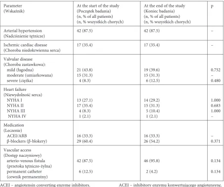
Ischemic heart disease was diagnosed in 17/48 (35.4%) cases. There were 5 patients (10.4%) who had undergone myocardial infarction (inferior infarction – 3 cases, inferior-anterior infarction – 1 case, anterior infarction – 1 case).
Percutane-ous or surgical revascularization of coronary ar-teries had been performed on 2 patients (4.2%). Percutaneous ablation had been performed on 1 patient (2.1%) due to recurrent atrioventricular nodal reentrant tachycardia [21] and due to ven-tricular preexcitation in another (2.1%). Arterial hypertension was shown in 42 patients (87.5%). The frequency of these conditions did not change during the study period. Other clinical data are shown in Table 2.
The HD adequacy was satisfactory (Table 3). The UFV was 2115 ± 1010 mL at the beginning of the study and 2275 ± 1014 mL at the end of the study (not a significant difference). Laboratory parameters did not change significantly (data not shown).
There was no significant difference in the fre-quency of systolic LVD, which was about 6% both at the beginning and at the end of the study. Dia-stolic LVD was noted in 91.7% of the patients dur-ing the entire study. There were no significant dif-ferences over the 6-month period in the frequency of patients with different degrees of diastolic LVD in the echocardiographicstudies performed before HD sessions (echo 1 vs. echo 3) or after HD ses-sions (echo 2 vs. echo 4). However, even with the significantly reduced preload after HD sessions, mild diastolic LVD was diagnosed at the begin-ning of the study (echo 2) in 60.4% of the patients, moderate in 29.2% and severe in 2.1%, but at the end of the study (echo 4) mild diastolic LVD was diagnosed in54.2% of patients, moderate in 33.3% and severe in 4.2%. In 5 patients left ventricular function improved; in 31 patients there was no change in classification of diastolic LVD, whereas in 12 patients a more advanced degree of diastolic LVD was found.
Table 1. Values – median, lower and upper quartiles) of echocardiographic parameters obtained before a hemodialysis ses-sion in patients who survived the six-month study and in those who died before the end of the study
Tabela 1. Wartości – mediana, kwartyle górny i dolny) parametrów echokardiograficznych uzyskanych przed zabiegiem
hemodializy u chorych, którzy przeżyli okres 6-miesięcznego badania i u tych, którzy zmarli przed zakończeniem badania
Parameter
(Parametr) Patients who survived(Chorzy, którzy przeżyli) n = 49
Patients who died (Chorzy, którzy zmarli) n = 7
p
LVEDd – mm
LVESd – mm
LVEF – %
LA – mm
RA area – cm2
Pole RA – cm2
LA area – cm2
Pole LA – cm2
IVC diameter – mm Wymiar IVC – mm E – m/s
A – m/s
E/A
DT – ms
IVRT – ms
S – m/s
D – m/s
Ar – m/s
S/D
E` – m/s
A` – m/s
E`/A` E/E` 49.0 46.0–53.0 34.0 30.0–38.0 65.0 58.0–72.0 42.0 37.0–44.0 15.0 13.1–17.5 19.4 16.3–21.8 23.0 21.0–24.0 0.64 0.70–1.06 0.83 0.68–1.07 0.94 0.76–1.35 224.0 176.0–278.0 85.0 75.0–94.0 0.65 0.55–0.78 0.64 0.50–0.84 0.42 0.36–0.53 1.03 0.78–1.36 0.06 0.05–0.07 0.08 0.07–0.10 0.67 0.58–0.80 13.50 10.22–18.40 52.0 49.5–56.0 38.0 34.5–47.5 70.0 45.5–70.5 48.0 45.0–49.5 22.6 18.4–27. 3 22.2 20.2–24.6 23.0 21.0–25.0 1.05 0.87–1.10 0.91 0.76–0.99 1.04 0.95–1.38 251.0 216.5–291.0 89.0 74.5–96.0 0.76 0.53–0.88 0.58 0.49–0.70 0.40 0.38–0.46 1.10 0.89–1.33 0.06 0.03–0.07 0.10 0.05–0.11 0.60 0.56–0.69 17.50 12.43–35.17 0.206 0.112 0.664 0.004 0.001 0.076 0.901 0.215 0.795 0.249 0.450 0.654 0.560 0.611 0.795 0.872 0.269 0.940 0.413 0.134
A – mitral late diastolic velocity.
A` – mitral annular late diastolic velocity. Ar – atrial reversal.
D – diastolic pulmonary vein velocity. DT – deceleration time.
E – mitral early diastolic velocity.
E` – mitral annular early diastolic velocity. IVC – inferior vena cava.
IVRT – isovolumetric relaxation time. LA – left atrium.
LVEDd – left ventricular end-diastolic diameter. LVESd – left ventricular end-systolic diameter. LVEF – left ventricular ejection fraction. RA – right atrium.
S – systolic pulmonary vein velocity.
A – prędkość maksymalna napływu mitralnego w czasie skurczu przedsionka.
A` – prędkość ruchu pierścienia mitralnego po skurczu przedsionka.
Ar – faza przedsionkowa przepływu wstecznego w żyle płucnej.
D – faza rozkurczowa przepływu w żyle płucnej. DT – czas deceleracji fali wczesnego napływu mitralnego. E – prędkość maksymalna fali wczesnego napływu mitral-nego.
E` – wczesnorozkurczowa maksymalna prędkość ruchu pierścienia mitralnego.
Table 2. Comparison of clinical data of 48 patients participating in the study
Tabela 2. Porównanie danych klinicznych 48 chorych uczestniczących w badaniu
Parameter
(Wskaźnik) At the start of the study (Początek badania) (n, % of all patients) (n, % wszystkich chorych)
At the end of the study (Koniec badania) (n, % of all patients) (n, % wszystkich chorych)
p
Arterial hypertension
(Nadciśnienie tętnicze) 42 (87.5) 42 (87.5) –
Ischemic cardiac disease
(Choroba niedokrwienna serca) 17 (35.4) 17 (35.4) – Valvular disease
(Choroba zastawkowa): mild (łagodna)
moderate (umiarkowana) severe (ciężka)
21 (43.8) 15 (31.3) 4 (8.3)
19 (39.6) 15 (31.3) 6 (12.5)
0.752 – 0.480 Heart failure
(Niewydolność serca) NYHA I
NYHA II NYHA III NYHA IV
13 (27.1) 17 (35.4) 4 (8.3) 1 (2.1)
14 (29.2) 15 (31.3) 5 (10.4) 1 (2.1)
1.000 0.683 1.000 – Medication
(Leczenie) ACEI/ARB
β-blockers (β-blokery) 16 (33.3)29 (60.4) 16 (33.3)26 (54.2) –0.371 Vascular access
(Dostęp naczyniowy) arterio-venous fistula (przetoka tętniczo-żylna) permanent catheter (cewnik permanentny)
42 (87.5)
6 (12.5)
46 (95.8)
2 (4.2)
0.134
0.134
ACEI – angiotensin converting enzyme inhibitors. ARB – angiotensin receptor blockers.
NYHA – New York Heart Association.
ACEI – inhibitory enzymu konwertującego angiotensynę. ARB – blokery receptora angiotensyny.
NYHA – Nowojorskie Towarzystwo Kardiologiczne.
Table 3. Hemodialysis adequacy parameters (mean ± standard deviation) during the 6-month study
Tabela 3. Wskaźniki adekwatności hemodializy (średnia ± odchylenie standardowe) podczas 6-miesięcznego badania
Parameter
(Wskaźnik) Month (Miesiąc) The entire study (Całe badanie)
1 2 3 4 5 6
URR 0.74 ± 0.05 0.75 ± 0.04 0.75 ± 0.04 0.75 ± 0.04 0.73 ± 0.04 0.76 ± 0.03 0.75 ± 0.03 Urea Kt/V
(Kt/V mocznika) 1.56 ± 0.26 1.63 ± 0.22 1.65 ± 0.20 1.64 ± 0.20 1.55 ± 0.17 1.62 ± 0.17 1.62 ± 0.17 Online Kt/V
(Kt/V w czasie zabiegu)
1.30 ± 0.17 1.32 ± 0.15 1.36 ± 0.14 1.39 ± 0.13 1.41 ± 0.12 1.42 ± 0.13 1.37 ± 0.12
Kt/V – clearance normalized to distribution volume.
URR – urea reduction ratio. Kt/V – klirens normalizowany względem objętości dys-trybucji. URR – wskaźnik wydializowania mocznika.
cava diameter was similar at the beginning and at the end of the study (22.8 ± 2.6 mm vs. 22.7 ± 2.9 mm, p = 0.764).
num-Table 4. Significant differences (median, lower and upper quartiles) in echocardiographic parameters before (echo 1 and echo 3) and after (echo 2 and echo 4) hemodialysis session in patients whose left ventricular diastolic function category did not change during the 6-month study (n = 31)
Tabela 4. Znamienne różnice (mediana, kwartyle górny i dolny) w parametrach echokardiograficznych przed zabiegiem
hemodializy (echo 1 i echo 3) i po nim (echo 2 i echo 4) u chorych, u których klasyfikacja czynności rozkurczowej lewej komory mięśnia sercowego nie zmieniła się w czasie 6-miesięcznego badania (n = 31)
Echocardiographic parameter
(Wskaźnik echokar-diograficzny)
Difference between echocardiographic parameters
(Różnica między wskaźnikami echokardiograficznymi) Difference between echo 1 and echo 2 vs. difference between echo 3 and echo 4 (Różnica między echo 1 i echo 2 vs różnica między echo 3 i echo 4)
in echo 1 and echo 3
(w echo 1 i echo 3) in echo 2 and echo 4(w echo 2 i echo 4)
result of the dif-ference (wynik różnicy)
p result of the dif-ference (wynik różnicy)
P result of the dif-ference (wynik różnicy)
p
Two-dimensional presentation (Prezentacja dwuwymiarowa) LA – mm –2.0
–5.0 – 0.00 0.002551 –1.0 –2.5 – 1.0 0.075428 –1.0 –4.0 – 1.0 0.013580 LA area – cm2)
(Pole LA – cm2) –1.69 –4.12 – 0.29 0.012819 –0.04 –1.85 – 1.89 0.968736 –1.18 –4.30 – 0.80 0.028178
RA area – cm2
(Pole RA – cm2) –1.48 –2.61 – 0.38 0.065465 0.02 –0.87 – 2.07 0.798912 –1.44 –2.43 – 0.91 0.039626
LA – left atrium. LA – lewy przedsionek. RA – right atrium. RA – prawy przedsionek.
ber of patients theser findings were not statistically significant for LA area (Table 5).
Discussion
As expected, in the entire group of 48 patients, in whom HD adequacy was in accordance with Eu-ropean Renal Best Practice [22], there were no sig-nificant differences in the prevalence of LVD dur-ing the 6-month study period, and the entire group of patients was considered stable. However, at the end of the study the numbers of patients with mod-erate and severe diastolic LVD, NYHA functional class III and severe valvular disease were slightly higher than at the study beginning (although not significantly so), because the number of patients with mild pathology decreased. Moreover, the di-vision of the patients into subgroups whose dia-stolic LVD classification had changed and those whose classification had not changed showed that even patients whose degree of diastolic LVD did not deteriorate had greater LA dimensions before HD sessions, and the HD-induced differences in LA and RA diameters were significantly greater at the end of the study compared to the respective results at the beginning.
Increasing atrial diameters before HD sessions are usually attributed to increasing preload from
Table 5. Significant differences (median, lower and upper quartiles) in echocardiographic parameters before (echo 1 and echo 3) and after (echo 2 and echo 4) hemodialysis session in patients whose left ventricular diastolic function category dete-riorated during the 6-month study (n = 12)
Tabela 5. Znamienne różnice (mediana, kwartyle górny i dolny) w parametrach echokardiograficznych przed zabiegiem
hemodializy (echo 1 i echo 3) i po nim (echo 2 i echo 4) u chorych, u których klasyfikacja czynności rozkurczowej lewej komory mięśnia sercowego pogorszyła się w czasie 6-miesięcznego badania (n = 12)
Echocardiographic parameter
(Wskaźnik echokar-diograficzny)
Difference between echocardiographic parameters
(Różnica między wskaźnikami echokardiograficznymi) Difference between echo 1 and echo 2 vs. difference between echo 3 and echo 4 (Różnica między echo 1 i echo 2 vs różnica między echo 3 i echo 4)
in echo 1 and echo 3
(w echo 1 i echo 3) in echo 2 and echo 4(w echo 2 i echo 4)
result of the dif-ference (wynik różnicy)
p result of the dif-ference (wynik różnicy)
P result of the dif-ference (wynik różnicy)
p
Two-dimensional presentation (Prezentacja dwuwymiarowa) LA – mm –3.0
–5.0 – 2.00 0.049861 0.00 –2.25 – 2.0 0.953 –1.0 –5.0 – 0.00 0.097 RA area – cm2
(Pole RA – cm2) –3.06 –6.24 – –0.13 0.034 0.12 –1.41 – 1.22 0.182 –2.7–6.1 – –0.18 0.018
Pulsed Doppler (Doppler pulsacyjny) E – m/s –0.22
–0.38 – –0.14 < 0.001 –0.05–0.15 – 0.15 0.724 –0.16 –0.23 – –0.05 0.008
E/A –0.39
0.66– –0.32 0.002 0.03 –0.25 – 0.10 0.583 –0.36 –0.66 – –0.18 0.006 IVRT – ms 16.0
–1.25 – 21.0 0.062 0.0 –14.5 – 0.25 0.330 16.0 7.0 – 21.75 0.049861
S/D –0.01
–0.12 – 0.07 1.000 0.24 0.05 – 0.37 0.041 –0.17 –0.27 – –0.09 0.136 Pulsed/Tissue Doppler
(Doppler pulsacyjny/tkankowy)
E/E` –2.14
–6.47 – – 1.07 0.012 0.22 –4.55 – 2.83 0.530 –3.69 –5.26 – –0.4 0.041
only differences were higher LA and RA dimen-sions. As noted above, differences in LA and RA diameters (among others) were also observed in 12 patients whose left ventricular function classi-fication worsened during the study period. How-ever, the authors cannot say which of this group’s echocardiographic parameters deteriorated earlier and which later. A continuation of the study with repeated echocardiographic evaluations of patients with stable left ventricular function could be help-ful to verify the hypothesis that increases in atrial dimensions may be an early sign of deterioration of left ventricular function.
Zoccali et al. concluded that “periodic echocar-diographic studies of systolic function are useful for monitoring asymptomatic dialysis patients” [23]. The authors of the current study suggest that in
stratifying cardiovascular risk in HD patients, special attention should be paid to the repeated evaluation of LA dimensions immediately before HD sessions. It is worth noting that early signs of LVD progression can be detected by simple two-dimensional echocardiography when performed repeatedly at intervals of a few months.
that “in the subgroup [of patients] that had been on dialysis for > 3.7 years, randomization to high-flux dialysis was associated with lower risks of [...] cardiac deaths (RR 0.63; 95% CI 0.43 to 0.92; p = 0.016) compared with low-flux dialysis”. Those data and the results of the present study indicate a need to revise the standard modality of HD treat-ment and to replace it with cardioprotective tech-niques, possibly by implementing high-flux dialyz-ers and hemodiafiltration. Additionally, a greater emphasis on renal transplantation is advocated, as this form of renal replacement therapy provides
the best cardioprotection [25]. As Kaplan et al. [26] noted “Cardiovascular annual adjusted death rates were more than 7-fold higher after graft loss as compared to during transplantation (43.1 vs. 6.9 per 100 patient years)”.
In conclusion, systolic and diastolic LVD does not deteriorate significantly during the period of six months of adequate HD treatment in stable HD patients, but increasing atrial diameters under hypervolemic conditions may be considered an early predictive sign of progression of LVD.
References
[1] Foley RN, Parfrey PS, Sarnak MJ: Clinical epidemiology of cardiovascular disease in chronic renal disease. Am J Kidney Dis 1998, 32 Suppl 3, 112–119.
[2] Feigenbaum H: Echocardiography. Lea and Febiger, Philadelphia 1994, 5th ed.
[3] Rakowski H, Appleton C, Chan KL, Dumesnil JG, Honos G, Jue J, Koilpillai C, Lepage S, Martin RP, Mercier LA, O’Kelly B, Prieur T, Sanfilippo A, Sasson Z, Alvarez N, Pruitt R, Thompson C, Tomlinson C: Canadian consensus recommendations for the measurement and reporting of diastolic dysfunction by echocardiography: from the Investigators of Consensus on Diastolic Dysfunction by Echocardiography. J Am Soc Echocardiogr 1996, 9, 736–760.
[4] Dorosz JL, Lehmann KG, Stratton JR: Compartion of tissue Doppler and propagation velocity to invasive meas-ures for measuring left ventricular filling pressmeas-ures. Am J Cardiol 2005, 95, 1017–1020.
[5] Sengupta PP, Mohan JC, Mehta V, Arora R, Pandian NG, Khandheria BK: Accuracy and pitfalls of early diasto-lic motion of the mitral annulus for diagnosing constrictive pericarditis by tissue Doppler imaging. Am J Cardiol 2004, 93, 886–890.
[6] Sohn DW, Chai IH, Lee DJ, Kim HC, Kim HS, Oh BH, Lee MM, Park YB, Choi YS, Seo JD, Lee YW: Assessment of mitral annulus velocity by Doppler tissue imaging in the evaluation of left ventricular diastolic function. J Am Coll Cardiol 1997, 30, 474–480.
[7] Grzegorzewska AE, Ratajewska A, Wiesiołowska A: Factors influencing the tissue Doppler echocardiography indices of systolic and diastolic function of left myocardial ventricle in patients treated with intermittent hemodi-alysis. Adv Clin Exp Med 2010, 19, 469–480.
[8] Vahanian A, Baumgartner H, Bax J, Butchart E, Dion R, Filippatos G, Flachskampf F, Hall R, Iung B, Kasprzak J, Nataf P, Tornos P, Torracca L, Wenink A: Guidelines on the management of valvular heart disease. The Task Force on the Management of Valvular Heart Disease of the European Society of Cardiology. Eur Heart J 2007, 28, 230–268.
[9] Marantz PR, Tobin JN, Wassertheil-Smoller S, Steingart RM, Wexler JP, Budner N, Lense L, Wachspress J: The relationship between left ventricular systolic function and congestive heart failure diagnosed by clinical criteria. Circulation 1988, 77, 607–612.
[10] The Criteria Committee for the New York Heart Association. Nomenclature and Criteria for Diagnosis of Diseases of the Heart and Great Vessels. Little, Brown & Co, Boston 1994, 9th ed.
[11] Daugirdas JT, Schneditz D: Overestimation of hemodialysis dose (delta Kt/V) depends on dialysis efficiency (Kt/V) by regional blood flow and conventional 2-pool urea kinetic analyses. ASAIO J 1995, 41, 719–724.
[12] Daugirdas JT: Second generation logarithmic estimates of single-pool variable volume Kt/V: An analysis of error. J Am Soc Nephrol 1993, 4, 1205–1213.
[13] Bella JN, Palmieri V, Roman MJ, Liu JE, Welty TK, Lee ET, Fabsitz RR, Howard BV, Devereux RB: Mitral ratio of peak early to late diastolic filling velosity as a predictor of mortality in middle – aged elderly adults: the Strong Heart Study. Circulation 2002, 105, 1928–1933.
[14] Klein AL, Tajik AJ: Doppler assessment of pulmonary venous flow in healthy subjects and patients with heart disease. J Am Soc Echocardiogr 1991, 4, 379–392.
[15] Rossvoll O, Hatle LK: Pulmonary venous flow velocities recorded by transthoracic Doppler, relations to LV diastolic pressures. J Am Coll Cardiol 1993, 21, 1687–1696.
[16] Hashimoto I, Bhat AH, Li X, Jones M, Davies C, Swanson J, Schindera S, Sahn D: Tissue Doppler – derived myo-cardial acceleration for evaluation of left ventricular diastolic function. J Am Coll Cardiol 2004, 44, 1459–1466.
[17] Hsiao SH, Huang WC, Sy CL, Lin SK, Lee TY, Liu CP: Doppler tissue imaging and color M-mode flow propaga-tion velocity: are they really preload independent? J Am Soc Echocardiogr 2005, 18, 1277–1284.
[18] ESC Guidelines for the diagnosis and treatment of acute and chronic heart failure 2008. Eur Heart J 2008, 29, 2388–2442.
[20] Yamada H, Goh PP, Sun JP, Odabashian J, Garcia MJ, Thomas JD, Klein AL: Prevalence of left ventricular diastolic dysfunction by Doppler echocardiography: clinical application of the Canadian consensus guidelines. J Am Soc Echocardiogr 2002, 15, 1238–1244.
[21] Ratajewska A, Banachowicz W, Grzegorzewska AE: Recurrent atrioventricular nodal re-entrant tachycar-dia treated with percutaneous ablation in a 75-year old patient undergoing intermittent hemotachycar-dialysis. Int Urol Nephrol 2009, 41, 225–230.
[22] European best practice gudelines for hemodialysis; section II: hemodialysis adequacy. Nephrol Dial Transplant 2002, 17, 17–31.
[23] Zoccali C, Benedetto FA, Tripepi G, Mallamaci F, Rapisarda F, Seminara G, Bonanno G, Malatino LS: Left ventricular systolic function monitoring in asymptomatic dialiysis patients: a prospective cohort study. J Am Soc Nephrol 2006, 17, 1460–1465.
[24] Cheung AK, Levin NW, Greene T, Agodoa L, Bailey J, Beck G, Clark W, Levey AS, Leypoldt JK, Ornt DB, Rocco MV, Schulman G, Schwab S, Teehan B, Eknoyan G: Effect of high-flux hemodialysis on clinical outcomes: result of the HEMO study. J Am Soc Nephrol 2003, 14, 3251–3263.
[25] Casas-Aparicio G, Castillo-Martínez L, Orea-Tejeda A, Abasta-Jiménez M, Keirns-Davies C, Rebollar-González V: The effect of successful kidney transplantation on ventricular dysfunction and pulmonary hyperten-sion. Transplant Proc 2010, 42, 3524–3528.
[26] Kaplan B, Meier-Kriesche H-U: Death after graft loss: an important late study endpoint in kidney transplanta-tion. Am J Transplant 2002, 2, 970–974.
Address for correspondence:
Alicja E. Grzegorzewska Department of Nephrology
Poznan University of Medical Sciences Przybyszewskiego 49
60-355 Poznań Poland
Tel.: +48 61 86 91 700, 48 696 084 487 E-mail: [email protected]
Conflict of interest: None declared.