R E S E A R C H
Open Access
Downregulation of miR-204 expression
defines a highly aggressive subset of Group
3/Group 4 medulloblastomas
Harish Shrikrishna Bharambe
1,10†, Raikamal Paul
1,10†, Pooja Panwalkar
1,10†, Rakesh Jalali
4, Epari Sridhar
5,
Tejpal Gupta
2, Aliasgar Moiyadi
6, Prakash Shetty
6, Sadaf Kazi
1, Akash Deogharkar
1,10, Shalaka Masurkar
1,10,
Kedar Yogi
1,10, Ratika Kunder
1,10, Nikhil Gadewal
3, Atul Goel
8, Naina Goel
9, Girish Chinnaswamy
7,
Vijay Ramaswamy
11*and Neelam Vishwanath Shirsat
1,10*Abstract
Genome-wide expression profiling studies have identified four core molecular subgroups of medulloblastoma: WNT, SHH, Group 3 and Group 4. Molecular markers are necessary for accurate risk stratification in the non-WNT subgroups due to the underlying heterogeneity in genetic alterations and overall survival. MiR-204 expression was evaluated in molecularly classified 260 medulloblastomas from an Indian cohort and in 763 medulloblastomas from the MAGIC cohort, SickKids, Canada. Low expression of miR-204 in the Group 3 / Group 4 tumors identify a highly aggressive subset of tumors having poor overall survival, in the two independent cohorts of medulloblastomas. Downregulation of miR-204 expression correlates with poor survival within the Group 4 as well indicating it as a valuable risk-stratification marker in the subgroup. Restoration of miR-204 expression in multiple medulloblastoma cell lines was found to inhibit their
anchorage-independent growth, invasion potential and tumorigenicity. IGF2R was identified as a novel target of miR-204. MiR-204 expression resulted in downregulation of both M6PR and IGF2R that transport lysosomal proteases from the Golgi apparatus to the lysosomes. Consistent with this finding, miR-204 expression resulted in reduction in the levels of the lysosomal proteases in medulloblastoma cells. MiR-204 expression also resulted in inhibition of autophagy that is known to be dependent on the lysosomal degradation pathway and LC3B, a known 204 target. Treatment with HDAC inhibitors resulted in upregulation of miR-204 expression in medulloblastoma cells, suggesting therapeutic role for these inhibitors in the treatment of medulloblastomas. In summary, miR-204 is not only a valuable risk stratification marker in the combined cohort of Group 3 / Group 4 medulloblastomas as well as in the Group 4 itself, that has paucity of good prognostication markers, but also has therapeutic potential as indicated by its tumor suppressive effect on medulloblastoma cells.
Keywords: Medulloblastoma, MiR-204, Risk stratification, Tumor-suppression, Autophagy
© The Author(s). 2019Open AccessThis article is distributed under the terms of the Creative Commons Attribution 4.0 International License (http://creativecommons.org/licenses/by/4.0/), which permits unrestricted use, distribution, and reproduction in any medium, provided you give appropriate credit to the original author(s) and the source, provide a link to the Creative Commons license, and indicate if changes were made. The Creative Commons Public Domain Dedication waiver (http://creativecommons.org/publicdomain/zero/1.0/) applies to the data made available in this article, unless otherwise stated. * Correspondence:vijay.ramaswamy@sickkids.ca;nshirsat@actrec.gov.in
†Harish Shrikrishna Bharambe, Raikamal Paul and Pooja Panwalkar
contributed equally to this work.
11Division of Haematology/Oncology, Department of Paediatrics, Hospital for
Sick Children and University of Toronto, 555 University Ave, Toronto, ON M5G 1X8, Canada
1Shirsat Laboratory, Advanced Centre for Treatment, Research & Education in
Introduction
Brain tumors are the second most common cancers in children and the leading cause of cancer-related mortal-ity in this age group [40]. Medulloblastoma, a highly ma-lignant tumor of the posterior fossa region of the brain, is the single most common pediatric malignant brain tumor. Genome-wide expression profiling studies have identified four core molecular subgroups of medulloblas-tomas: WNT, SHH, Group 3 and Group 4 that are not only distinct in their underlying genetic alterations but also differ in clinical characteristics like age, gender re-lated incidence, incidence of metastasis and overall sur-vival rates [36]. WNT subgroup medulloblastomas that are characterized by the activation of the canonical WNT signaling pathway have excellent (> 95%) long
term survival [44]. SHH subgroup medulloblastomas
having activated Sonic Hedgehog signaling expression profile, have intermediate survival rates with those
harboring mutation in theTP53 tumor suppressor gene
or amplification of MYCN oncogene having poor
sur-vival [44]. The two non-WNT, non-SHH subgroups have
some overlap in their expression profiles with a number of transcription factors involved in neural development
being overexpressed in both the subgroups [37]. The
two subgroups are distinguished based on the preferen-tial expression of proliferation related genes, retina-spe-cific genes in the Group 3 tumors and neuronal differentiation related genes in the Group 4 tumors [37]. Group 3 tumors have the worst survival rates among all the four subgroups while Group 4 tumors have
inter-mediate survival rate. NRL and CRX, the two
retina-specific transcription factors have been found to be master regulators of photoreceptor signaling program
in the Group 3 medulloblastomas [9]. MYC
amplifica-tions are restricted to Group 3 [38]. Structural variants
leading to aberrant induction of GFI1/GFI1B oncogenes
and MYCN amplifications are found in both Group 3
and Group 4. Pathway analysis of recurrent genetic alter-ations have found overrepresentation of genes involved
in the TGFβ and Notch signaling pathway in Group 3
and chromatin modifiers in Group 4 [34].
Surgery followed by radiation therapy and chemother-apy is the standard multimodal treatment for medulloblas-toma [1]. Long term sequelae of the intense treatment include neurocognitive impairment, endocrine dysfunc-tion, psychiatric, developmental deficits and in some cases secondary malignancies [17]. Accurate risk stratification of medulloblastomas is therefore necessary to spare the children having low risk of recurrence from excessive treatment to the developing brain. On the other hand, sur-vival of high risk medulloblastoma cases can be improved by more aggressive treatment. Considerable heterogeneity exists in each of the three non-WNT subgroups for which molecular markers are necessary so that accurate risk
stratification can be done for effective treatment with least side effects [44].
MicroRNAs are small non-coding molecules that have been shown to regulate a wide array of cell functions, ranging from cell proliferation, differentiation, cell death and stress resistance. Since the first report of miR-15/ miR-16 deletion in B cell chronic lymphocytic leukemia, large number of studies have reported microRNA dys-regulation in cancer including medulloblastoma [5, 56]. We have earlier reported differential expression profiles of microRNAs in the molecular subgroups of
medullo-blastomas [13]. Further, we have developed an assay
based on the microRNA profile that has 97% accuracy for molecular classification of medulloblastomas and is particularly useful for formalin-fixed, paraffin-embedded (FFPE) tumor tissues [19]. In the present study, miR-204 expression was analyzed in 260 medulloblastomas from an Indian cohort and in 763 medulloblastomas from the MAGIC (Medulloblastoma Advanced Genomics Inter-national Consortium) cohort [2]. A subset of Group 3 / Group 4 medulloblastomas having low expression of miR-204 was found to have significantly poor survival. The role of miR-204 expression in medulloblastoma biology was investigated by restoring miR-204 expres-sion in established medulloblastoma cell lines and study-ing its effect on growth and malignant behavior of medulloblastoma cells.
Materials and methods Human tissue samples
Medulloblastoma tumor tissues either as fresh frozen or FFPE tissues were obtained after acquiring informed consent from the patients. The study was approved by the Institutional Ethics Committee of the Tata Memorial Centre. The tumor tissues were snap-frozen in liquid ni-trogen immediately after surgical resection and stored at
−80 °C. The histopathological diagnosis and grading of the tumor tissues was done as per the World Health Organization 2007 classification of tumors of the Central Nervous System [25] and only the tumors diagnosed as medulloblastomas were included in the study. Normal human brain tissues were obtained from the Human Brain Tissue Repository at the National Institute of Mental Health and Neurosciences, Bengaluru, India.
Analysis of miR-204 expression
Molecular classification of 260 medulloblastomas from the Indian cohort was carried out using real time RT-PCR (Reverse Transcription-Polymerase Chain Reaction) assay as described before [19]. MiR-204 expression was
deter-mined by the Taqman assay. RNU48 was used as a
house-keeping small RNA control. Relative Quantity (RQ) was estimated as RQ = 2- (Cttest – Ctcontrol) X 100. In the
analyzed across 763 primary medulloblastoma samples, profiled on the Affymetrix Gene 1.1 ST array as described
previously, normalized using the RMA (Robust
Multi-array Average) method and, subgrouped / subtyped using similarity network fusion (GSE85217) [2]. Differ-ences across subgroups and subtypes were evaluated using ANOVA (Analysis of variance) in the R statistical environ-ment (v3.4.2). Survival was measured from the time of ini-tial diagnosis to the date of death or last follow up. Survival distribution was estimated according to the Kaplan–Meier method using optimal cut-off selection and log-rank statistics using the survival package (v2.40–1) in the R statistical environment (v3.4.2).Pvalues < 0.01 were considered to be statistically significant.
Cell culture
Human medulloblastoma cell line D283 was obtained from ATCC (American Type Culture Collection), Manas-sas, VA, USA. Authenticity of the cell lines was confirmed by the Short Tandem Repeat (STR) marker profiling be-fore initiating the experiments. Medulloblastoma cell lines D425, D341 are kind gifts from Dr. Darell Bigner, Duke University Medical Centre, Durham, NC, USA. HD-MB03 cell line is a kind gift from Dr. Till Milde, German Cancer Research Centre, Germany. All the cell lines were checked for the presence of mycoplasma contamination by PCR based assay [53]. The cells were grown in Dulbecco’s Modified Eagle Medium: Nutrient Mixture F-12 (DMEM/ F-12) supplemented with 10% Fetal Bovine Serum (FBS) in a humidified atmosphere of 5% CO2.
Restoration of miR-204 expression in medulloblastoma cells
Genomic region encoding miR-204 was amplified from nor-mal human lymphocyte DNA by PCR and cloned in pTRIPZ lentiviral vector downstream of doxycyline-inducible min-imal Cytomegalo virus (CMV) promoter (Additional file 1: Table S1). The medulloblastoma cell lines were transduced with the pTRIPZ-miR-204 lentiviral particles and stable poly-clonal populations were selected in the presence of puro-mycin. The cells transduced with lentiviral particles of empty pTRIPZ vector (Dharmacon, Lafayette, CO, USA) were used as vector control.
Effect of miR-204 expression on proliferation and anchorage-independent growth
Growth of miR-204 expressing cells and control cells was studied by the MTT reduction assay as described before [32, 59]. 2000 cells of the medulloblastoma cell lines were seeded per well of a 96-well micro-titer plate. Cell growth was followed over a period of
10-12 days with replenishment of medium every 3rd
day. For studying anchorage-independent growth by soft agar colony formation assay, 2000 cells were
seeded in DMEM/F12 medium supplemented with 10% FBS containing 0.3% agarose over a basal layer of 1% agarose in DMEM-F12/10% FBS. The cells were incubated for about 1-2 weeks and the colonies formed were counted.
Invasion assay
75,000 cells of D283 / HD-MB03 cell line were seeded in
200μl of serum-free DMEM / F12 medium in the upper
chamber of 8-μm pore size transwell inserts (BD
Biosci-ences, San Hose, CA, USA) coated with Matrigel™,
placed in a 24 well micro-titre plate. 750μl of the
medium supplemented with 10% FBS was added to the lower chamber. The cells were allowed to migrate for 56 h to 72 h depending upon the cell line and then labeled with Calcein-AM (Life technologies, Carlsbad, CA, USA), a fluorescent dye, 30 min prior to terminating the invasion. Non-invaded cells from the upper chamber were removed by wiping the upper portion of the insert with a cotton bud. The inserts were photographed using a Zeiss Axiovert 200 M fluorescence microscope. Fluor-escence intensity of the Calcein-AM labeled cells on the lower side of the insert was measured using a Mithras LB940 multimode reader (Berthhold Technologies, Bad Wildbad, Germany) using excitation wavelength of 485 nm and emission wavelength of 535 nm.
Tumorigenicity assay
The experimental protocols were approved by the Insti-tutional Animal ethics committee. Medulloblastoma cells were transduced with lentiviral particles of pCS-CG vector (a gift from Inder Verma, Addgene plasmid
#12154 [30]) expressing firefly luciferase cDNA FL2
(from pCAG-luciferase vector, a gift from Snorri Thorgeirsson, Addgene plasmid #55764 [21]) under the
CMV promoter. 2 X 10⁵doxycycline-induced cells were
injected into the cerebellum of NOD/SCID mice
(NOD.CB17-Prkdcscid/NCrCrl, Charles River, USA)
through 0.5 mm burr hole in the midline, 2 mm poster-ior to lambda at 2 mm depth, using small animal
stereo-taxic frame under anesthesia [59]. Tumor growth was
Transcriptome sequencing
Libraries were prepared using the Truseq RNA sample prep kit V2 as per the manufacturer’s protocol (Illumina, San Diego, USA) from the total RNA extracted from the medulloblastoma cells and subjected to 100 nucleotides deep sequencing using the Illumina HiSeq 2500 sequen-cing system to get a minimum of 10 million reads per li-brary. The reads were aligned to the reference human
genome hg19 using the TopHat version 2.0.13 (http://
ccb.jhu.edu/software/tophat) with default parameters.
Raw counts for the reads aligned to the gene intervals were produced by the python package HTSeq version
0.6.1 (www-huber.embl.de/users/anders/HTSeq) using
the default union-counting mode. The data was normal-ized by variance stabilizing transformation using the DESeq software that takes into account RNA-seq data size of each sample (http://bioconductor.org/packages/
release/bioc/html/DESeq.html). Gene Set enrichment
analysis of the genes differentially expressed upon miR-204 expression was done using the GSEA (Gene Set Enrichment Analysis) software (software.broadinstitute.
org/gsea/index.jsp). Downregulation of expression of
known miR-204 target genes upon miR-204 expression in medulloblastoma cell lines was validated by SYBR green real time RT-PCR assay using gene-specific primers (Additional file1: Table S1).
Western blotting
Total protein extracted from the medulloblastoma cells was separated by SDS-PAGE electrophoresis, blotted onto a PVDF membrane (Merck Millipore, Berlington, MA, USA) and probed with the primary antibody as per the manufacturer’s protocol. The images were captured using the ChemiDoc gel imaging system (Biorad Hercules, CA, USA) or by autoradiography. The cap-tured images were quantified using the Image Lab soft-ware (Bio-Rad, Hercules, CA, USA) or ImageJ softsoft-ware (imajeJ.nih.gov.in). The antibodies used for the Western blotting experiments are listed below.
I. Anti-LC3B (#2775), anti-p62/SQSTM1 (#8025), anti-Cathepsin D (#2284), anti-Cathepsin B (#31718) and, anti-IGF2R (#14364) antibodies from the Cell signaling technology, Boston, MA, USA. II. Anti-GAPDH antibody (SC 47724) from Santa Cruz
Biotechnology, Dallas, TX, USA.
III. Anti-Histone H3 (acetyl K9) antibody (ab10812) from Abcam, Cambridge, UK.
Luciferase reporter assay
Firefly luciferase cDNA was cloned in the pcDNA 3.0 vector (Invitrogen, Carlsbad, CA, USA) downstream of the CMV promoter to generate‘pLuc’reporter vector. 3′-UTR regions of the miR-204 target genes were amplified from the
genomic DNA of normal human lymphocytes and cloned downstream of the firefly luciferase cDNA in the‘pLuc’ vec-tor. Putative miR-204 binding sites in the 3′-UTRs were mu-tated by site-directed mutagenesis using primers having 4 nucleotides corresponding to the binding site altered [59]. Luciferase activity was assessed from the HEK293FT cells transfected with the luciferase reporter plasmid, miR-204 ex-pressing plasmid/vector control pcDNA4 (Invitrogen, Carls-bad, CA, USA), and a plasmid vector expressing EGFP fluorescent protein. Luciferase activity was assessed from the total protein extracted from the transfected HEK293FT cells and was normalized against the EGFP fluorescence mea-sured using the BioTek Cytation Hybrid Multimode Reader, Winooski, VT, USA.
TRPM3/MIR-204promoter methylation analysis and upregulation of miR-204 expression upon treatment with histone deacetylase inhibitors
Genomic DNA was isolated from the medulloblastoma cell lines using QIAamp DNA mini kit (Qiagen, GmbH, Hilden, Germany) as per the manufacturer’s instructions. Bisulfite conversion of 500 ng of the genomic DNA was performed using EZ DNA Methylation-Gold Kit from Zymo Research, Irvine, CA, USA, as per the manufac-turer’s instructions. The 203 bp region covering - 200 to + 3 with respect to the known transcription start site of the TRPM3 gene was PCR amplified using the primers de-signed to amplify bisulfate converted DNA and sequenced (Additional file 1: Table S1). Medulloblastoma cells were treated with HDAC inhibitors Sodium valproate (6 mM) and Trichostatin A (400 nM) for a period of 16 h. Histone acetylation status was evaluated by separating total protein extracts from the treated cells by SDS-PAGE and probing the Western blot using anti-H3K9 acetylation antibody .
All experiments were performed at least three times and the Student’s t-test was used for evaluating statistical signifi-cance of the difference in the test as compared to the con-trol. Error bars indicate standard error of the mean/median.
Results
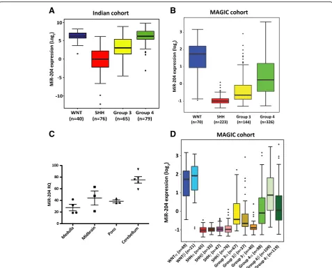
MiR-204 downregulation identifies a highly aggressive subset of Group 3 / Group 4 medulloblastomas having poor survival
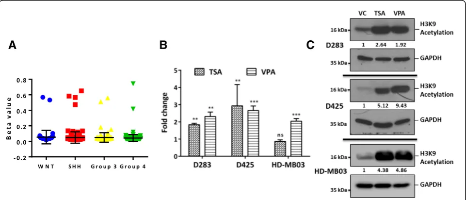
subgroups of medulloblastoma [11], was found to range from RQ = 21 to 93 (Fig. 1c). Thus, miR-204 expression is downregulated in the SHH subgroup and in a subset of the Group 3/Group 4 medulloblastomas. Integrated analysis of the genome wide DNA methylation data, ex-pression data and copy number alterations data, has identified 12 subtypes corresponding to the four core subgroups of medulloblastomas [2]. MiR-204 expression levels were found to be high in both the WNT subtypes, low in all four SHH subtypes, low in all three Group 3 subtypes and low to moderate in two out of 3 subtypes of Group 4 (Fig. 1d). Group 3γ having the worst
out-come [2] has the least miR-204 expression among the
Group 3 / Group 4 subtypes. Highly integrative analysis that integrated somatic mutation data analysis in addition to the genome wide methylation, transcriptome
and copy number variation data has reported 8 subtypes
within the Group 3/Group 4 medulloblastomas [34].
Analysis of MiR-204 expression in these 8 subtypes showed that the expression levels vary across the 8 subtypes with the least expression in the 3 subtypes (ii,
iii and iv) that contain only Group 3 tumors
(Additional file2: Figure S1). The subtype ii enriched for MYC amplification has the least miR-204 expression levels. MYC amplification is a known marker for poor prognosis in the Group 3 medulloblastomas [44].
Group 3 / Group 4 medulloblastomas in the Indian co-hort having metastasis at diagnosis were found to have significantly (p= 0.024) lower miR-204 expression (Fig.2a). In the MAGIC cohort as well, miR-204 expression levels are lower in the Group 3 / Group 4 tumors having metas-tasis at diagnosis, although the difference is not
A
B
C
D
statistically significant (Fig. 2b). Low miR-204 expression in the Group 3 / Group 4 medulloblastomas was found to correlate with poor overall survival in the Indian cohort (n= 126) as well as in the larger MAGIC cohort (n= 377) (Fig.2c, d). Five year overall survival of the‘miR-204 low’ subset in the Indian cohort is 33% (95% CI, 16.8-50.2%) as compared to 69.4% (95% CI, 54.1-80.4%) of the ‘miR-204
high’ subset. In the MAGIC cohort as well, five year
survival of the ‘miR-204 low’ subset is lower at 56.3% (95% CI 48.1% - 65. 9%) as compared to 78.9% (95%
CI 73.1-85.2%) of the ‘miR-204 high’ subset. Thus,
low miR-204 levels identify a subset of Group 3/ Group 4 tumors having poor overall survival both in the Indian cohort and in the larger MAGIC cohort.
A
B
C
D
E
F
Fig. 2Correlation of miR-204 expression with metastasis at diagnosis and overall survival. MiR-204 expression in Group 3 / Group 4
medulloblastomas having presence (M+) or absence (M0) of metastasis at diagnosis in the Indian cohort (a) and in the MAGIC cohort (b). Kaplan Meier survival analysis comparing overall survival of‘MiR-204 low’subset with that of‘MiR-204 high’subset of the Group 3 / Group 4
In the MAGIC cohort within the Group 3 tumors, low expression levels of miR-204 have a trend to-wards worse survival, although it does not reach stat-istical significance due to lower fraction of ‘miR-204 high’ tumors (Fig. 2e). In the Indian cohort, low ex-pression levels of miR-204 correlate with poor
sur-vival within the Group 3 as well (Additional file 3:
Figure S2). Due to small proportion of ‘miR-204 low
subset’ in the Group 4 tumors of the Indian cohort,
survival analysis for the Group 4 was done only for the MAGIC cohort. The five year survival of the
Group 4 ‘miR-204 high’ subset is 80% (95% CI
74-86.6%) while that of the ‘miR-204 low’ subset is
59.7% (95% CI 47.2-75.4%) in the MAGIC cohort (Fig. 2f ). Low miR-204 expression levels thus, identify a subset having poor overall survival within the Group 4 itself as well.
Restoration of miR-204 expression inhibits anchorage-independent growth, and tumorigenicity of
medulloblastoma cells
MiR-204 expression levels in the established medullo-blastoma cell lines D341, D425 and HD-MB03 were
found to be in the range of RQ = 0.02 to 0.14 (Fig. 3a). D341, D425 and the recently established HD-MB03 cell line belong to the Group 3 [16,29]. MiR-204 expression in the D283 cell line which has characteristics intermediate between Group 3 and Group 4 is at RQ = 9.4 ± 1.0 (Fig. 3a) [16]. The cell lines were transduced with the pTRIPZ lentiviral vector expressing miR-204 in a doxycycline inducible manner. Stable polyclonal popu-lations of the four medulloblastoma cell lines express miR-204 at levels (RQ = 25 to 70) comparable to that in the normal brain tissues after induction with doxy-cycline (Fig. 3a). Effect of miR-204 expression on the proliferation and anchorage-independent growth of these cell lines was studied by the MTT assay and soft agar colony formation assay respectively. While miR-204 expres-sion inhibited proliferation of D283 and D425 cells by 25 to 40%, it did not affect proliferation of D341 and HD-MB03 medulloblastoma cells (Fig.3b). MiR-204 expression resulted in significant inhibition of soft agar colony formation cap-acity (35 to 55%,p< 0.001) of all the four medulloblastoma cell lines studied (Fig.3c, d).
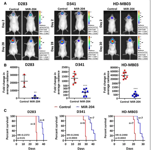
In order to study the effect of miR-204 expression on tumorigenic potential, D283, D341, HD-MB03 cells as
well as their miR-204 expressing polyclonal population cells were engineered to express firefly luciferase and, were injected stereotactically in cerebellum of NOD/ SCID mice after doxycycline induction. MiR-204 expres-sion was found to significantly (p< 0.002 to 0.0001) de-crease tumorigenicity of all the 3 medulloblastoma cell
lines as judged by the in vivo imaging of the orthotopic
tumors (Fig. 4a). The tumor volume decreased by
8.8-fold to 25-fold upon miR-204 expression (Fig. 4b). Further, survival of the tumor bearing mice increased by 26 to 34% upon miR-204 expression (Fig. 4c) in all the three medulloblastoma cell lines studied.
A
B
C
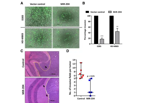
MiR-204 expression inhibits invasion potential of medulloblastoma cells in vitro and in vivo
Effect of miR-204 expression on the invasion potential of medulloblastoma cells was studied by evaluating inva-sion of the cells through Matrigel™coated membranes in
transwell inserts. Figure 5a shows images of the
CalceAM labeled medulloblastoma cells that have in-vaded the matrigel coated membrane after 56 h to 72 h. Evaluation of the fluorescence intensity of the
in-vaded cells showed 60–80% reduction in the invasion
potential of D283 and HD-MB03 cells upon miR-204
expression (Fig. 5b). Furthermore, invasive capacity
of the medulloblastoma cells as judged by their in vivo invasion across the cerebellar folia boundary was found to be reduced upon miR-204 expression
(Fig. 5c, d). Thus, miR-204 expression reduced
inva-sion potential of the medulloblastoma cells both in vitro and in vivo.
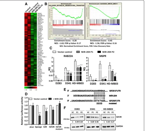
MiR-204 expression downregulatesM6PR,IGF2Rgenes involved in the lysosomal pathway and inhibits autophagy of medulloblastoma cells
In order to delineate the molecular mechanism under-lying the tumor suppressive role of miR-204 in medullo-blastoma cells, genes significantly differentially expressed upon miR-204 expression were identified by the
RNA-seq analysis. Figure 6a shows a heat map of the
top 60 genes downregulated upon miR-204 expression in HD-MB03 cells. GSEA analysis identified most signifi-cant enrichment of Epithelial Mesenchymal Transition
A
C
D
B
genes in the genes downregulated upon miR-204 expres-sion (Fig.6b). GSEA analysis using the microRNA target motif database identified most significant enrichment of miR-204 targets in the genes downregulated upon miR-204 expression (Fig.6b). Known validated targets of
miR-204 like RAB22A, M6PR are at the top of the list
whose downregulation upon miR-204 expression was fur-ther confirmed by real time RT-PCR in the three medullo-blastoma cell lines (Fig.6c). IGF2R was one of the putative miR-204 targets identified by the GSEA analysis. 3′-UTR
A
B
D
E
F
C
regions of IGF2Rand the known target EZR were cloned downstream of the luciferase cDNA in the pcDNA3.0 vec-tor. Luciferase reporter assay showed inhibition of
lucifer-ase activity upon co-transfection of these 3′-UTR
constructs with the vector expressing miR-204 in
HEK293FT cells suggesting IGF2R as a direct target of
miR-204 (Fig.6d). MiR-204 mediated inhibition of the lu-ciferase activity was lost upon site-directed mutagenesis of the miR-204 binding site in the 3′-UTR ofIGF2R, validat-ing it as a direct target of miR-204 (Fig. 6d, e). Further-more, downregulation of IGF2R protein levels upon miR-204 expression was confirmed by the western blot-ting in all the three medulloblastoma cell lines (Fig.6f ).
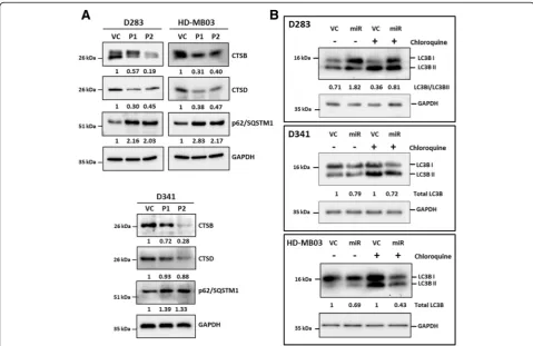
Both cation-dependent and cation-independent man nose-6-phosphate receptors i.e. M6PR and IGF2R respectively are known to be involved in trafficking of lysosomal proteases from the Golgi apparatus to lysosomes [26]. Therefore, effect of miR-204 expression on the levels of lysosomal enzymes Cathepsin B and Ca-thepsin D in medulloblastoma cells was studied by western blotting. MiR-204 expression resulted in considerable
downregulation of these lysosomal enzymes in all three me-dulloblastoma cell lines D283, D341 and HD-MB03 (Fig.7a). Lysosomal degradation pathway plays a major role in autophagy [12]. Besides, LC3B is a known target of miR-204 that plays a crucial role in autophagy [28]. Effect of miR-204 expression on autophagy was studied by evalu-ating LC3B flux. Upon autophagy induction, LC3BI isoform gets converted to LC3BII as a result of conjugation with phosphatidylethanolamine [12]. LC3BII levels however, de-crease upon fusion of autophagosome to lysosome due to degradation by lysosomal enzymes. LC3B turnover is there-fore studied in the presence and absence of an inhibitor of lysosomal degradation like chloroquine to evaluate LC3B flux [18]. MiR-204 expression resulted in high LC3BI / LC3BII ratio in D283 cells both before and after chloro-quine treatment indicating low LC3B flux and thereby
au-tophagy inhibition (Fig. 7b). In D341 and HD-MB03
medulloblastoma cells, total levels of LC3B decrease upon miR-204 expression both before and after treatment with chloroquine, indicating lower LC3B turnover and thereby autophagy inhibition (Fig. 7b). Autophagy inhibition is
known to be accompanied by increase in the levels of p62/ SQSTM1 adapter protein [12]. Expression levels of p62/ SQSTM1 increased upon miR-204 expression in the three medulloblastoma cell lines further confirming autophagy inhibition (Fig.7a). Thus, miR-204 expression inhibits au-tophagy mediated degradation pathway in medulloblastoma cells.
TRPM3/MIR204promoter methylation analysis and upregulation of miR-204 expression upon treatment with HDAC inhibitors
MiR-204 is located within the cancer associated genomic region at 9q21.1-q22.3 that exhibits high frequency of loss of heterozygosity in various cancers [55]. Loss of chromo-some 9q is however, not frequent in Group 3 / Group 4 medulloblastomas with less than 20% and 5% loss of chr 9q arm in Group 3, Group 4 tumors respectively based on the analysis of structural variations in 1000 medulloblastomas [38]. Downregulation of miR-204 expression has also been reported to occur as a result of promoter methylation [58].
CpG island at theTRPM3/MIR204 promoter locus seems
to be not methylated in Group 3 / Group 4 medulloblasto-mas based on the data from the Illumina 450 K array and bisulfite sequence analysis done on Group 3 medulloblas-toma cell lines for the CpG island in theTRPM3/MIR204 promoter region (Fig. 8a; Additional file 4: Figure S3). HDAC inhibitors have been reported to inhibit growth of medulloblastoma cells particularly that of MYC-driven
Group 3 cell lines [29,43]. Treatment of medulloblastoma cell lines D283, D425 and HD-MB03 with Trichostatin A and Sodium valproate, the HDAC inhibitors resulted in 2 to 4 fold increase in expression levels of miR-204 (Fig.8b) accompanied by the increased histone acetylation (Fig.8c). Thus, treatment of medulloblastoma patients with HDAC inhibitors could help in upregulation of miR-204 that has tumor suppressive effect.
Discussion
In a large scale study on 3312 tumors and 1107 non-malignant tissues contributed by 51 different cancer types, miR-204-211 family was found to be the top de-leted microRNA family in cancer, suggesting its crucial role as a tumor suppressive miRNA in multiple cancer types [55]. In the present study, miR-204 was found to be differentially expressed in the four core molecular subgroups of medulloblastomas, with almost all SHH and a subset of Group 3/Group 4 tumors showing downregulation of miR-204 expression. Despite the complexity of the heterogeneity and overlap present in the copy number variations, methylation profiles and somatic mutation profiles in the Group 3 / Group 4 me-dulloblastomas [2], miR-204 expression levels identify a subset of these tumors having poor survival in the In-dian as well as in the large MAGIC cohort. This finding is consistent with lower expression of miR-204 correlat-ing with poor survival in breast cancer [23], non-small
A
B
C
cell lung cancer [15], and neuroblastoma [47]. Integrated genomic studies have identified novel molecular sub-types within the four core subgroups of medulloblasto-mas [2,49]. Among the three Group 3 subtypes, subtype 3γ was found to have the worst 5 year survival rate of 41.9% as compared to that of subtype 3αand subtype 3β at 66.2 and 55.8% respectively [2]. Group 3γ having the worst survival showed the least miR-204 expression among the 3 subtypes of Group 3. The Group 4 subtypes do not show significant difference in their overall sur-vival [2]. MiR-204 expression on the other hand, identi-fied a subset of Group 4 medulloblastomas having significantly poor survival of 59.7% as compared to 80% of the ‘miR-204 high’subset. Group 3 poor
prognostica-tion markers likeMYCamplification and isochrome 17q
do not have prognostication value in Group 4 [44].
FSTL5 immunopositivity serves as a marker for poor prognostication in both Group 3 and Group 4 medullo-blastomas [45]. Adult Group 4 patients have also been
reported to have poor survival rates [46]. Loss of
chromosome 11 or gain of chromosome 17 identify a small subset of Group 4 patients who have excellent sur-vival [51]. Biology underlying these cytogenetic alter-ations is however, not understood. Thus, low miR-204 expression serves as a marker of poor prognosis in Group 4 that has paucity of markers for prognostication. Integrated genomic analysis is expensive as well as tech-nically demanding and thus cannot be used in routine clinical practice for risk stratification. MiR-204, a single microRNA on the other hand, can be easily combined with the Nanostring assay that classifies medulloblasto-mas into the four molecular subgroups [39]. Further-more, miR-204 due its small size resists degradation during formalin fixation and thus would be a reliable marker even in poor quality FFPE tissues.
Downregulation of miR-204 expression with poor sur-vival is consistent with its tumor-suppressive effect in me-dulloblastoma cell lines. Restoration of miR-204 expression in multiple established Group 3 medulloblastoma cell lines was found to inhibit their anchorage-independent growth, invasion potential and tumorigenicity. Tumor suppressive effect of miR-204 in theMYCamplified Group 3 cell lines is remarkable since other microRNAs downregulated in medulloblastoma like miR-206 for instance, fail to inhibit tumorigenicity of these cell lines [41]. MiR-204 has been shown to inhibit invasion and tumorigenicity of various cancer cells including glioma, colorectal cancer, endomet-rial cancer and cervical cancer cells [3, 27, 57, 58]. Thus, the tumor suppressive role of miR-204 in medulloblastoma cells is consistent with its role in other cancers.
MiR-204 has been reported to target a number of genes
including RAB22A, FOXC1, EZR, BCL2L2, M6PR, BCL2,
MCL1, FOXA1, FOXM1, EPHB2[22,42,50,52,57]. Tran-scriptome sequencing / real time RT-PCR / western blot
analysis showed downregulation of RAB22A, M6PR, EZR,
EPHB2,upon miR-204 expression in medulloblastoma cells as well.IGF2Rwas identified and validated as a novel target of miR-204. MiR-204 expression in medulloblastoma cells resulted in downregulation of both M6PR and IGF2R that mediate transport of lysosomal enzymes from the Golgi ap-paratus to lysosomes [26]. Furthermore, reduction in the levels of lysosomal enzymes Cathepsin B and Cathepsin D upon miR-204 expression in medulloblastoma cells sug-gests impairment of the lysosomal degradation pathway. Autophagy brings about p62/SQSTM1 mediated degrad-ation of its cargo by lysosomal degraddegrad-ation pathway [12]. MiR-204 is known to target LC3B, a crucial mediator of au-tophagy [28]. In the present study as well, miR-204 expres-sion in medulloblastoma cells resulted in reduction in the LC3B flux and increase in the levels of p62/SQSTM1 indi-cating autophagy inhibition. Autophagy has been shown to play role in tumor promotion by sustaining survival in stress, by reducing oxidative stress and, maintaining meta-bolic homeostasis [14]. Inhibition of tumor growth upon miR-204 expression is consistent with these reports on the role of autophagy in tumor promotion. Autophagy has also been reported to promote invasion by activating Epithelial Mesenchymal Transition of hepatocellular carcinoma cells [48], by promoting secretion of factors like IL6, MMP2 [24] and by activating the MAP kinase signaling pathway in glio-blastoma cells [8]. Consistent with the inhibition of inva-sion capacity of medulloblastoma cells upon miR-204 expression, downregulation of miR-204 expression was found to be associated with higher incidence of metastasis at diagnosis in Group 3 / Group 4 medulloblastomas. Thus, poor survival of Group 3 / Group 4 medulloblastomas hav-ing low miR-204 expression is likely due to their higher in-vasive capacity and higher malignant potential.
Several microRNAs whose expression is deregulated in medulloblastoma are known to play role in embryonic brain development [56]. MiR-9 and miR-124a that play crucial role in the onset of neurogenesis by targeting transcription factors like SOX9, FOXG1 and MEIS1, are
downregulated in medulloblastoma [6]. MiR-9 and
miR-199b-5p target HES1, thereby silence Notch signal-ing pathway at the onset of neuronal differentiation [7, 10]. Low expression of miR-9 and miR-199b-5p has been found to correlate with poor survival in medulloblas-toma and their expression in medulloblasmedulloblas-toma cell lines
promotes growth arrest [7, 10]. MiR-17-92 cluster
microRNAs are overexpressed predominantly in the
SHH subgroup medulloblastomas [35]. Knock-out of
this microRNA cluster brings about reduction in size of cerebellum and inhibits medulloblastoma formation in
Ptch knock-out mouse model of SHH subgroup
medul-loblastomas indicating role of these microRNAs in
nor-mal development and tumorigenesis [33]. MiR-204 has
development by targeting MEIS2 transcription factor in Medaka fish [4]. MiR-204 expression has been found to be upregulated during aging in mouse hippocampus and target Ephrin B2 that plays role in axon guidance [31]. MiR-204 has also been reported to control neuronal mi-gration and cortical morphogenesis in mouse embryos presumably by targeting Doublecortin that is known to play role in neuronal migration [54]. Effect of miR-204 on invasive capacity of medulloblastoma cells is consist-ent with the role of miR-204 in neuronal migration. Thus, miR-204 appears to play role in both normal brain development and tumorigenesis like several other
miR-NAs that are known to be deregulated in
medulloblastoma.
Delineating the molecular mechanism underlying downregulation of miR-204 expression would suggest ways to increase its expression, thereby improving sur-vival rate of medulloblastoma patients. Group 3 medul-loblastoma cells treated with HDAC inhibitors showed modest 2 to 4 fold increase in the miR-204 expression levels. Treatment with HDAC inhibitors has been re-ported to inhibit medulloblastoma cell growth in several studies [20, 29, 43]. Thus, HDAC inhibitors appear to
have therapeutic potential in the treatment of
medulloblastoma.
Conclusions
In summary, downregulation of miR-204 expression cor-relates with poor survival in the Group 3 / Group 4 me-dulloblastomas. Furthermore, within the Group 4 itself, low expression of miR-204 identifies a subset having sig-nificantly poor survival, making it a valuable marker for risk stratification in the subgroup that has paucity of prognostication markers. Restoration of miR-204 expres-sion leading to reduction in the invasive capacity and tumorigenic potential of medulloblastoma cells suggests therapeutic potential of miR-204 in the treatment of me-dulloblastomas. Upregulation of miR-204 expression upon treatment with HDAC inhibitors, although modest, suggests a role of these inhibitors in the treatment of medulloblastomas.
Additional files
Additional file 1:Table S1. The nucleotide sequences of the primers used in the study. All sequences are given in 5′to 3′direction. (DOCX 15 kb)
Additional file 2:Figure S1. MiR-204 expression levels in the 8 subtypes of Group 3 / Group 4 medulloblastomas from Northcott et al. [34] data. (PPTX 39 kb)
Additional file 3:Figure S2. Kaplan Meier Survival Analysis of Group 3 medulloblastomas from the Indian cohort comparing overall survival of
‘miR-204 high’subset with that of‘miR-204 low’subset. (PPTX 69 kb)
Additional file 4:Figure S3. Methylation analysis of the CpG island from the promoter region ofTRPM3/MIR204. A 203 bp region of the CpG
island from the promoter region of theTRPM3gene was PCR amplified from bisulfite converted genomic DNA of the medulloblastoma cell lines. Representative nucleotide sequence of this PCR product from the indicated medulloblastoma cell line is shown. Arrows indicate the CpG residues in the DNA sequence and their sequence in the bisulfite converted (BSP) DNA from the medulloblastoma cells. (PPTX 121 kb)
Abbreviations
3′-UTR:3′- untranslated region; ANOVA: Analysis of variance; ATCC: American Type Culture Collection; CI: Confidence interval; DMEM: Dulbecco’s Modified Eagle Medium; FBS: Fetal Bovine Serum; FFPE: Formalin- Fixed, Paraffin-Embedded; GSEA: Gene Set Enrichment Analysis; HDAC: Histone deacetylase; MAGIC: Medulloblastoma Advanced Genomics International Consortium; RMA: Robust Multi-array Average; RQ: Relative Quantity; RT-PCR: Reverse Transcription-Polymerase Chain Reaction; SDS-PAGE: Sodium Dodecyl Sulphate-Polyacrylamide Gel Electrophoresis; STR: Short Tandem Repeat
Acknowledgements
We thank Mr. Anant Sawant for technical assistance and Ms. Nazia Bano, Ms. Amita Wawdekar, clinical trial coordinators. We thank Prof. S. K. Shankar, NIMHANS, Bengaluru for making normal brain tissues available for the study.
Funding
We thank Department of Biotechnology, India for the financial support. Dr. V. Ramaswamy is supported by operating grants from the Canadian Institutes for Health Research, the Brain Tumor Foundation of Canada, Meagan’s Walk and the American Brain Tumor Association.
Availability of data and materials
The datasets used during the current study are available from the corresponding author on reasonable request.
Authors’contributions
HB, RP and PP have done the major experimental work involving Group 3 medulloblastoma cell lines. AD, SK, RK, KY have contributed to the experimental work that includes MicroRNA profile, molecular classification of medulloblastomas and evaluation of tumorigenicity. SM and Nikhil G have done the transcriptome sequencing and bioinformatics analysis. RJ, ES, TG, AM, AG, NG, PS, GC have contributed by recruitment of medulloblastoma cases, diagnosis and clinical data including treatment given and follow-up. NVS has contributed to the experimental design, implementation, analysis and, interpretation. VR has contributed to the Experimental design, analysis and interpretation. All authors read and approved the final manuscript.
Ethics approval and consent to participate
The study was approved by the Institutional Ethics Committee of the Tata Memorial Centre. Tumor tissues were procured after getting informed consent from the patients. Animal study was approved by the Institutional Animal Ethics Committee.
Consent for publication
Not applicable.
Competing interests
The authors declare that they have no competing interests.
Publisher’s Note
Springer Nature remains neutral with regard to jurisdictional claims in published maps and institutional affiliations.
Author details
1Shirsat Laboratory, Advanced Centre for Treatment, Research & Education in
Cancer, Tata Memorial Centre, Kharghar, Navi Mumbai 410210, India. 2Department of Radiation Oncology, Tata Memorial Centre, Kharghar, Navi
Mumbai 410210, India.3Bioinformatics Centre, Advanced Centre for Treatment, Research & Education in Cancer, Tata Memorial Centre, Kharghar, Navi Mumbai 410210, India.4Department of Radiation Oncology, Tata Memorial Hospital, Tata Memorial Centre, Parel, Mumbai 400012, India. 5Department of Pathology, Tata Memorial Hospital, Tata Memorial Centre,
Memorial Hospital, Tata Memorial Centre, Parel, Mumbai 400012, India. 7Department of Medical Oncology, Tata Memorial Hospital, Tata Memorial
Centre, Parel, Mumbai 400012, India.8Department of Neurosurgery, Seth G. S. Medical College & K. E. M. Hospital, Parel, Mumbai 400012, India.
9Department of Pathology, Seth G. S. Medical College & K. E. M. Hospital,
Parel, Mumbai 400012, India.10Homi Bhabha National Institute, Training School Complex, Anushakti Nagar, Mumbai 400085, India.11Division of Haematology/Oncology, Department of Paediatrics, Hospital for Sick Children and University of Toronto, 555 University Ave, Toronto, ON M5G 1X8, Canada.
Received: 30 January 2019 Accepted: 10 March 2019
References
1. Bourdeaut F, Miquel C, Alapetite C, Roujeau T, Doz F (2011)
Medulloblastomas: update on a heterogeneous disease. Curr Opin Oncol 23: 630–637
2. Cavalli FMG, Remke M, Rampasek L, Peacock J, Shih DJH, Luu B et al (2017) Intertumoral heterogeneity within Medulloblastoma subgroups. Cancer Cell 31:737–754 e736.https://doi.org/10.1016/j.ccell.2017.05.005
3. Chung TK, Lau TS, Cheung TH, Yim SF, Lo KW, Siu NS et al (2012) Dysregulation of microRNA-204 mediates migration and invasion of endometrial cancer by regulating FOXC1. Int J Cancer 130:1036–1045.
https://doi.org/10.1002/ijc.26060
4. Conte I, Carrella S, Avellino R, Karali M, Marco-Ferreres R, Bovolenta P et al (2010) miR-204 is required for lens and retinal development via Meis2 targeting. Proc Natl Acad Sci U S A 107:15491–15496.https://doi.org/10. 1073/pnas.0914785107
5. Di Leva G, Croce CM (2013) miRNA profiling of cancer. Curr Opin Genet Dev 23:3–11.https://doi.org/10.1016/j.gde.2013.01.004
6. Ferretti E, De Smaele E, Po A, Di Marcotullio L, Tosi E, Espinola MS et al (2009) MicroRNA profiling in human medulloblastoma. Int J Cancer 124: 568–577
7. Fiaschetti G, Abela L, Nonoguchi N, Dubuc AM, Remke M, Boro A et al (2014) Epigenetic silencing of miRNA-9 is associated with HES1 oncogenic activity and poor prognosis of medulloblastoma. Br J Cancer 110:636–647.
https://doi.org/10.1038/bjc.2013.764
8. Galavotti S, Bartesaghi S, Faccenda D, Shaked-Rabi M, Sanzone S, McEvoy A et al (2013) The autophagy-associated factors DRAM1 and p62 regulate cell migration and invasion in glioblastoma stem cells. Oncogene 32:699–712.
https://doi.org/10.1038/onc.2012.111
9. Garancher A, Lin CY, Morabito M, Richer W, Rocques N, Larcher M et al (2018) NRL and CRX define photoreceptor identity and reveal subgroup-specific dependencies in Medulloblastoma. Cancer Cell 33:435–449 e436.
https://doi.org/10.1016/j.ccell.2018.02.006
10. Garzia L, Andolfo I, Cusanelli E, Marino N, Petrosino G, De Martino D et al (2009) MicroRNA-199b-5p impairs cancer stem cells through negative regulation of HES1 in medulloblastoma. PLoS One 4:e4998.https://doi.org/ 10.1371/journal.pone.0004998
11. Gibson P, Tong Y, Robinson G, Thompson MC, Currle DS, Eden C et al (2010) Subtypes of medulloblastoma have distinct developmental origins. Nature 468:1095–1099.https://doi.org/10.1038/nature09587
12. Glick D, Barth S, Macleod KF (2010) Autophagy: cellular and molecular mechanisms. J Pathol 221:3–12.https://doi.org/10.1002/path.2697
13. Gokhale A, Kunder R, Goel A, Sarin R, Moiyadi A, Shenoy A et al (2010) Distinctive microRNA signature of medulloblastomas associated with the WNT signaling pathway. J Cancer Res Ther 6:521–529.https://doi.org/10. 4103/0973-1482.77072
14. Guo JY, Xia B, White E (2013) Autophagy-m.ediated tumor promotion. Cell 155:1216–1219.https://doi.org/10.1016/j.cell.2013.11.019
15. Guo W, Zhang Y, Zhang Y, Shi Y, Xi J, Fan H, et al (2015) Decreased expression of miR-204 in plasma is associated with a poor prognosis in patients with non-small cell lung cancer. Int J Mol Med 36: 1720–1726 Doi
https://doi.org/10.3892/ijmm.2015.2388
16. Ivanov DP, Coyle B, Walker DA, Grabowska AM (2016) In vitro models of medulloblastoma: choosing the right tool for the job. J Biotechnol 236:10– 25.https://doi.org/10.1016/j.jbiotec.2016.07.028
17. King AA, Seidel K, Di C, Leisenring WM, Perkins SM, Krull KR et al (2017) Long-term neurologic health and psychosocial function of adult survivors of childhood
medulloblastoma/PNET: a report from the childhood Cancer survivor study. Neuro Oncol 19:689–698.https://doi.org/10.1093/neuonc/now242
18. Klionsky DJ, Abdelmohsen K, Abe A, Abedin MJ, Abeliovich H, Acevedo Arozena A et al (2016) Guidelines for the use and interpretation of assays for monitoring autophagy (3rd edition). Autophagy 12:1–222.https://doi. org/10.1080/15548627.2015.1100356
19. Kunder R, Jalali R, Sridhar E, Moiyadi A, Goel N, Goel A et al (2013) Real-time PCR assay based on the differential expression of microRNAs and protein-coding genes for molecular classification of formalin-fixed paraffin embedded medulloblastomas. Neuro-Oncology 15:1644–1651.https://doi. org/10.1093/neuonc/not123
20. Lee SJ, Krauthauser C, Maduskuie V, Fawcett PT, Olson JM, Rajasekaran SA (2011) Curcumin-induced HDAC inhibition and attenuation of
medulloblastoma growth in vitro and in vivo. BMC Cancer 11:144.https:// doi.org/10.1186/1471-2407-11-144
21. Lee YH, Andersen JB, Song HT, Judge AD, Seo D, Ishikawa T et al (2010) Definition of ubiquitination modulator COP1 as a novel therapeutic target in human hepatocellular carcinoma. Cancer Res 70:8264–8269.https://doi. org/10.1158/0008-5472.CAN-10-0749
22. Li G, Luna C, Qiu J, Epstein DL, Gonzalez P (2011) Role of miR-204 in the regulation of apoptosis, endoplasmic reticulum stress response, and inflammation in human trabecular meshwork cells. Invest Ophthalmol Vis Sci 52:2999–3007.https://doi.org/10.1167/iovs.10-6708
23. Li W, Jin X, Zhang Q, Zhang G, Deng X, Ma L (2014) Decreased expression of miR-204 is associated with poor prognosis in patients with breast cancer. Int J Clin Exp Pathol 7:3287–3292
24. Lock R, Kenific CM, Leidal AM, Salas E, Debnath J (2014) Autophagy-dependent production of secreted factors facilitates oncogenic RAS-driven invasion. Cancer Discov 4:466–479.https://doi.org/10.1158/2159-8290.CD-13-0841
25. Louis DN, Ohgaki H, Wiestler OD, Cavenee WK, Burger PC, Jouvet A et al (2007) The 2007 WHO classification of tumours of the central nervous system. Acta Neuropathol 114:97–109.https://doi.org/10.1007/s00401-007-0243-4
26. Luzio JP, Pryor PR, Bright NA (2007) Lysosomes: fusion and function. Nat Rev Mol Cell Biol 8:622–632.https://doi.org/10.1038/nrm2217
27. Mao J, Zhang M, Zhong M, Zhang Y, Lv K (2014) MicroRNA-204, a direct negative regulator of ezrin gene expression, inhibits glioma cell migration and invasion. Mol Cell Biochem 396:117–128.https://doi.org/10.1007/ s11010-014-2148-6
28. Mikhaylova O, Stratton Y, Hall D, Kellner E, Ehmer B, Drew AF et al (2012) VHL-regulated MiR-204 suppresses tumor growth through inhibition of LC3B-mediated autophagy in renal clear cell carcinoma. Cancer Cell 21:532– 546.https://doi.org/10.1016/j.ccr.2012.02.019
29. Milde T, Lodrini M, Savelyeva L, Korshunov A, Kool M, Brueckner LM et al (2012) HD-MB03 is a novel group 3 medulloblastoma model demonstrating sensitivity to histone deacetylase inhibitor treatment. J Neuro-Oncol 110: 335–348.https://doi.org/10.1007/s11060-012-0978-1
30. Miyoshi H, Blomer U, Takahashi M, Gage FH, Verma IM (1998) Development of a self-inactivating lentivirus vector. J Virol 72:8150–8157
31. Mohammed CP, Rhee H, Phee BK, Kim K, Kim HJ, Lee H, et al (2016) miR-204 downregulates EphB2 in aging mouse hippocampal neurons. Aging Cell 15: 380–388 Doihttps://doi.org/10.1111/acel.12444
32. Mosmann T (1983) Rapid colorimetric assay for cellular growth and survival: application to proliferation and cytotoxic assays. J Immunol Methods 65:55–63 33. Murphy BL, Obad S, Bihannic L, Ayrault O, Zindy F, Kauppinen S et al (2013)
Silencing of the miR-17~92 cluster family inhibits medulloblastoma progression. Cancer Res 73:7068–7078.https://doi.org/10.1158/0008-5472. CAN-13-0927
34. Northcott PA, Buchhalter I, Morrissy AS, Hovestadt V, Weischenfeldt J, Ehrenberger T et al (2017) The whole-genome landscape of
medulloblastoma subtypes. Nature 547:311–317.https://doi.org/10.1038/ nature22973
35. Northcott PA, Fernandez LA, Hagan JP, Ellison DW, Grajkowska W, Gillespie Y et al (2009) The miR-17/92 polycistron is up-regulated in sonic hedgehog-driven medulloblastomas and induced by N-myc in sonic hedgehog-treated cerebellar neural precursors. Cancer Res 69:3249–3255.https://doi.org/10. 1158/0008-5472.CAN-08-4710
36. Northcott PA, Korshunov A, Pfister SM, Taylor MD (2012) The clinical implications of medulloblastoma subgroups. Nat Rev Neurol 8:340–351 37. Northcott PA, Korshunov A, Witt H, Hielscher T, Eberhart CG, Mack S et al
38. Northcott PA, Shih DJ, Peacock J, Garzia L, Morrissy AS, Zichner T et al (2012) Subgroup-specific structural variation across 1,000 medulloblastoma genomes. Nature 488:49–56
39. Northcott PA, Shih DJ, Remke M, Cho YJ, Kool M, Hawkins C et al (2012) Rapid, reliable, and reproducible molecular sub-grouping of clinical medulloblastoma samples. Acta Neuropathol 123:615–626
40. Ostrom QT, Gittleman H, Fulop J, Liu M, Blanda R, Kromer C et al (2015) CBTRUS statistical report: primary brain and central nervous system tumors diagnosed in the United States in 2008-2012. Neuro-Oncology 17(Suppl 4): iv1–iv62.https://doi.org/10.1093/neuonc/nov189
41. Panwalkar P, Moiyadi A, Goel A, Shetty P, Goel N, Sridhar E et al (2015) MiR-206, a cerebellum enriched miRNA is downregulated in all Medulloblastoma subgroups and its overexpression is necessary for growth inhibition of Medulloblastoma cells. J Mol Neurosci 56:673–680.https://doi.org/10.1007/ s12031-015-0548-z
42. Paylakhi SH, Moazzeni H, Yazdani S, Rassouli P, Arefian E, Jaberi E et al (2013) FOXC1 in human trabecular meshwork cells is involved in regulatory pathway that includes miR-204, MEIS2, and ITGbeta1. Exp Eye Res 111:112– 121.https://doi.org/10.1016/j.exer.2013.03.009
43. Pei Y, Liu KW, Wang J, Garancher A, Tao R, Esparza LA et al (2016) HDAC and PI3K antagonists cooperate to inhibit growth of MYC-driven Medulloblastoma. Cancer Cell 29:311–323.https://doi.org/10.1016/j.ccell. 2016.02.011
44. Ramaswamy V, Remke M, Bouffet E, Bailey S, Clifford SC, Doz F et al (2016) Risk stratification of childhood medulloblastoma in the molecular era: the current consensus. Acta Neuropathol 131:821–831.https://doi.org/10.1007/ s00401-016-1569-6
45. Remke M, Hielscher T, Korshunov A, Northcott PA, Bender S, Kool M et al (2011) FSTL5 is a marker of poor prognosis in non-WNT/non-SHH medulloblastoma. J Clin Oncol 29:3852–3861.https://doi.org/10.1200/JCO. 2011.36.2798
46. Remke M, Hielscher T, Northcott PA, Witt H, Ryzhova M, Wittmann A et al (2011) Adult medulloblastoma comprises three major molecular variants. J Clin Oncol 29:2717–2723.https://doi.org/10.1200/JCO.2011.34.9373
47. Ryan J, Tivnan A, Fay J, Bryan K, Meehan M, Creevey L et al (2012) MicroRNA-204 increases sensitivity of neuroblastoma cells to cisplatin and is associated with a favourable clinical outcome. Br J Cancer 107:967–976.
https://doi.org/10.1038/bjc.2012.356
48. Sausen M, Leary RJ, Jones S, Wu J, Reynolds CP, Liu X et al (2013) Integrated genomic analyses identify ARID1A and ARID1B alterations in the childhood cancer neuroblastoma. Nat Genet 45:12–17.https://doi.org/10.1038/ng.2493
49. Schwalbe EC, Lindsey JC, Nakjang S, Crosier S, Smith AJ, Hicks D et al (2017) Novel molecular subgroups for clinical classification and outcome prediction in childhood medulloblastoma: a cohort study. Lancet Oncol 18: 958–971.https://doi.org/10.1016/S1470-2045(17)30243-7
50. Shen SQ, Huang LS, Xiao XL, Zhu XF, Xiong DD, Cao XM et al (2017) miR-204 regulates the biological behavior of breast cancer MCF-7 cells by directly targeting FOXA1. Oncol Rep 38:368–376.https://doi.org/10.3892/or. 2017.5644
51. Shih DJ, Northcott PA, Remke M, Korshunov A, Ramaswamy V, Kool M et al (2014) Cytogenetic prognostication within medulloblastoma subgroups. J Clin Oncol 32:886–896.https://doi.org/10.1200/JCO.2013.50.9539
52. Sun Y, Yu X, Bai Q (2015) miR-204 inhibits invasion and epithelial-mesenchymal transition by targeting FOXM1 in esophageal cancer. Int J Clin Exp Pathol 8:12775–12783
53. Uphoff CC, Drexler HG (2014) Detection of mycoplasma contamination in cell cultures. Curr Protoc Mol Biol 106: 28 24:21–14.https://doi.org/10.1002/ 0471142727.mb2804s106
54. Veno MT, Veno ST, Rehberg K, van Asperen JV, Clausen BH, Holm IE et al (2017) Cortical morphogenesis during embryonic development is regulated by miR-34c and miR-204. Front Mol Neurosci 10:31.https://doi.org/10.3389/ fnmol.2017.00031
55. Volinia S, Galasso M, Costinean S, Tagliavini L, Gamberoni G, Drusco A et al (2010) Reprogramming of miRNA networks in cancer and leukemia. Genome Res 20:589–599.https://doi.org/10.1101/gr.098046.109
56. Wang X, Holgado BL, Ramaswamy V, Mack S, Zayne K, Remke M et al (2018) miR miR on the wall, who’s the most malignant medulloblastoma miR of them all? Neuro-Oncology 20:313–323.https://doi.org/10.1093/neuonc/nox106
57. Yin Y, Zhang B, Wang W, Fei B, Quan C, Zhang J et al (2014) miR-204-5p inhibits proliferation and invasion and enhances chemotherapeutic
sensitivity of colorectal cancer cells by downregulating RAB22A. Clin Cancer Res 20:6187–6199.https://doi.org/10.1158/1078-0432.CCR-14-1030
58. Ying Z, Li Y, Wu J, Zhu X, Yang Y, Tian H et al (2013) Loss of miR-204 expression enhances glioma migration and stem cell-like phenotype. Cancer Res 73:990–999.https://doi.org/10.1158/0008-5472.CAN-12-2895