R E V I E W A R T I C L E
Analysis of DFS70 pattern and impact on ANA screening using
a novel HEp-2 ELITE/DFS70 knockout substrate
Kishore Malyavantham1•Lakshmanan Suresh1
Received: 27 February 2017 / Accepted: 7 March 2017 / Published online: 17 March 2017
ÓThe Author(s) 2017. This article is an open access publication
Abstract Indirect immunofluorescence (IIF) using human epithelial cell (HEp-2) substrate is a widely used and the recommended method for screening of antinuclear anti-bodies (ANA). Dense fine speckled (DFS70) pattern on HEp-2 has been widely reported in various healthy and disease groups. Interpretation of DFS70 pattern can be challenging on a conventional HEp-2 substrate due to its similarity to some of the disease associated patterns. The high prevalence of DFS70 autoantibodies in normal pop-ulation, lack of association with a particular disease group and a general negative association with systemic and ANA associated autoimmune rheumatic diseases (SARD/AARD) necessitates the confirmation of DFS70 pattern. Results using available commercial assays for confirmation of DFS70 autoantibodies do not always agree with IIF screening results further complicating the lab work flow and ANA algorithms. In this review, we discuss the prevalence of DFS70 antibodies and factors affecting the performance of IIF and DFS70 specific confirmatory assays. Factors that contribute to disagreement between DFS70 suspicion by IIF and confirmatory assays will also be discussed. In addition, we also describe a novel IIF HEp-2 substrate, and its positive impact on DFS70 reporting and ANA screening-confirmation algorithm.
Keywords DFS70LEDGFANAs (antinuclear antibodies)HEp-2 IIF (indirect immunofluorescence)
Abbreviations
ANA Antinuclear antibody
CLIA Chemiluminescence immunoassay DFS70 Dense fine speckled 70
EIA/ELISA Enzyme-linked immunosorbent assay ICAP International consensus on ANA pattern IIF Indirect immunofluorescence
LEDGF Lens epithelium derived growth factor AARD ANA associated rheumatic diseases SARD Systemic autoimmune rheumatic diseases
Introduction
ANAs remain a hallmark of systemic autoimmune dis-eases. Patterns of ANA observed on HEp-2 cells by IIF provide the clinicians with insight into specificity of autoantibodies present, indications of disease likelihood and further implicate or rule out a clinical suspicion [1]. IIF by HEp-2 is a widely prevalent screening method among the techniques used for the determination of ANA. Despite advances in EIA/ELISA/multiplex methodologies for screening of ANAs, IIF-HEp-2 remains one of the most prevalent methods due to its diagnostic usefulness and cost effectiveness. HEp-2 cells are able to present a variety of autoantigens that result in a multitude of distinct patterns. Though this method has been widely used for more than 50 years, standardization of the quality of HEp-2 substrates (clones, growth phase, fixation method), strength and specificity of FITC-conjugates (fluorescein isothiocyanate), F/P ratio (fluorescein/protein molar ratio), anti-human IgG specificity (Heavy chain/light chain/Fc region), washing technique, buffers, counterstain, microscope setup (exci-tation light source, use of neutral density filters, narrow/ broad band emission filters, quality and specifications of
& Kishore Malyavantham [email protected]
1 Immco Diagnostics, A Trinity Biotech Company, 60
objectives) is lacking. In addition to this, technical exper-tise and human subjectivity of the readers can impact accurate interpretation of IIF [2]. In an effort to standardize the IIF, International consensus on ANA patterns (ICAP) workshops recommend a consensus nomenclature for the HEp-2 patterns and provide training and description of the nuclear/cytoplasmic/mitosis stage specific patterns on HEp-2 and their antigen/disease associations [3]. The ICAP committee described 28 distinct patterns on HEp-2 and assigned each pattern an AC (Anti-cell) number of 1–28 [3]. DFS70 AC-02 pattern has received the most scrutiny in the field in recent years due to its high rates of prevalence in healthy and ANA positive populations and negative association with SARD/AARD [4,5].
DFS70 pattern: background
The DFS70 pattern resulting from autoantibodies binding to the ubiquitously expressed protein called lens epithelium derived growth factor (LEDGF) or p75 or psip1 gene product is frequently observed during routine ANA screening by IIF-HEp-2. DFS70 pattern and autoantibodies were originally described by Ochs et al. [8] and were later confirmed in higher frequencies in patients with atopic dermatitis and asthma [6,7] (Fig.1). DFS70 is a unique pattern characterized by dense and heterogeneous fine speckled staining of the nucleoplasm in interphase, and speckled staining tightly associated with chromatin during mitosis [7–9]. Independent efforts by various groups have unraveled the identity of the gene encoding this antigen and
resulted in characterization of the role of LEDGF/psip1/ p75 [7, 10–13]. DFS70/LEDGF/p75 is a ubiquitously expressed growth/transcription factor that localizes to the cell nucleus. The N-terminus has a high affinity for chro-matin binding due to which the autoantigen remains tightly associated with chromatin during entire cell cycle [7, 14–16]. Epitope mapping analysis of the DFS70 autoantigen revealed a conformational autoepitope on the C-terminus of the antigen which was responsible for majority of the DFS70 autoantibody binding [17]. DFS70 pattern resulting from LEDGF/p75 gained major attention of the diagnostic field when Watanabe and colleagues reported that 11.6% (64) of the 597 healthy hospital workers in Japan were positive for DFS70 pattern [18]. Role of DFS70/LEDGF/p75 antigen as a transcription factor, cellular co-factor of HIV-1 integration, m-RNA splicing, cell-stress survival factor, its potential interaction with STAT3 in IL-6/STAT3 inflammatory pathway has been reviewed by Casiano and co-workers [19–25]. The mechanism underlying the appearance and the clinical impact of DFS70 antibodies is not yet clear but these have been reported by various groups across the world in both healthy and disease populations. It is still unknown if the DFS70 autoantibodies are natural and protective or pathogenic. Inaccurate interpretation and reporting of AC-02 as one of the disease associated ANA patterns (homo-geneous/AC-01, fine speckled/AC-04, speckled/AC-05 or a combination of AC-01/AC-04/AC-05) can lead to unnec-essary testing and negatively impact patient care. Due to their high prevalence in ANA screening population and lower association with SARD/AARD, ICAP committee recommends all clinical labs to report the DFS70 pattern [3].
DFS70 pattern: impact on ANA screening
and reporting
With the increased demand for ANA testing, many labs have switched to newer solid phase and multiplex methodologies for screening of ANAs [2]. Although these methods are automation friendly and reduce subjectivity in the interpretation of results, they are based on a limited number of purified recombinant/native autoantigens and do not equate in performance to HEp-2 IIF [2]. The American College of Rheumatology(ACR) position statement describes IIF-HEp-2 as the gold standard method for ANA testing [26,27]. The HEp-2 cell represents at least 100–150 autoantigens in native configuration which provide the unique pattern and titer. This information is of useful value to clinical labs in determining the positive/negative ANA status and selection of appropriate solid phase confirmatory assays [26, 27]. In addition to the ACR, the European
• Discovery of autoantibodies recognizing a nuclear dense fine speckled pattern in patients with interstitial cystitis
1994
•Detecon of cytotoxic an-LEDGF autoanbodies in atopic dermas 1999
•Characterizaon of DFS70 paern (LEDGF autoangen) using anbodies from paents with atopic dermas & DFS70 autoanbodies are present at low frequencies in SARD paents
2000
•Characterizaon of DFS70/LEDGF/p75 autoangen 1999-2004
•DFS70 autoanbodies are present in apparently healthy individuals 2004
•Availability of various commercial DFS70 specific confirmatory assays •DFS70 autoanbodies are frequent in general and ANA posive populaon •DFS70 autoanbodies are rarely diagnosed in SARD and when present, they
accompany a disease specific paern/anbody along with DFS70 in majority of the cases
•DFS70 proposed as an exclusion marker for SARD 2005-present
DFS70/LEDGF/p75 autoantibodies - Background and significance
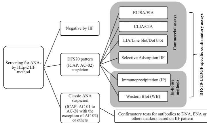
autoimmunity standardization initiative group also recog-nizes the IIF-HEp-2 as reference method despite identify-ing some of the advantages offered by solid phase/multiplex assays [2]. IIF-HEp-2 is the first step of the routine screening of ANAs and if the IIF result is negative, the samples are not tested further unless there is a strong clinical suspicion (Figs.2,3). IIF positive results are ana-lyzed for pattern and titer. Classic disease associated ANA patterns, AC-01 to AC-28 with the exception of AC-02 are further confirmed on appropriate solid phase assays (Figs.2,3). Due to the efforts of ICAP and work of experts in the field, clinical laboratories around the world are gaining an understanding of DFS70/AC-02 pattern. DFS70 specific commercial assays are now available for routine use in the form of ELISA/EIA, CLIA/CIA, line blot or dot blot and modified IIF (selective adsorption IIF) procedures (Fig.2). For research and confirmation of suspected sam-ples, some labs are also using IP (immunoprecipitation) and Western blot assays with cell lysates (HeLa, HEpG2, HEp-2, Jurkat or PC3 cell lines) known to express ample levels of LEDGF/DFS70 protein [25,28,29].
Prevalence of DFS70 autoantibodies
ANA-IIF-HEp-2 is being increasingly requested not only on clinical suspicion of AARD but also for differential diagnosis from AARD. In many clinical laboratories, ANA-IIF referrals come from rheumatologists, hepatolo-gists, neurolohepatolo-gists, dermatolohepatolo-gists, allergy/immunologists and increasingly from general practitioners to rule out SARD. Based on this trend, the complexity and
heterogeneity of ANA screening populations change sig-nificantly from one clinical lab to the other. Systematic review for DFS70 autoantibody positivity has been per-formed by multiple groups [4, 6, 30]. The majority of
In-house methods
Commercial assays
Screening for ANAs by HEp-2 IIF
method
DFS70 pattern (ICAP: AC-02)
suspicion
ELISA/EIA
CLIA/CIA
LIA/Line blot/Dot blot
Selective Adsorption IIF
Immunoprecipitation (IP)
Western Blot (WB) Classic ANA
suspicion (ICAP: AC-01 to
AC-28 with the exception of AC-02)
or others
Confirmatory tests for antibodies to DNA, ENA or others markers based on IIF pattern Negative by IIF
Current state of diagnostic assays for screening/confirmation of DFS70 pattern
DFS70
-LEDGF specific
confirmatory
assa
ys
Fig. 2 Schematic summarizes the current generation of diagnostic assays used for confirmation of DFS70 pattern and how they are used in the context of ANA screening algorithm
Screening by convenonal HEp2-IIF
Posive
Classic ANA paern posive
DFS70/Homogeneous /Speckled/Mixed ?
Confirm by DFS70 specific assays (Figure 2)
Negave Posive Confirm for AARD
anbodies using ELISA/EIA/LIA/Mulplex
Negave: No follow up
Posive: Follow up
Current diagnostic algorithm using conventional HEp-2 IIF method
Negave: No follow up
clinical studies have used IIF-HEp2 for establishing a suspicion of DFS70 pattern and the rates of positivity for DFS70 autoantibodies in each group varied widely between studies [7,9,18, 21, 28,31–44]. DFS70 antibodies have been reported in high titers from cohorts of healthy indi-viduals, blood donors, patients being screened for ANA, patients with various autoimmune disorders and various non-autoimmune disorders including cancers [4, 6, 30]. These studies have shown that DFS70 autoantibodies lack distinct clinical association, with most disease groups, except for certain inflammatory conditions of eyes and skin [4,6,7,18,30,44,45]. The method of screening, selection, and composition of study cohorts may also influence the reported rates of DFS70 autoantibody positivity. A study by Bizzaro et al. [30] using a highly specific commercial DFS70-CLIA method as the first screening step, reported significant variability in DFS70 positivity in clinically defined cases of anti-phospholipid syndrome (60%), Hashimoto’s thyroiditis (47.8%), rheumatoid arthritis (11.1%), Sjogren’s syndrome (4.3%), systemic lupus ery-thematosus (15.4%), and undifferentiated connective tissue disease (40%) [30]. One hypothesis for this phenomenon is that routine ANA screening by IIF method may not reveal the low levels of DFS70 autoantibodies when disease associated autoantibodies co-exist. Other theories include the challenges associated with setting up an appropriate clinical cut-off value for the confirmatory method.
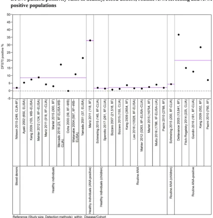
Based on published studies, a great majority of the routine ANA screening population is negative for all ANAs and a large subset of the positive ANA group has DFS70 autoantibodies alone or in combination with other disease associated ANAs. Due to these factors, this review focuses on 20 studies from research group around the world that have reported the frequency of DFS70 suspected cases (Fig.4). The selected studies provide results from 78,399 cases from various patient cohort types, including blood donors (249), healthy individuals (adult: 2793; pediatric: 406), routine ANA screening populations (adult: 59,444; pediatric: 200), ANA positive healthy individuals (118), and routine ANA screening cohorts with ANA positive status (n=15,189; Fig. 4). The majority of the studies used IIF-HEp-2 as the screening step, with a few using the CLIA or ELISA methods. A detailed review of the results from the selected studies found the rate of DFS70 positivity to be 0–5% in blood donors, healthy children, and in rou-tine ANA screening populations. In contrast, cohorts con-sisting of healthy individuals that have not been differentiated as pediatric or adults, and ANA positive cases (healthy or routine ANA screening populations) have a higher DFS70 pattern positivity ranging from 0 to 37% (Fig.4). Group mean for each cohort is indicated by purple lines in Fig.3 but due to the heterogeneous nature of screening populations, geographic diversity, inter-lab
variations in IIF interpretation and accuracy of DFS70 suspicion, the statistics for this data may be of limited value. However, it is clear from the data that DFS70 autoantibodies are highly prevalent in both healthy and disease states where SARD is unlikely. Over and under estimation of DFS70 positivity can have serious impact on patient care and management and clinical labs are obli-gated to run a number of reflex tests prior to ruling out a suspicion of SARD/AARD (Fig.3). Many reviews by experts in the field suggested the importance of confirming DFS70 suspicion using specific methods and evaluate its overall impact on ANA screening algorithm and associated costs [1, 4,6,30, 34,39, 46–48]. As per certain studies, approximately a third of the positive ANA cases were positive for DFS70 pattern [9, 33]. Due to these com-plexities associated with DFS70 autoantibodies, the use of current method of screening significantly increase the number of confirmatory reflex tests run by labs and the financial burden for patients and the system.
Gap between DFS70 suspicion by IIF-HEp-2
and confirmatory assays
run a panel of reflex assays (ENAs, Anti-DNA, Anti-Nu-cleosome, Anti-Histone assays among the others) for DFS70 pattern suspect cases irrespective of the DFS70 solid phase assay results prior to ruling out the absence of classic ANAs (Fig.3). Recently proposed selective absorption IIF method (NovaLite, HEp-2 Select, INOVA Diagnostics, USA) uses a high concentration of recombi-nant truncated LEDGF antigen to cross adsorb DFS70 specific autoantibodies in the sample prior to IIF reaction [51]. Users are expected to implement selective adsorption
procedure on DFS70 suspect samples and evaluate the relative reduction in the intensity of DFS70 pattern. While this method attempts to address some of the deficiencies of other solid phase assays, it is an extra IIF assay step and there is a likelihood of incomplete adsorption due to high levels of DFS70 autoantibodies in serum. This possibility reduces the level of confidence for confirming a mono-specific DFS70 reaction and may warrant the use of a second confirmation step for DFS70 and/or multiple con-firmatory assays for other ANAs.
Fig. 4 Results from 20 different studies pertaining to reported rates of DFS70 suspicion by IIF/ELISA/CLIA in blood donor, healthy, ANA screening and ANA positive cohorts are depicted. First authors of the study, year of publication is followed by samples size and
Screening for classic ANAs, detection
and confirmation of DFS70 antibodies in one step
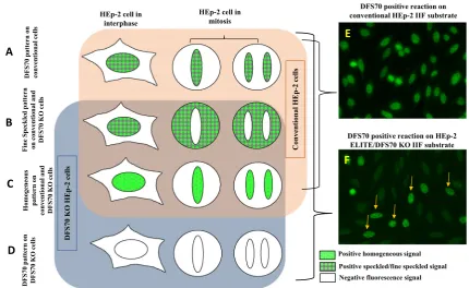
Here, we introduce a novel HEp-2 IIF substrate (HEp-2 ELITE/DFS70 KO, Immco Diagnostics-Trinity Biotech USA) that presents a mixture of natural HEp-2 cells and genetically engineered HEp-2 cells that do not express DFS70/LEDGF/psip1/p75 antigen (referred to as DFS70 KO cells) in 1:9 ratio on glass slide wells. The new IIF substrate retains all the capabilities of conventional HEp-2 substrates for screening of ANAs and further is able to simultaneously detect and confirm with high confidence both mixed and mono-specific/isolated DFS70 patterns (Fig.5). Figure5a–c illustrates how, conventional HEp-2 cells (interphase and mitosis) present classic homogeneous, speckled and DFS70 patterns in natural pattern as expected. Figure5d shows that the DFS70 KO cells (interphase and mitosis) present only on the novel substrate do not react with DFS70 autoantibodies (Fig.5d). Therefore, when the substrate is reacted with mono-specific DFS70 sera, a typical pattern with 10% brightly labelled nuclei (derived from conventional HEp-2) and 90% negatively stained nuclei (derived from DFS70 KO cells) is observed. This substrate eliminates the need for evaluation of mitotic
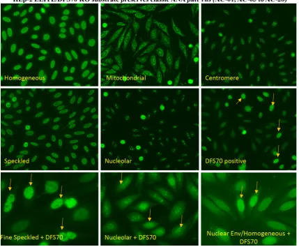
pattern to distinguish DFS70 from classic patterns (ho-mogeneous/speckled). Typical reactions obtained using a DFS70 mono-specific sample on conventional HEp-2 IIF substrate (Fig.5e) and novel HEp-2 ELITE/DFS70 KO substrate (Fig. 5f) emphasize the differences and ease of interpretation. Fine speckled and homogeneous patterns are most frequent in ANA positive cases and are associated with AARD/SARD. These patterns can be distinguished by granular vs. smooth staining of interphase nuclei and negative vs. smooth positive staining of mitotic chromatin. Cases where both speckled and homogeneous patterns co-occur are challenging to distinguish from the DFS70 pat-tern. HEp-2 ELITE/DFS70 KO substrate is able to present all classic ANA patterns (AC-01 to AC-28 with exception of AC-02) similar to conventional substrates. Representa-tive results from internal studies using HEp-2 ELITE/ DFS70 KO substrate produced identical classic ANA pat-terns when reacted with control sera for respective patpat-terns (Fig.6). Differential staining was observed only for mono-specific DFS70 (AC-02) pattern and mixed reactions. In case of DFS70 mono-specific reaction, the engineered cells are negative for DFS70 compared to natural HEp-2 cells which show a strong reaction (Fig.6). In a few cases, the novel substrate revealed classic ANAs that were concealed
HEp-2 cell in interphase
HEp-2 cell in mitosis
Positive homogeneous signal
Positive speckled/fine speckled signal
Negative fluorescence signal
DFS70 positive reaction on conventional HEp-2 IIF substrate
Interpretation of DFS70 pattern using conventional vs.engineered HEp-2 substrate
DFS70 positive reaction on HEp-2 ELITE/DFS70 KO IIF substrate
Fine Speckled
pattern
on conventional and
DFS70 KO
cells
DFS70 pattern
on
conventional cells
Homogeneou
s
pattern on
conventional and DFS70 KO
cells
DFS70 pattern
on
DFS70 KO
cells
A
B
C
D
E
F
Conventional HEp
-2 cells
DFS70 KO
HEp-2 cells
Fig. 5 A schematic represents the design of the novel HEp-2 ELITE/ DFS70 KO substrate.a–dSchematics for common patterns (DFS70, homogeneous and speckled) on both interphase and mitotic HEp-2 nuclei in conventional and DFS70 KO cells is described.eExample of a DFS70 mono-specific reaction on conventional HEp-2 substrate.
under the intense DFS70 pattern (Fig.6: examples of fine speckled, nucleolar and nuclear envelope/homogeneous reactions co-occurring with DFS70 reaction). The new method simplifies the interpretation of DFS70 pattern even in challenging cases presenting low titers of antibodies and mixed patterns.
The preliminary evaluation of HEp-2 ELITE/DFS70 KO substrate was performed by the Laboratory of Clinical Pathology at the San Antonio Hospital located in Tol-mezzo, Italy, using a total of 746 cases across five different cohorts. The study included 148 cases suspected of having DFS70 autoantibodies, which were initially identified by conventional HEp-2 IIF (Inova Diagnostics, USA). The other cases evaluated include healthy donors (100), infec-tious disease positive patients (118), patients diagnosed with an autoimmune disease (138 total; 108 ANA positive and 30 ANA negative), and a routine ANA screening
population (242) (unpublished results). The 148 cases suspected of DFS70 pattern by conventional HEp-2 IIF were analyzed using a CLIA assay (QUANTA FlashÒ DFS70, Inova Diagnostics) and IIF using HEp-2 ELITE/ DFS70 KO substrate. The CLIA assay determined 61% (90) of the 148 cases to be positive and 39% (58) as neg-ative. The HEp-2 ELITE/DFS70 KO analysis confirmed 65% (96) of the 148 cases to be positive. New IIF substrate produced a 94% (85) positive agreement with the 90 CLIA positive cases. In addition, the new substrate confirmed approximately a fifth (19%) of the 58 CLIA negative cases to be positive for DFS70 autoantibodies. The new HEp-2 ELITE/DFS KO substrate produced an improved overall sensitivity of 65% compared to 61% obtained with CLIA. The other study cohorts were also tested for DFS70 pres-ence using the HEp-2 ELITE/DFS70 KO substrate. The routine ANA screening population had five cases (2%)
Fig. 6 Shows examples of homogeneous, mitochondrial, centromere, speckled, nucleolar and DFS70 (mono-specific) reactions on the new HEp-2 ELITE/DFS70 KO substrates.Arrowsrepresent conventional HEp-2 cell nuclei intensely stained with DFS70 reactive serum. For classic ANA patterns both conventional and engineered HEp-2 cells show identical reactions. Bottom panel shows examples of mixed
identified to be DFS70 positive and the healthy donor population had two cases (2%) as positive. Infectious dis-ease (118) and autoimmune cases (both ANA positive and negative) did not identify any DFS70 positive cases using this improved IIF substrate.
Conclusion
DFS70 autoantibodies have been reported by numerous groups not only in various autoimmune and non-autoim-mune disease states but also in healthy population. DFS70 autoantibodies present a unique interpretation challenge for clinical labs that use the recommended HEp-2 IIF for screening of ANAs. Currently available commercial assays for the confirmation of DFS70 autoantibodies do not always agree with DFS70 suspicion by IIF. Over and under estimation of DFS70 pattern using conventional IIF com-plicates the ANA screening work flow by increasing the number of reflex tests which further increases the cost of implementing the diagnostic algorithm (Fig.3). The novel HEp-2 ELITE/DFS70 KO substrate presented here sim-plifies the interpretation of DFS70 pattern (Fig.5) and improves the overall accuracy of the ANA screening algorithm by revealing classic ANA reactions masked by DFS70. This new substrate can screen and confirm mono-specific or isolated DFS70 positive cases in one step while adhering to the standard IIF methodology and not com-promising on the abilities of a conventional HEp-2 IIF method (Fig.6). A major subset of the routine ANA
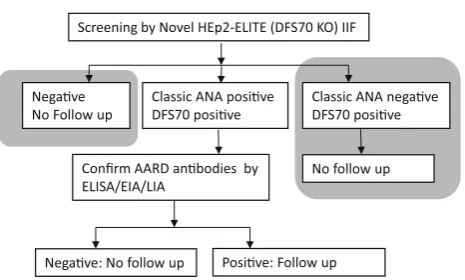
screening population consists of ANA negative and DFS70 positive cases which if confirmed with confidence do not need a clinical follow-up (Fig.7). The current generation of DFS70 specific confirmatory assays neither provide high levels of agreement with IIF results nor are able to confirm the mono-specific/isolated DFS70 positivity, thereby complicating the ANA screening and confirmation algo-rithm (Fig.3). Therefore, to eliminate suspicion of SARD/ AARD, clinical labs rely on a large panel of ANA specific assays even in cases of DFS70 suspicion (Fig.3). Imple-mentation of the newly described HEp-2 ELITE/DFS70 KO substrate as the first step IIF, significantly improves and simplifies the ANA screening and confirmation algo-rithm (Fig.6). The new HEp-2 ELITE/DFS70 KO sub-strate overcomes the limitations associated with accurate interpretation of DFS70 pattern and increases the overall accuracy of the HEp-2 IIF method for screening of ANAs.
Compliance with ethical standards
Conflict of interest K. Malyavantham and L. Suresh are employed at Immco Diagnostics, A Trinity Biotech Company, providers of autoimmune diagnostic kits and services.
Ethical approval For this type of study formal consent is not required.
Human and animal rights This article does not contain any studies with human participants performed by any of the authors.
Informed consent Informed consent was obtained from all individ-ual participants included in the study.
Open Access This article is distributed under the terms of the Creative Commons Attribution 4.0 International License (http://crea tivecommons.org/licenses/by/4.0/), which permits unrestricted use, distribution, and reproduction in any medium, provided you give appropriate credit to the original author(s) and the source, provide a link to the Creative Commons license, and indicate if changes were made.
References
1. Tan EM (1982) Autoantibodies to nuclear antigens (ANA): their immunobiology and medicine. Adv Immunol 33:167–240 2. Agmon-Levin N, Damoiseaux J, Kallenberg C, Sack U, Witte T,
Herold M, Bossuyt X, Musset L, Cervera R, Plaza-Lopez A, Dias C, Sousa MJ, Radice A, Eriksson C, Hultgren O, Viander M, Khamashta M, Regenass S, Andrade LE, Wiik A, Tincani A, Ronnelid J, Bloch DB, Fritzler MJ, Chan EK, Garcia-De La Torre I, Konstantinov KN, Lahita R, Wilson M, Vainio O, Fabien N, Sinico RA, Meroni P, Shoenfeld Y (2014) International recom-mendations for the assessment of autoantibodies to cellular antigens referred to as anti-nuclear antibodies. Ann Rheum Dis 73(1):17–23. doi:10.1136/annrheumdis-2013-203863
3. Chan EK, Damoiseaux J, Carballo OG, Conrad K, de Melo CW, Francescantonio PL, Fritzler MJ, Garcia-De La Torre I, Herold M, Mimori T, Satoh M, von Muhlen CA, Andrade LE (2015) Report of the first international consensus on standardized Screening by Novel HEp2-ELITE (DFS70 KO) IIF
Negave No Follow up
Classic ANA posive DFS70 posive
Classic ANA negave DFS70 posive
Confirm AARD anbodies by ELISA/EIA/LIA
No follow up
Negave: No follow up Posive: Follow up
IIF by HEp-2 ELITE/DFS70 KO simplifies the ANA screening and confirmation algorithm
nomenclature of antinuclear antibody HEp-2 cell patterns 2014–2015. Front Immunol 6:412. doi:10.3389/fimmu.2015. 00412
4. Seelig CA, Bauer O, Seelig HP (2016) Autoantibodies against DFS70/LEDGF exclusion markers for systemic autoimmune rheumatic diseases (SARD). Clin Lab 62(4):499–517
5. Conrad K, Rober N, Andrade LE, Mahler M (2016) The clinical relevance of anti-DFS70 autoantibodies. Clin Rev Allergy Immunol. doi:10.1007/s12016-016-8564-5
6. Ochs RL, Mahler M, Basu A, Rios-Colon L, Sanchez TW, Andrade LE, Fritzler MJ, Casiano CA (2016) The significance of autoantibodies to DFS70/LEDGFp75 in health and disease: integrating basic science with clinical understanding. Clin Exp Med 16(3):273–293. doi:10.1007/s10238-015-0367-0
7. Ochs RL, Muro Y, Si Y, Ge H, Chan EK, Tan EM (2000) Autoantibodies to DFS 70 kd/transcription coactivator p75 in atopic dermatitis and other conditions. J Allergy Clin Immunol 105(6 Pt 1):1211–1220
8. Ochs RL, Stein TW Jr, Peebles CL, Gittes RF, Tan EM (1994) Autoantibodies in interstitial cystitis. J Urol 151(3):587–592 9. Dellavance A, Viana VS, Leon EP, Bonfa ES, Andrade LE, Leser
PG (2005) The clinical spectrum of antinuclear antibodies asso-ciated with the nuclear dense fine speckled immunofluorescence pattern. J Rheumatol 32(11):2144–2149
10. Ge H, Si Y, Roeder RG (1998) Isolation of cDNAs encoding novel transcription coactivators p52 and p75 reveals an alternate regulatory mechanism of transcriptional activation. EMBO J 17(22):6723–6729. doi:10.1093/emboj/17.22.6723
11. Ge H, Si Y, Wolffe AP (1998) A novel transcriptional coacti-vator, p52, functionally interacts with the essential splicing factor ASF/SF2. Mol Cell 2(6):751–759
12. Shinohara T, Singh DP, Chylack LT Jr (2000) Review: age-re-lated cataract: immunity and lens epithelium-derived growth factor (LEDGF). J Ocul Pharmacol Ther 16(2):181–191. doi:10. 1089/jop.2000.16.181
13. Itoh Y, Hamada H, Imai T, Seki T, Igarashi T, Yuge K, Fukunaga Y, Yamamoto M (1997) Antinuclear antibodies in children with chronic nonspecific complaints. Autoimmunity 25(4):243–250 14. Llano M, Vanegas M, Hutchins N, Thompson D, Delgado S,
Poeschla EM (2006) Identification and characterization of the chromatin-binding domains of the HIV-1 integrase interactor LEDGF/p75. J Mol Biol 360(4):760–773. doi:10.1016/j.jmb. 2006.04.073
15. Nishizawa Y, Usukura J, Singh DP, Chylack LT Jr, Shinohara T (2001) Spatial and temporal dynamics of two alternatively spliced regulatory factors, lens epithelium-derived growth factor (ledgf/p75) and p52, in the nucleus. Cell Tissue Res 305(1):107–114
16. Turlure F, Maertens G, Rahman S, Cherepanov P, Engelman A (2006) A tripartite DNA-binding element, comprised of the nuclear localization signal and two AT-hook motifs, mediates the association of LEDGF/p75 with chromatin in vivo. Nucleic Acids Res 34(5):1653–1665. doi:10.1093/nar/gkl052
17. Ogawa Y, Sugiura K, Watanabe A, Kunimatsu M, Mishima M, Tomita Y, Muro Y (2004) Autoantigenicity of DFS70 is restricted to the conformational epitope of C-terminal alpha-helical domain. J Autoimmun 23(3):221–231. doi:10.1016/j.jaut.2004.07.003
18. Watanabe A, Kodera M, Sugiura K, Usuda T, Tan EM, Takasaki Y, Tomita Y, Muro Y (2004) Anti-DFS70 antibodies in 597 healthy hospital workers. Arthritis Rheum 50(3):892–900. doi:10. 1002/art.20096
19. Ganapathy V, Daniels T, Casiano CA (2003) LEDGF/p75: a novel nuclear autoantigen at the crossroads of cell survival and apoptosis. Autoimmun Rev 2(5):290–297
20. Ganapathy V, Casiano CA (2004) Autoimmunity to the nuclear autoantigen DFS70 (LEDGF): what exactly are the
autoantibodies trying to tell us? Arthritis Rheum 50(3):684–688. doi:10.1002/art.20095
21. Daniels T, Zhang J, Gutierrez I, Elliot ML, Yamada B, Heeb MJ, Sheets SM, Wu X, Casiano CA (2005) Antinuclear autoanti-bodies in prostate cancer: immunity to LEDGF/p75, a survival protein highly expressed in prostate tumors and cleaved during apoptosis. Prostate 62(1):14–26. doi:10.1002/pros.20112
22. Brown-Bryan TA, Leoh LS, Ganapathy V, Pacheco FJ, Medi-avilla-Varela M, Filippova M, Linkhart TA, Gijsbers R, Debyser Z, Casiano CA (2008) Alternative splicing and caspase-mediated cleavage generate antagonistic variants of the stress oncoprotein LEDGF/p75. Mol Cancer Res 6(8):1293–1307. doi:10.1158/ 1541-7786.MCR-08-0125
23. Leoh LS, van Heertum B, De Rijck J, Filippova M, Rios-Colon L, Basu A, Martinez SR, Tungteakkhun SS, Filippov V, Christ F, De Leon M, Debyser Z, Casiano CA (2012) The stress oncoprotein LEDGF/p75 interacts with the methyl CpG binding protein MeCP2 and influences its transcriptional activity. Mol Cancer Res 10(3):378–391. doi:10.1158/1541-7786.MCR-11-0314
24. Dai L, Li J, Ortega R, Qian W, Casiano CA, Zhang JY (2014) Preferential autoimmune response in prostate cancer to cyclin B1 in a panel of tumor-associated antigens. J Immunol Res 2014:827827. doi:10.1155/2014/827827
25. Basu A, Sanchez TW, Casiano CA (2015) DFS70/LEDGFp75: an enigmatic autoantigen at the interface between autoimmunity, AIDS, and cancer. Front Immunol 6:116. doi:10.3389/fimmu. 2015.00116
26. American College of Rheumatology Position Statement (2011) Methodology of testing for antinuclear antibodies.www.rheuma tologyorg/practice/ana_position_stmtpdf. Approved by Board of Directors: Aug 2011
27. Meroni PL, Schur PH (2010) ANA screening: an old test with new recommendations. Ann Rheum Dis 69(8):1420–1422. doi:10.1136/ard.2009.127100
28. Mercado MV, Gomez-Banuelos E, Navarro-Hernandez RE, Pizano-Martinez O, Saldana-Millan A, Chavarria-Avila E, Gon-zalez-Rosas L, Andrade-Ortega L, Saavedra MA, Vera-Lastra OL, Jara LJ, Medrano-Ramirez G, Cruz-Reyes C, Garcia-De la Torre I, Escarra-Senmarti M, Anjos LM, Basu A, Albesa R, Mahler M, Casiano CA (2017) Detection of autoantibodies to DSF70/LEDGFp75 in Mexican Hispanics using multiple com-plementary assay platforms. Auto Immun Highlights 8(1):1. doi:10.1007/s13317-016-0089-7
29. Basu A, Woods-Burnham L, Ortiz G, Rios-Colon L, Figueroa J, Albesa R, Andrade LE, Mahler M, Casiano CA (2015) Specificity of antinuclear autoantibodies recognizing the dense fine speckled nuclear pattern: preferential targeting of DFS70/LEDGFp75 over its interacting partner MeCP2. Clin Immunol 161(2):241–250. doi:10.1016/j.clim.2015.07.014
30. Bizzaro N, Tonutti E, Tampoia M, Infantino M, Cucchiaro F, Pesente F, Morozzi G, Fabris M, Villalta D (2015) Specific chemoluminescence and immunoasdorption tests for anti-DFS70 antibodies avoid false positive results by indirect immunofluo-rescence. Clin Chim Acta 451(5):271–277. doi:10.1016/j.cca. 2015.10.008
31. Bizzaro N, Tonutti E, Visentini D, Alessio MG, Platzgummer S, Morozzi G, Antico A, Villalta D, Piller-Roner S, Vigevani E (2007) Antibodies to the lens and cornea in anti-DFS70-positive subjects. Ann N Y Acad Sci 1107:174–183. doi:10.1196/annals. 1381.019
33. Kang SY, Lee WI (2009) Clinical significance of dense fine speckled pattern in anti-nuclear antibody test using indirect immunofluorescence method. Korean J Lab Med 29(2):145–151. doi:10.3343/kjlm.2009.29.2.145
34. Lee H, Kim Y, Han K, Oh EJ (2016) Application of anti-DFS70 antibody and specific autoantibody test algorithms to patients with the dense fine speckled pattern on HEp-2 cells. Scand J Rheumatol 45(2):122–128. doi:10.3109/03009742.2015.1060260
35. Mariz HA, Sato EI, Barbosa SH, Rodrigues SH, Dellavance A, Andrade LE (2011) Pattern on the antinuclear antibody-HEp-2 test is a critical parameter for discriminating antinuclear anti-body-positive healthy individuals and patients with autoimmune rheumatic diseases. Arthritis Rheum 63(1):191–200. doi:10.1002/ art.30084
36. Marlet J, Ankri A, Charuel JL, Ghillani-Dalbin P, Perret A, Martin-Toutain I, Haroche J, Amoura Z, Musset L, Miyara M (2015) Thrombophilia associated with Anti-DFS70 autoantibod-ies. PLoS One 10(9):e0138671. doi:10.1371/journal.pone. 0138671
37. Muro Y, Sugiura K, Morita Y, Tomita Y (2008) High concomi-tance of disease marker autoantibodies in anti-DFS70/LEDGF autoantibody-positive patients with autoimmune rheumatic dis-ease. Lupus 17(3):171–176. doi:10.1177/0961203307086311
38. Muro Y, Sugiura K, Nakashima R, Mimori T, Akiyama M (2013) Low prevalence of anti-DFS70/LEDGF antibodies in patients with dermatomyositis and other systemic autoimmune rheumatic diseases. J Rheumatol 40(1):92–93. doi:10.3899/jrheum.121168
39. Mutlu E, Eyigor M, Mutlu D, Gultekin M (2016) Confirmation of anti-DFS70 antibodies is needed in routine clinical samples with DFS staining pattern. Cent Eur J Immunol 41(1):6–11. doi:10. 5114/ceji.2016.58812
40. Okamoto M, Ogawa Y, Watanabe A, Sugiura K, Shimomura Y, Aoki N, Nagasaka T, Tomita Y, Muro Y (2004) Autoantibodies to DFS70/LEDGF are increased in alopecia areata patients. J Autoimmun 23(3):257–266. doi:10.1016/j.jaut.2004.07.004
41. Pazini AM, Fleck J, dos Santos RS, Beck ST (2010) Clinical relevance and frequency of cytoplasmic and nuclear dense fine speckled patterns observed in ANA-HEp-2. Rev Bras Reumatol 50(6):655–660
42. Schmeling H, Mahler M, Levy DM, Moore K, Stevens AM, Wick J, McMillan JD, Horneff G, Assassi S, Charles J, Salazar G, Mayes MD, Silverman ED, Klien-Gitelman M, Lee T, Brunner
HI, Reed AM, Fritzler MJ (2015) Autoantibodies to dense fine speckles in pediatric diseases and controls. J Rheumatol. doi:10. 3899/jrheum.150567
43. Sperotto F, Seguso M, Gallo N, Plebani M, Zulian F (2017) Anti-DFS70 antibodies in healthy schoolchildren: a follow-up analysis. Autoimmun Rev. doi:10.1016/j.autrev.2017.01.001
44. Yamada K, Senju S, Shinohara T, Nakatsura T, Murata Y, Ishi-hara M, Nakamura S, Ohno S, Negi A, Nishimura Y (2001) Humoral immune response directed against LEDGF in patients with VKH. Immunol Lett 78(3):161–168
45. Ayaki M, Ohoguro N, Azuma N, Majima Y, Yata K, Ibaraki N, Singh DP, Ko V, Shinohara T (2002) Detection of cytotoxic anti-LEDGF autoantibodies in atopic dermatitis. Autoimmunity 35(5):319–327
46. Bizzaro N, Pesente F, Cucchiaro F, Infantino M, Tampoia M, Villalta D, Fabris M, Tonutti E (2016) Anti-DFS70 antibodies detected by immunoblot methods: a reliable tool to confirm the dense fine speckles ANA pattern. J Immunol Methods 436:50–53. doi:10.1016/j.jim.2016.06.008
47. Bentow C, Rosenblum R, Correia P, Karayev E, Karayev D, Williams D, Kulczycka J, Fritzler MJ, Mahler M (2016) Devel-opment and multi-center evaluation of a novel immunoadsorption method for anti-DFS70 antibodies. Lupus 25(8):897–904. doi:10. 1177/0961203316641773
48. Conrad K, Ro¨ber N, Rudolph S, Mahler M (2015) DFS70 anti-bodies—biomarkers for the exclusion of ANA-associated autoimmune rheumatic diseases. LaboratoriumsMedizin 38(6). doi:10.1515/labmed-2015-0040
49. Miyara M, Albesa R, Charuel JL, El Amri M, Fritzler MJ, Ghillani-Dalbin P, Amoura Z, Musset L, Mahler M (2013) Clinical phenotypes of patients with DFS70/LEDGF anti-bodies in a routine ANA referral cohort. Clin Dev Immunol 2013:703759. doi:10.1155/2013/703759
50. Bentow C, Fritzler MJ, Mummert E, Mahler M (2016) Recog-nition of the dense fine speckled (DFS) pattern remains chal-lenging: results from an international internet-based survey. Auto Immun Highlights 7(1):8. doi:10.1007/s13317-016-0081-2