R E S E A R C H A R T I C L E
Open Access
Accuracy of microRNAs as markers for the
detection of neck lymph node metastases in
patients with head and neck squamous cell
carcinoma
Ana Carolina de Carvalho
1,2, Cristovam Scapulatempo-Neto
3, Danielle Calheiros Campelo Maia
1,
Adriane Feijó Evangelista
2, Mariana Andozia Morini
3, André Lopes Carvalho
4*and André Luiz Vettore
1,5*Abstract
Background:The presence of metastatic disease in cervical lymph nodes of head and neck squamous cell carcinoma (HNSCC) patients is a very important determinant in therapy choice and prognosis, with great impact in overall survival. Frequently, routine lymph node staging cannot detect occult metastases and the post-surgical histologic evaluation of resected lymph nodes is not sensitive in detecting small metastatic deposits. Molecular markers based on tissue-specific microRNA expression are alternative accurate diagnostic markers. Herein, we evaluated the feasibility of using the expression of microRNAs to detect metastatic cells in formalin-fixed paraffin-embedded (FFPE) lymph nodes and in fine-needle aspiration (FNA) biopsies of HNSCC patients.
Methods:An initial screening compared the expression of 667 microRNAs in a discovery set comprised by metastatic and non-metastatic lymph nodes from HNSCC patients. The most differentially expressed microRNAs were validated by qRT-PCR in two independent cohorts: i) 48 FFPE lymph node samples, and ii) 113 FNA lymph node biopsies. The accuracy of the markers in identifying metastatic samples was assessed through the analysis of sensitivity, specificity, accuracy, negative predictive value, positive predictive value, and area under the curve values.
Results:Seven microRNAs highly expressed in metastatic lymph nodes from the discovery set were validated in FFPE lymph node samples. MiR-203 and miR-205 identified all metastatic samples, regardless of the size of the metastatic deposit. Additionally, these markers also showed high accuracy when FNA samples were examined.
Conclusions:The high accuracy of miR-203 and miR-205 warrant these microRNAs as diagnostic markers of neck metastases in HNSCC. These can be evaluated in entire lymph nodes and in FNA biopsies collected at different time-points such as pre-treatment samples, intraoperative sentinel node biopsy, and during patient follow-up. These markers can be useful in a clinical setting in the management of HNSCC patients from initial disease staging and therapy planning to patient surveillance.
Keywords:Diagnostic markers, Fine-needle aspiration biopsies, Head and neck cancers, MicroRNAs, miR-200 family, miR-203, miR-205, Neck metastases
* Correspondence:alopescarvalho@uol.com.br;andre.vettore@gmail.com 4Department of Head and Neck Surgery, Barretos Cancer Hospital, Rua
Antenor Duarte Vilela, 1331, Barretos, SP 14784-400, Brazil
1Laboratory of Cancer Molecular Biology, Department of Biological Sciences,
Diadema Campus, Federal University of São Paulo, Rua Pedro de Toledo, 669, São Paulo, SP 04039-032, Brazil
Full list of author information is available at the end of the article
Background
Head and neck squamous cell carcinoma (HNSCC) is one of the most common cancers in the world, with an incidence of approximately 600,000 new cases per year [1]. Despite different strategies employed in the treat-ment of HNSCC patients, including surgery, radiother-apy, and chemotherradiother-apy, late diagnosis and the frequent development of loco-regional recurrences frequently contribute to treatment failure and low rates of overall survival observed [2].
The involvement of neck lymph nodes in HNSCC pa-tients significantly reduces the odds for disease control. The 5-year survival rate for patients without lymph node metastases is 63% to 86%, while patients with neck me-tastases have rates of 20% to 36% [3-6]. Besides being an important prognostic indicator, the presence of meta-static disease in lymph nodes also influences the choice of the adjuvant therapy used to reduce disease recur-rence [7-9]. Therefore, assessment of the neck has be-come an integral part of the treatment planning for HNSCC patients.
There is a well-accepted strategy for the treatment of patients with clinically positive lymph nodes, which includes therapeutic neck dissection followed by postop-erative radiotherapy with or without concurrent chemo-therapy. However, the treatment choice for patients without clinical evidence of neck metastases remains controversial [10,11]. Initial tumors are highly curable by surgery or radiotherapy alone. For this reason, a ‘ wait-and-see’approach, in which the neck is not treated and the patient is followed and monitored with special atten-tion to the evoluatten-tion of disease in cervical lymph nodes is acceptable. Nevertheless, studies have shown that 30% to 60% of cases with initial tumors will present cervical metastases [12-14]. On the other hand, the use of elect-ive neck dissection for all patients with early tumors and clinical and radiological N0 necks at high risk of harbor-ing subclinical disease in the lymph nodes (mainly tongue and floor of the mouth sites, T2 tumors with high depth of invasion) is also defended [15]. Although some studies showed a better loco-regional control and longer regional disease-free survival for patients submit-ted to elective neck dissection [16,17], only 20% to 50% will actually harbor metastatic disease in their lymph nodes. In other words, 50% to 80% of these patients were subjected to the morbidity of unnecessary surgical treatment [12-14,16,18].
A limitation for the correct evaluation of neck metas-tases in HNSCC patients is the lack of sufficient sensitiv-ity of the postoperative assessment of lymph nodes by formalin-fixed tissue sections stained with hematoxylin and eosin (H&E), especially regarding small metastatic deposits. Studies show that 10% to 40% of HNSCC pa-tients with histopathologically negative neck lymph
nodes eventually develop regional metastases, suggesting that metastatic cells present in the lymph nodes could not be detected at diagnosis [12,19,20]. In line with this, 5% to 20% of lymph nodes previously classified as metastasis-free by routine histopathological evaluation present positivity for cytokeratins in immunohistochemi-cal (IHC) analysis [21-25]. To overcome this problem, molecular detection of metastases seems to be one of the most promising methods for definitive lymph node evaluation. Through this approach, metastatic deposits in lymph nodes can be detected in a more sensitive, ac-curate, and less time-consuming manner. Toward this end, the identification of molecular markers capable of detecting the presence of metastatic cells in a back-ground of lymphatic cells is mandatory.
MicroRNAs are small non-coding RNA molecules of approximately 22 nucleotides, described as regulators of gene expression in a variety of multicellular organisms [26,27]. These small molecules interact mainly with the 3′-untranslated regions of specific messenger RNAs (mRNA), inducing their degradation or inhibiting their translation [26,27]. Specific microRNA expression has been observed in different cancer types and at distinct differentiation stages [28-31]. These molecules also play an important role in the development of head and neck cancers. HNSCC-specific microRNA expression profiles and key microRNAs known to orchestrate gene and protein expression levels in these tumors have been established [32]. Recent studies have highlighted differ-ent applications of these molecules as biomarkers in HNSCC, such as in the detection of HNSCC cells in sal-iva samples, in the identification of human papilloma virus-positive oropharyngeal tumors, or as prognostic markers associated with disease progression [32-36]. Furthermore, Fletcher et al. [37] found that the expres-sion of miR-205 was specific to metastatic lymph nodes of HNSCC patients.
In this article, we report the identification of micro-RNAs capable of detecting metastatic cells in formalin-fixed paraffin-embedded (FFPE) lymph nodes and in fine-needle aspiration (FNA) biopsies of HNSCC pa-tients with high sensitivity, specificity, and accuracy.
Methods
Population cohorts
evidence of metastases in cervical lymph nodes at diag-nosis; submitted to surgery as the first therapeutic modality for treatment of the primary tumor; plus elective supraomohyoid neck dissection or sentinel lymph node evaluation. The use of these samples was approved by the Barretos Cancer Hospital Institutional Review Board.
The prospective cohort was comprised by FNA biop-sies of resected lymph nodes from 79 patients who received treatment between 2013 and 2014 at the Department of Head and Neck Surgery of the Barretos Cancer Hospital, Barretos, SP, Brazil. This cohort in-cluded HNSCC patients, regardless of tumor site or stage, who underwent neck dissection during the sur-gery of the primary tumor, as a salvage treatment after organ preservation protocol, or for the treat-ment of patients who developed neck metastases dur-ing follow-up. Informed consent was obtained from each individual prior to tissue collection and the study protocol was approved by Barretos Cancer Hospital Institutional Review Board.
Study design
This study can be divided into three distinct phases. Firstly, a‘discovery set’comprising metastatic and non-metastatic FFPE lymph nodes from patients with tumors in the oral cavity was evaluated in order to select prom-ising markers to be tested in a larger sample cohort. This analysis allowed the selection of good candidates to be evaluated in a‘validation set’containing an additional group of metastatic and non-metastatic FFPE lymph nodes from patients presenting oral cavity tumors. This analysis identified microRNAs with high sensitivity and specificity in detecting the presence of metastatic cells in
lymph nodes. Later, the accuracy of these markers was evaluated in an independent prospective cohort of FNA samples collected from negative and positive lymph nodes from HNSCC patients harboring tumors in the oral cavity, pharynx, and larynx.
FFPE samples processing and RNA purification
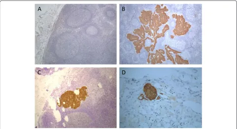
H&E sections corresponding to paraffin blocks contain-ing the samples of interest were reviewed by a patholo-gist to confirm the diagnosis and for characterization of the cellular components present in the samples.
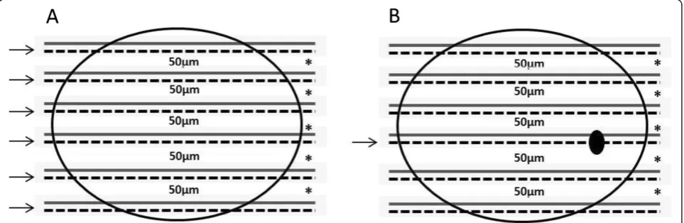
To confirm the absence of metastatic deposits in his-tologically free-of-metastases cases, the blocks contain-ing all lymph nodes resected from each patient were step-sectioned at 50μm intervals and six sections with a thickness of 5 μm each were obtained from each level (Figure 1). The first section was examined by IHC ana-lysis for the expression of cytokeratins, while the remaining five sections were used in molecular analyses. Samples were finally classified as negative when the specimen contained no tumor cells (no positivity for cytokeratins) and positive when isolated tumor cells (<0.2 mm), micrometastasis (0.2–2.0 mm), or macrome-tastasis (>2.0 mm) were detected.
We also challenged the selected markers to detect the presence of metastatic cells in the leftover material removed from each level (intervals of 50 μm) of the step-sectioned FFPE lymph nodes examined in the IHC analyses (Figure 1).
For RNA extraction from FFPE positive lymph node cases, macrodissection was applied to five paraffin-embedded sections to collect the metastatic deposit prior to RNA extraction. For the non-metastatic FFPE cases, total RNA was extracted from sections obtained
from different levels of the lymph nodes. Total RNA was isolated using the Recoverall Total Nucleic Acid Isolation kit (Ambion, Austin, TX, USA), which contains a DNAse treatment step. Total RNA was quantified in the Qubit fluorometer (Invitrogen, Carlsbad, CA, USA) and stored at−80°C until use.
Due to the scarcity of RNA quantity yielded from FFPE samples, no step for evaluation of the RNA purity or integrity was performed. The amplification of the internal controls (U6 and U47) by qPCR was used as indicative of the high quality of the RNA samples. It is important to mention that all samples included in the study showed amplification of both internal controls at early cycles.
Fine-needle aspiration biopsies processing and RNA purification
The material collected through FNA of the resected lymph nodes was used to perform a smear, and the slides were stained to enable the cytological diagnostic of the lymph nodes as‘positive’or‘negative’for the pres-ence of metastatic cells. The remaining material on the needle was washed in 200 μL of sterile saline solution and ethylenediaminetetraacetic acid, snap frozen, and stored at−80°C until RNA extraction.
The extraction of total RNA from FNA samples was performed using the Trizol reagent (Invitrogen, Carlsbad, CA, USA) as previously described [38]. Total RNA was quantified in the Qubit fluorometer (Invitrogen) and stored at−80°C until use.
Global microRNA profiling by TaqMan human MicroRNA arrays
In the‘discovery set’, a global microRNA expression pro-filing was performed in lymph nodes from six FFPE oral tongue SCC samples (four with macrometastases and two with non-metastatic nodes) using TaqMan Human MicroRNA Arrays (Applied Biosystems, Foster City, CA, USA).
Total RNA (40 ng) from each lymph node sample were reverse transcribed into cDNA using the TaqMan microRNA Kit and Megaplex RT Primers (both from Applied Biosystems). After synthesis, the cDNA was pre-amplified using the TaqMan PreAmp Master Mix Kit and Megaplex PreAmp Primers (Applied Biosys-tems). The amplified-cDNA was then transferred to the TaqMan Human MicroRNA Array plates and the ampli-fication was carried out in an Applied Biosystems 7900HT Real-Time PCR system.
The data obtained was analyzed using the software DataAssist v3.0 (Applied Biosystems). The fold-change difference between metastatic and non-metastatic lymph node samples was calculated using the 2-ΔΔCt method [39]. The small nuclear RNA U6 was used as an
endogenous control and the non-metastatic group was assigned as a reference since this was the most stable control in the assay.
Validation of differentially expressed microRNAs by real-time PCR
The ‘validation set’included 48 FFPE lymph node sam-ples and 113 FNA lymph node biopsies from HNSCC patients. Each assay was conducted using the Taqman MicroRNA Reverse Transcription kit (Applied Biosystems) according to the manufacturer’s protocols. Briefly, 10 ng of total RNA was reverse-transcribed to cDNA using a MultiScribe Reverse Transcriptase and a stem-loop primer (Applied Biosystems) specific for each selected microRNA according to manufacturer’s instructions. Quantitative real-time PCR (qRT-PCR) was performed using a TaqMan PCR kit on a 7500 Fast Real-Time PCR System (Applied Biosystems). All reactions were performed in triplicate. To evaluate the differential expression of each microRNA between metastatic and non-metastatic lymph nodes, the 2-ΔΔCtmethod was employed [39]. The ratio between the average Ct values of each microRNA and the internal con-trols (U6 and U47) in 10 non-metastatic lymph node sam-ples was used as reference in the 2-ΔΔCtformula.
Statistical analysis
In the discovery set, to search for differentially expressed microRNAs in metastatic and non-metastatic lymph nodes, a global microRNA expression profiling was con-ducted. In this analysis,ΔCt values from each microRNA were evaluated by the non-parametric Rank Products test using the RankProd R package [40]. The Rank Products test is a robust tool to perform ranking lists with high performance on biological validation. This method did not give any assumption on the data dis-tribution, but provided frequency-based ranking scores. In the validation sets (FFPE and FNA), differentially expressed microRNAs were identified using the Mann– Whitney U test.
adopted to simplify the analysis and allow better repro-ducibility of the tests.
The area under the ROC curve (AUC) was able to identify optimal sensitivity and specificity levels to dis-tinguish metastatic samples from non-metastatic ones. Sensitivity, specificity, accuracy, and positive and nega-tive predicnega-tive values (PPV and NPV) of each individual microRNA in distinguishing metastatic from non-metastatic samples were also calculated along with 95% confidence intervals (95% CI). For FNA samples, the agreement between molecular findings with cyto-logical and histocyto-logical diagnostic was assessed using the Kappa test.
All two-tailed P values were derived from statistical tests, using a computer-assisted program (IBM SPSS Statistics, Version 19), and considered statistically signifi-cant atP<0.05.
Results
Patient characteristics
Clinical and histopathological data of the patients en-rolled in this study are presented in Additional file 1: Tables S1 and S2.
For the FFPE series, of the 48 patients profiled in this cohort, 47.9% were smokers, 79.2% were males, and the age ranged from 43 to 84 years (median 60 years). Pri-mary tumor sites were oral tongue (58.3%), floor of mouth (31.3%), alveolar ridge (8.3%), and lower gum (2.1%) and most were cT2 (62.5%). All patients in this cohort underwent surgery as the primary modality of treatment, and 23 (47.9%) received adjuvant radiation or chemo-radiation therapy.
For the FNA cohort, of the 79 patients included in this cohort, 76% were smokers and 88.6% were males, with age ranging from 29 to 78 years (median 57 years). The primary tumor sites were oral cavity (59.5%), oropharynx (19.0%), larynx (15.2%), and hypopharynx (6.3%) and 78.5% had advanced disease (III–IV). All patients in this cohort underwent neck dissection either during the sur-gery of the primary tumor (69.6%), as a salvage treat-ment after organ preservation protocol (24.1%), or for the treatment of patients who developed neck metasta-ses during follow-up (6.3%).
Identification of metastatic cell deposits in FFPE and FNA lymph node samples
A total of 356 lymph nodes resected from the 48 pa-tients included in the FFPE cohort were examined through H&E to provide the histologic diagnostic of the lymph nodes for the presence of metastatic cells. All histologically-free of metastases lymph nodes were fur-ther step-sectioned and submitted to IHC for cytokera-tins to confirm the absence of metastatic cells and to identify possible small metastatic deposits. Therefore, of
the 48 patients included in the FFPE cohort, 25 harbored metastatic lymph nodes (18 with macrometastases, 5 with micrometastases, and 2 with isolated tumor cells) and 23 samples had metastases-free lymph nodes (Figure 2).
Overall, 113 FNA biopsies were collected from lymph nodes resected from 79 patients submitted to neck dis-section. During the collection, whenever possible, FNA biopsies were conducted in macroscopically positive and negative lymph nodes from the same patient. These samples were further classified as ‘positive’or ‘negative’ according to the cytological examination of the lymph node biopsies. Moreover, the resected lymph nodes were processed according to the routine of the Department of Pathology and H&E sections were assessed to provide the histologic diagnostic of the lymph nodes as‘positive’ or ‘negative’ for the presence of metastatic cells. For three of the 113 FNA samples, there was a disagreement between histological and cytological diagnostics. While the cytological smears were classified as negative in all three samples, the histological assessment found micro-metastatic deposits in two cases and isolated tumor cells in the third one. Thus, according to the cytological evaluation, 42 (37.2%) of the FNA samples collected were classified as positive and 71 (62.8%) as negative; in the histological evaluation, 45 (39.8%) were classified as positive and 68 (60.2%) were classified as negative for the presence of metastatic epithelial cells.
Global microRNA profiling in lymph node samples
Comprehensive microRNA profiles were generated for metastatic (n = 4) and non-metastatic (n = 2) lymph nodes from HNSCC patients using a quantitative RT-PCR array platform. From a total of 667 microRNAs, 439 were detected in at least two samples, regardless of the group, thereby serving as the pool of data for further analyses. From those, 61 presented aP<0.05 in the Rank Products test with 47 showing at least two-fold upregu-lation in all four metastatic samples. A non-supervised hierarchical clustering analysis using the ΔCt values of these 47 microRNAs displayed two distinct clusters formed by metastatic and non-metastatic lymph nodes (Figure 3). Finally, seven microRNAs (200a, miR-200c, miR-203, miR-205, miR-382, miR-628-5p, miR-758), presenting more than 100-fold increment in the expression level in metastatic lymph nodes, were selected for further analyses (Table 1).
Evaluation of the differentially expressed microRNAs in FFPE samples
expression level of the seven microRNAs selected in the
‘discovery set’ (miR-200a, miR-200c, miR-203, miR-205, miR-382, miR-628-5p, and miR-758) was examined in 19 positive lymph nodes (14 macrometastases and 5 micro-metastases) and in 13 negative lymph nodes (of the 23 patients with negative lymph nodes included in this study, 10 were used as a control reference, as indicated in Methods section).
This analysis showed that miR-628-5p, miR-758, and miR-382, were only able to detect 26.3%, 31.6%, and 52.6% of the metastatic samples, respectively, reflecting a low sensitivity (Additional file 1: Table S3, Additional file 2: Figure S1) and were excluded from the study. On the other hand, 200a, 200c, 203, and miR-205 presented maximum specificity (100%) and high sensitivity (84.2%, 94.7%, 100%, and 100%, respectively) (Additional file 1: Table S3). Therefore, in the second series, these four microRNAs were tested in the ex-panded cohort of samples (25 metastatic and 23 non-metastatic lymph nodes).
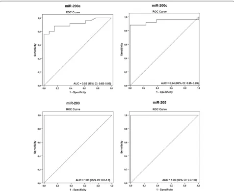
The Youden index obtained from ROC curves was used to determine the cutoff values for upregulation of miR-200a, miR-200c, miR-203, and miR-205, which var-ied from 1.54 to 5.96 (Table 2, Figure 4). The expression profile of these four microRNAs in the metastatic and non-metastatic FFPE lymph node samples showed they were highly associated with the presence of metastatic
cells in the neck lymph nodes of HNSCC cases (miR-200a, 76%; miR-200c, 88%; miR-203, 100%; miR-205, 100%; Figure 5, Table 2). However, 200a and miR-200c barely detected the presence of micrometastases (40% and 80%, respectively), and failed in detecting cases with isolated tumor cells. On the other hand, miR-203 and miR-205 displayed high sensitivity levels (100%) and were also able to correctly classify lymph nodes contain-ing macrometastases, as well as micrometastases or isolated tumor cells (Table 2).
Diagnostic accuracy of microRNAs in FFPE lymph node samples
The diagnostic accuracy of 200a, 200c, miR-203, and miR-205 in differentiating lymph nodes with and without metastases was assessed. The accuracy of miR-200a was 84.2% (95% CI, 68.1–93.4%), the positive predictive value was 100%, and the negative predictive value was 68.4% (95% CI, 43.5–87.3%). The accuracy of miR-200c was 92.1%, the positive and negative predictive values were of 100% and 81.2% (95% CI, 77.5–97.9%), re-spectively. For miR-203 and miR-205, both positive and negative predictive values, as well as the accuracy levels, were of 100% and AUC of 1.0 (Table 3).
Since miR-203 and miR-205 showed a high accuracy in identifying metastatic lymph nodes, the ability of these markers in detecting metastatic cells in a background of
lymphoid tissue was also evaluated. The presence of these microRNAs was examined in the leftover material removed from the step-sectioned lymph nodes during IHC analyses. According to immunostaining, these samples harbored macrometastases (n = 5), micrometastases (n = 4), and isolated tumor cells (n = 2). As shown in Additional file 2: Figure S2, expression levels above the cutoff value could be detected in all lymph nodes carrying metastatic cells, while the expression in the non-metastatic samples was always bellow the cutoff. These results suggested that the evaluation of miR-203 and miR-205 expression still presents high sensitivity and specificity even when the metastatic deposit is‘diluted’in a background of lymphoid tissue.
Validation and diagnostic accuracy of miR-203 and miR-205 expression in FNA samples
Given the high accuracy of miR-203 and miR-205 in tecting metastases in FFPE lymph node samples, we de-cided to evaluate these markers in FNA biopsies of lymph nodes from HNSCC patients.
The accuracy of both markers in detecting metastatic deposits in the FNA biopsies was compared with the re-sults obtained from cytological assessment, which had detected 42 positive FNA samples and 71 negative ones for the presence of epithelial cells. This analysis showed a complete concordance between the cytological and
Figure 3Heatmap representations of the 47 differentially expressed microRNAs with fold-change≥2 andPvalue <0.05 (Rank Products) in the comparison between four metastatic lymph nodes (M: 7A, 1A, 8A, and 9A) and two non-metastatic lymph nodes (NM: 4C and 5C) resected from patients with T2N0 tongue squamous cell carcinomas. Non-supervised hierarchical clustering plotted based on the average
ΔCt values. Up-regulated and down-regulated microRNAs are shown as red and blue, respectively. The columns represent samples and the microRNAs differentially expressed are shown in the lines.
Table 1 Seven microRNAs highly upregulated in metastatic lymph node samples collected from HNSCC patients according to the global microRNA profiling using the TaqMan human microRNA array cards
microRNAs Mean fold-change Pvalue*
miR-200a 594 0.008
miR-200c 628 0.006
miR-203 2169 0.001
miR-205 1648 0.0001
miR-382 82984 <0.00001
miR-628-5p 1564 0.01
miR-758 182 0.01
molecular approaches in the detection of positive and negative nodes with a Kappa value of 1.000. All 42 positive and 71 negative FNA on the cytological assess-ment were correctly identified by both markers with a sensitivity rate of 100% (42/42, CI 95%, 91.5–100) and a specificity level of 100% (71/71, CI 95%, 94.9–100) (Table 2; Figure 6A). A high accuracy of both miR-203 and miR-205 was observed with an AUC of 1.00 (Table 3; Figure 7A), negative and positive predictive values of 100% (95% CI, 94.9–100 and 95% CI, 91.5– 100, respectively), and accuracy levels of 100% (95% CI, 96.05–100) (Table 3).
Next, the results obtained from the histological evalu-ation (after lymph node excision), which is considered the definitive classification of lymph nodes according to the metastatic status, were compared with the molecular analyses of the FNA samples using both miR-203 and miR-205. The histological analyses found 45 positive FNA and 68 negative biopsies. There was an agreement of 100% between both approaches in detecting negative lymph nodes and of 93.3% in classifying positive ones (Kappa value = 0.944). Only three metastatic samples (6.7%) were not detected in FNA samples using the molecular markers, the same ones harboring micro-metastases and isolated tumor cells identified only in the H&E sections (after lymph node excision) and not detected by cytological assessment. This result suggests that small clusters of cells cannot be repre-sented during the aspiration, which would preclude its detection through cytological and molecular methods. All in all, the sensitivity rate for both markers
was 92.9% (39/42, CI 95%, 80.5–98.4), with a specificity level of 100% (71/71, CI 95%, 94.9–100) (Table 2; Figure 6B). A high accuracy of both 203 and miR-205 in distinguishing positive and negative FNA with AUC of 0.963 and 0.966, respectively, and accuracy levels of 97.3% (95% CI, 92.1–99.4) were also observed (Table 3; Figure 7B). Moreover, negative predictive values were of 95.9% (95% CI, 88.6–99.1%) and positive predictive values of 100% (95% CI, 90.9–100%) for both microRNAs (Table 3).
Discussion
HNSCC is a heterogeneous group of tumors that arises from multiple factors that alter different pathways con-tributing to its development and progression. The pres-ence of lymph node metastases is determinant for prognosis and significantly reduces the effectiveness of disease control [7,9,42]. Thus, the accurate detection of lymph node metastases in HNSCC patients is of para-mount importance for its correct staging and more effective treatment planning.
Although the H&E-based pathologic examination of these tumors is very sensitive for macrometastases de-tection, smaller metastatic deposits are difficult to iden-tify and the correct diagnosis relies on the slide quality, operator’s proficiency, sensitivity of the method, and, often, the need for IHC. Although IHC is a more sensi-tive alternasensi-tive, it is a laborious and time-consuming analysis that demands the investigation of serial sections from different levels of each lymph node excised from the patient. Therefore, even though a more detailed
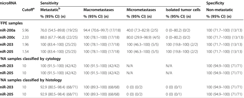
Table 2 Sensitivity and specificity values of microRNAs evaluated in discriminating metastatic and non-metastatic lymph nodes in FFPE and FNA biopsies from lymph node samples
microRNA Sensitivity Specificity
Cutoffa Metastaticb Macrometastases Micrometastases Isolated tumor cells Non-metastatic
% (95% CI) (n) % (95% CI) (n) % (95% CI) (n) % (95% CI) (n) % (95% CI) (n)
FFPE samples
miR-200a 5.96 76.0 (54.5–89.8) (19/25) 94.4 (70.6–99.7) (17/18) 40.0 (7.3–82.9) (2/5) 0 (0–80.2) (0/2) 100 (71.7–100) (13/13)
miR-200c 2.33 88.0 (67.7–96.8) (22/25) 100 (78.1–100) (17/18) 80.0 (29.9–98.9) (4/5) 0 (0–80.2) (0/2) 100 (71.7–100)) (13/13)
miR-203 1.96 100 (83.4–100) (25/25) 100 (78.1–100) (17/18) 100 (46.3–100) (5/5) 100 (19.8–100) (2/2) 100 (71.7–100) (13/13)
miR-205 1.54 100 (83.4–100) (25/25) 100 (78.1–100) (17/18) 100 (46.3–100) (5/5) 100 (19.8–100) (2/2) 100 (71.7–100) (13/13)
FNA samples classified by cytology
miR-203 10 100 (91.5–100) (42/42) 100 (91.5–100) (42/42) N/A N/A 100 (94.9–100) (71/71)
miR-205 10 100 (91.5–100) (42/42) 100 (91.5–100) (42/42) N/A N/A 100 (94.9–100) (71/71)
FNA samples classified by histology
miR-203 10 92.9 (80.5–98.4) (68/71) 100 (89.3–100) (68/68) 0 (0) (0/2) 0 (0) (0/1) 100 (94.9–100) (71/71)
miR-205 10 92.9 (80.5–98.4) (68/71) 100 (89.3–100) (68/68) 0 (0) (0/2) 0 (0) (0/1) 100 (94.9–100) (71/71)
FFPE, Formalin-fixed paraffin embedded; FNA, Fine-needle aspiration; PPV, Positive predictive value; NPV, Negative predictive value; AUC, Area under the ROC curve; CI, Confidence interval; N/A, Not applicable.a
The cutoff values for FFPE samples were determined according to the Youden index (value in which the difference between sensitivity and 1-specificity is maximum) obtained from the ROC curves.b
pathological examination of lymph nodes can provide more accurate information about metastases, this in-volves a long time for preparation of the specimens and a heavy workload for pathologists to examine them. Taken together, all these limitations highlight the need for new molecular markers to complement the patho-logical methods in the task of detecting metastatic cells in lymph nodes of HNSCC patients.
In an initial‘discovery set’analysis, the expression pat-tern of 667 microRNAs was compared between meta-static and non-metameta-static lymph nodes and, after filtering steps, seven microRNAs were chosen to be ex-amined in a FFPE‘validation set’. From those, miR-200a and miR-200c showed high levels of specificity and sen-sitivity, while miR-203 and miR-205 were able to detect even small metastatic deposits such as micrometastases or isolated tumor cells. Such high accuracy levels attest
the feasibility of using these markers in the clinical prac-tice as an additional tool to help pathologists in the correct assessment of lymph node status of HNSCC patients harboring sub-clinical neck metastases.
The sentinel node biopsy is a technique of cervical lymph node staging in patients with primary tumors, allowing a more detailed histological, immunohisto-chemical, and molecular search for occult neck metasta-ses. Usually, the histopathological evaluation of sentinel lymph nodes is based on immunohistochemistry analysis of serial sections, requiring a long time for the final nosis [43]. Consequently, the delay in the correct diag-nosis of the malignancy may lead to the need of additional surgeries, increasing the risk of post-operative complications and functional problems, as well as post-poning the beginning of the adjuvant treatment [44,45]. In the present study, miR-203 and miR-205 were able to
Figure 5Expression profile of miR-200a, miR-200c, miR-203, and miR-205 in lymph node samples containing macrometastases (Ma; n = 18), micrometastases/isolated tumor cells (Mi and isolated tumor cells; n = 7), or in non-metastatic lymph node specimens (NM; n = 13). The Y-axis shows the log10fold-change of the relative expression (2-ΔΔCt). ThePvalue (Mann–Whitney) from each comparison is provided. The dotted line indicates the
cutoff adopted according to the Youden index (value in which the difference between sensitivity and 1-specificity is maximum) obtained from the ROC curves analysis. The horizontal line indicated the median of fold-change values for each group.
Table 3 Accuracy characteristics of microRNAs in discriminating metastatic and non-metastatic lymph nodes in FFPE and FNA biopsies from lymph node samples
microRNA PPV NPV Accuracy AUC (95% CI)
% (95% CI) % (95% CI) % (95% CI)
FFPE samples
miR-200a 100 (82.2–100.0) 68.4 (43.5–87.3) 84.2 (68.1–93.4) 0.92 (0.83–0.99)
miR-200c 100 (84.4–100.0) 81.2 (54.34–95.73) 92.1 (77.5–97.9) 0.94 (0.85–1.0)
miR-203 100 (86.2–100) 100 (75.1–100) 100 (88.6–100) 1.0 (0–1.0)
miR-205 100 (86.2–100) 100 (75.1–100) 100 (88.6–100) 1.0 (0–1.0)
FNA samples classified by cytology
miR-203 100 (91.5–100) 100 (94.9–100) 100 (96.05–100) 1.0 (0–1.0)
miR-205 100 (91.5–100) 100 (94.9–100) 100 (96.05–100) 1.0 (0–1.0)
FNA samples classified by histology
miR-203 100 (90.9–100) 95.9 (88.6–99.1) 97.3 (92.1–99.4) 0.963 (0.921–1.0)
miR-205 100 (93.2–100) 94.6 (85.1–98.8) 96.7 (93.1–100) 0.966 (0.921–1.0)
detect the presence of metastatic deposits even in entire lymph node samples containing large amounts of lym-phocytes and lymphoid stroma. This highlights the high sensitivity of these markers in detecting epithelial cells in a lymphoid tissue background, allowing the correct detection of metastases in entire lymph nodes and avoid-ing false negative results related to partial lymph node assessment. Moreover, a methodology for automated qRT-PCR-based gene expression analysis in an average time of 35 minutes has been described recently [46]. Taken collectively, these results suggest the feasibility of performing microRNA marker analysis in sentinel lymph nodes in a time consistent with an intraoperative evalu-ation, which can be a relevant tool to assist the surgeon in deciding the best approach to be adopted in the neck treatment.
The biopsy based on cytological analysis of FNA sam-ples is a minimally invasive method of high accuracy for the diagnosis of lesions in various organs. Its diagnostic accuracy depends on several factors, such as site, type of injury, the experience of the professional collecting the sample, the quality of the preparation, and the diagnostic skills of the cytopathologist [47,48]. This study showed
that the comparison between cytology assessment, hist-ology examination, and molecular analysis presented high levels of agreement in detecting metastatic cells in lymph nodes. Both cytology and molecular assays could not detect the presence of tumor cells in three FNA samples containing micrometastases or isolated tumor cells. We believe that the misclassification of these three false-negative samples was due to sampling errors repre-sented by the absence of metastatic cells in the FNA specimen. Since metastatic cells were not aspirated, they were not present in the cytological smear and conse-quently were also absent in the sample obtained from washing the aspiration needle. Apart from those, all FFPE samples harboring micrometastases (according to pathological evaluation) could be correctly identified through the use of these molecular markers. Moreover, it is important to note that 30.4% of the FNA samples were collected from patients that have received previous radiotherapy treatment on the neck. Thus, the effects of radiation on lymph node tissues (hence, presence of necrosis and keratin granuloma) did not hinder the cor-rect identification of metastatic cells on these samples. Even though the molecular approach did not show an
Figure 6Expression profile of miR-203 and miR-205 in FNA biopsies from lymph nodes classified as positive (FNA+) or negative (FNA–) according to(A)cytological diagnostic (FNA+: n = 42; FNA–; n = 71) and(B)histological diagnostic (FNA+: n = 45; FNA–; n = 68). The Y-axis shows the log10
improvement in accuracy when compared to the cyto-logical assessment, we believe that its use is warranted given that, unlike molecular assessment, the accuracy of cytological assessments is highly dependent on the qual-ity of the preparation and the diagnostic skills of the cytopathologist. The results obtained with the analysis of FNA samples indicate a high specificity and sensitivity of miR-203 and miR-205 to detect the presence of meta-static cells in the FNA biopsy samples, suggesting their role in a future pre-treatment or follow-up test of suspi-cious lymph nodes in HNSCC patients.
Conclusions
To the best of our knowledge, this is the first study to demonstrate the usefulness of miR-203 and miR-205
expression as a sensitive, specific, and accurate mo-lecular approach for the diagnosis of cervical lymph node metastases. This molecular approach can be used i) at diagnosis, by analyzing samples collected by FNA biopsies, ii) in post-surgery analyses, as de-scribed in this work, as a tool to assist the histo-pathological examination, allowing the investigation of entire lymph nodes and reducing sampling bias, iii) during surgery as an alternative tool for examining sentinel lymph nodes, and iv) during patient follow-up for the testing of suspicious lymph nodes, again, evaluat-ing FNA biopsies.
Our results suggest that the evaluation of miR-203 and miR-205 expression could be an important tool for the management of HNSCC patients, assisting in the
stratification of patients that may harbor neck metasta-ses, aiding in therapy planning and patient surveillance, ultimately contributing to an improvement in quality of life and survival rates.
Additional files
Additional file 1: Table S1.Clinical features of the 48 HNSCC patients enrolled in the FFPE validation data set–FFPE lymph nodes from patients with cervical metastases and with negative lymph nodes were evaluated.Table S2.Clinical features of the 79 HNSCC patients enrolled in the FNA validation data set–113 FNA biopsies* were collected from patients who underwent neck dissection during the resection of the primary tumor, as a salvage treatment after organ preservation protocol or for the treatment of patients who developed neck metastases during follow-up.Table S3.Sensitivity and specificity values for the biomarkers evaluated in lymph node metastasis samples (n = 19, 14 macrometastases and 5 micrometastases), and in non-metastatic lymph nodes (n = 5).
Additional file 2: Figure S1.Expression profile of miR-628-5p, miR-758, and miR-382 in lymph node samples containing macrometastasis (Ma; n = 14) and micrometastases (Mi; n = 5), and in non-metastatic lymph nodes (NM; n = 5). The Y-axis shows the fold-change (2-ΔΔCt) relative expression.Figure S2.Expression profile of microRNAs miR-203 and miR-205 in lymph nodes containing macrometastases, micrometastases, or isolated tumor cells. The microRNAs were recovered from the metastatic cell obtained after macrodissection of five 5-mm sections or by addressing the leftover material after the processing of the entire lymph node from each case. Analysis of the entire non-metastatic (NM) lymph nodes was also included as a negative control. The Y-axis shows the average of the log10fold-change relative expression value (2-ΔΔCt) obtained after three
independent assays. The dotted line indicates the cutoff value determined according to the Youden index from ROC curves. Ma, Macrometastases; Mi, Micrometastases; ITC, Isolated tumor cells.
Abbreviations
AUC:Area under the ROC Curve; CI: Confidence interval; FFPE: Formalin-fixed, paraffin-embedded; FNA: Fine needle aspiration; H&E: Hematoxylin and Eosin; HNSCC: Head and neck squamous cell carcinoma;
IHC: Immunohistochemistry; miR: microRNA; mRNA: Messenger RNA; NPV: Negative predictive value; PCR: Polymerase chain reaction; PPV: Positive predictive value; qRT-PCR: Quantitative reverse transcription polymerase chain reaction; ROC: Receiver operating characteristic.
Competing interests
The authors declare that they have no competing interests.
Authors’contributions
ACC carried out the molecular biology studies, collected clinical data, analyzed data, and performed the statistical analysis. CSN recruited patients, collected tissue samples, conducted cytological and histological evaluations, and participated in study design and coordination. DCCM helped in the molecular biology studies, data analyzes, and helped to draft the manuscript. AFE analyzed PCR-array data. MAM recruited patients, collected tissue samples, and conducted cytological evaluations. CSN recruited patients, collected tissue samples, conducted cytological and histological evaluations, and participated in study design and coordination. ALC and ALV conceived of the study, participated in its design and coordination, and helped to draft the manuscript. All authors read and approved the final manuscript.
Acknowledgements
This study was funded by São Paulo Research Foundation (FAPESP), grant # 2012/14837-7. ACC was recipient of scholarship from São Paulo Research Foundation (FAPESP). ALC has a National Counsel of Technological and Scientific Development (CNPq) scholarship.
Author details
1
Laboratory of Cancer Molecular Biology, Department of Biological Sciences, Diadema Campus, Federal University of São Paulo, Rua Pedro de Toledo, 669,
São Paulo, SP 04039-032, Brazil.2Molecular Oncology Research Center, Barretos Cancer Hospital, Rua Antenor Duarte Vilela, 1331, Barretos, SP 14784-400, Brazil.3Department of Pathology, Barretos Cancer Hospital, Rua Antenor Duarte Vilela, 1331, Barretos, SP 14784-400, Brazil.4Department of Head and Neck Surgery, Barretos Cancer Hospital, Rua Antenor Duarte Vilela, 1331, Barretos, SP 14784-400, Brazil.5Cancer Stem Cell Biology Program, Duke-NUS Graduate Medical School, 8 College Road, Singapore 169857, Singapore.
Received: 23 December 2014 Accepted: 20 April 2015
References
1. Ferlay J, Shin HR, Bray F, Forman D, Mathers C, Parkin DM. Estimates of worldwide burden of cancer in 2008: GLOBOCAN 2008. Int J Cancer. 2010;127:2893–917.
2. Carvalho AL, Nishimoto IN, Califano JA, Kowalski LP. Trends in incidence and prognosis for head and neck cancer in the United States: a site-specific analysis of the SEER database. Int J Cancer. 2005;114:806–16. 3. Dias FL, Kligerman J, Matos de Sa G, Arcuri RA, Freitas EQ, Farias T, et al.
Elective neck dissection versus observation in stage I squamous cell carcinomas of the tongue and floor of the mouth. Otolaryngol Head Neck Surg. 2001;125:23–9.
4. Ferlito A, Rinaldo A, Robbins KT, Silver CE. Neck dissection: past, present and future? J Laryngol Otol. 2006;120:87–92.
5. Leemans CR, Tiwari R, Nauta JJ, van der Waal I, Snow GB. Recurrence at the primary site in head and neck cancer and the significance of neck lymph node metastases as a prognostic factor. Cancer. 1994;73:187–90. 6. Lim SC, Zhang S, Ishii G, Endoh Y, Kodama K, Miyamoto S, et al. Predictive
markers for late cervical metastasis in stage I and II invasive squamous cell carcinoma of the oral tongue. Clin Cancer Res. 2004;10:166–72. 7. Takes RP. Staging of the neck in patients with head and neck squamous
cell cancer: imaging techniques and biomarkers. Oral Oncol. 2004;40:656–67. 8. Shah JP. Patterns of cervical lymph node metastasis from squamous
carcinomas of the upper aerodigestive tract. Am J Surg. 1990;160:405–9. 9. Shores CG, Yin X, Funkhouser W, Yarbrough W. Clinical evaluation of a
new molecular method for detection of micrometastases in head and neck squamous cell carcinoma. Arch Otolaryngol Head Neck Surg. 2004;130:937–42.
10. Ferlito A, Rinaldo A, Silver CE, Gourin CG, Shah JP, Clayman GL, et al. Elective and therapeutic selective neck dissection. Oral Oncol. 2006;42:14–25.
11. Wei WI, Ferlito A, Rinaldo A, Gourin CG, Lowry J, Ho WK, et al. Management of the N0 neck–reference or preference. Oral Oncol. 2006;42:115–22.
12. Rhee D, Wenig BM, Smith RV. The significance of immunohistochemically demonstrated nodal micrometastases in patients with squamous cell carcinoma of the head and neck. Laryngoscope. 2002;112:1970–4. 13. Rodrigo JP, Shah JP, Silver CE, Medina JE, Takes RP, Robbins KT, et al.
Management of the clinically negative neck in early-stage head and neck cancers after transoral resection. Head Neck. 2011;33:1210–9.
14. Thomsen JB, Christensen RK, Sorensen JA, Krogdahl A. Sentinel lymph nodes in cancer of the oral cavity: is central step-sectioning enough? J Oral Pathol Med. 2007;36:425–9.
15. Elsheikh MN, Rinaldo A, Hamakawa H, Mahfouz ME, Rodrigo JP, Brennan J, et al. Importance of molecular analysis in detecting cervical lymph node metastasis in head and neck squamous cell carcinoma. Head Neck. 2006;28:842–9.
16. Kligerman J, Lima RA, Soares JR, Prado L, Dias FL, Freitas EQ, et al. Supraomohyoid neck dissection in the treatment of T1/T2 squamous cell carcinoma of oral cavity. Am J Surg. 1994;168:391–4.
17. Taylor SG. Head and neck cancer. Cancer Chemother Biol Response Modif. 2001;19:465–83.
18. Thomsen JB, Sorensen JA, Krogdahl A. Sentinel lymph nodes in cancer of the oral cavity–isolated tumour cells. J Oral Pathol Med. 2005;34:65–9. 19. Tu GY. Upper neck (level II) dissection for N0 neck supraglottic carcinoma.
Laryngoscope. 1999;109:467–70.
21. Barrera JE, Miller ME, Said S, Jafek BW, Campana JP, Shroyer KR. Detection of occult cervical micrometastases in patients with head and neck squamous cell cancer. Laryngoscope. 2003;113:892–6.
22. Ambrosch P, Brinck U. Detection of nodal micrometastases in head and neck cancer by serial sectioning and immunostaining. Oncology (Williston Park). 1996;10:1221–6. Discussion 1226, 1229.
23. Hamakawa H, Takemura K, Sumida T, Kayahara H, Tanioka H, Sogawa K. Histological study on pN upgrading of oral cancer. Virchows Arch. 2000;437:116–21.
24. Enepekides DJ, Sultanem K, Nguyen C, Shenouda G, Black MJ, Rochon L. Occult cervical metastases: immunoperoxidase analysis of the pathologically negative neck. Otolaryngol Head Neck Surg. 1999;120:713–7.
25. Woolgar JA. Micrometastasis in oral/oropharyngeal squamous cell carcinoma: incidence, histopathological features and clinical implications. Br J Oral Maxillofac Surg. 1999;37:181–6.
26. Bartel DP. MicroRNAs: genomics, biogenesis, mechanism, and function. Cell. 2004;116:281–97.
27. He L, Hannon GJ. MicroRNAs: small RNAs with a big role in gene regulation. Nat Rev Genet. 2004;5:522–31.
28. Lu J, Getz G, Miska EA, Alvarez-Saavedra E, Lamb J, Peck D, et al. MicroRNA expression profiles classify human cancers. Nature. 2005;435:834–8.
29. Olson P, Lu J, Zhang H, Shai A, Chun MG, Wang Y, et al. MicroRNA dynamics in the stages of tumorigenesis correlate with hallmark capabilities of cancer. Genes Dev. 2009;23:2152–65.
30. Ferracin M, Pedriali M, Veronese A, Zagatti B, Gafa R, Magri E, et al. MicroRNA profiling for the identification of cancers with unknown primary tissue-of-origin. J Pathol. 2011;225:43–53.
31. Rosenfeld N, Aharonov R, Meiri E, Rosenwald S, Spector Y, Zepeniuk M, et al. MicroRNAs accurately identify cancer tissue origin. Nat Biotechnol. 2008;26:462–9.
32. Nagadia R, Pandit P, Coman WB, Cooper-White J, Punyadeera C. miRNAs in head and neck cancer revisited. Cell Oncol (Dordr). 2013;36:1–7.
33. Childs G, Fazzari M, Kung G, Kawachi N, Brandwein-Gensler M, McLemore M, et al. Low-level expression of microRNAs let-7d and miR-205 are prognostic markers of head and neck squamous cell carcinoma. Am J Pathol. 2009;174:736–45.
34. Hui AB, Lenarduzzi M, Krushel T, Waldron L, Pintilie M, Shi W, et al. Comprehensive MicroRNA profiling for head and neck squamous cell carcinomas. Clin Cancer Res. 2010;16:1129–39.
35. Salazar C, Calvopina D, Punyadeera C. miRNAs in human papilloma virus associated oral and oropharyngeal squamous cell carcinomas. Expert Rev Mol Diagn. 2014;14:1033–40.
36. Salazar C, Nagadia R, Pandit P, Cooper-White J, Banerjee N, Dimitrova N, et al. A novel saliva-based microRNA biomarker panel to detect head and neck cancers. Cell Oncol (Dordr). 2014;37:331–8.
37. Fletcher AM, Heaford AC, Trask DK. Detection of metastatic head and neck squamous cell carcinoma using the relative expression of tissue-specific mir-205. Transl Oncol. 2008;1:202–8.
38. Corbi FC, Inaoka RJ, Felix RS, Andrade VC, Etto LY, Vettore AL, et al. Comparative expression of a set of genes to an internal housekeeping control in CDNA amplified and not amplified by PolyAPCR in non-Hodgkin’s lymphoma samples obtained from fine-needle aspiration cytology. Diagn Mol Pathol. 2010;19:40–4.
39. Livak KJ, Schmittgen TD. Analysis of relative gene expression data using real-time quantitative PCR and the 2(−Delta Delta C(T)) Method. Methods. 2001;25:402–8.
40. Hong F, Breitling R, McEntee CW, Wittner BS, Nemhauser JL, Chory J. RankProd: a bioconductor package for detecting differentially expressed genes in meta-analysis. Bioinformatics. 2006;22:2825–7.
41. Ruopp MD, Perkins NJ, Whitcomb BW, Schisterman EF. Youden index and optimal cut-point estimated from observations affected by a lower limit of detection. Biom J. 2008;50:419–30.
42. Shah JP. Cervical lymph node metastases–diagnostic, therapeutic, and prognostic implications. Oncology (Williston Park). 1990;4:61–9. Discussion 72, 76.
43. van Diest PJ, Torrenga H, Meijer S, Meijer CJ. Pathologic analysis of sentinel lymph nodes. Semin Surg Oncol. 2001;20:238–45.
44. Kuntz AL, Weymuller Jr EA. Impact of neck dissection on quality of life. Laryngoscope. 1999;109:1334–8.
45. Schiefke F, Akdemir M, Weber A, Akdemir D, Singer S, Frerich B. Function, postoperative morbidity, and quality of life after cervical sentinel node biopsy and after selective neck dissection. Head Neck. 2009;31:503–12. 46. Ferris RL, Xi L, Seethala RR, Chan J, Desai S, Hoch B, et al. Intraoperative
qRT-PCR for detection of lymph node metastasis in head and neck cancer. Clin Cancer Res. 2011;17:1858–66.
47. Heilo A, Sigstad E, Fagerlid KH, Haskjold OI, Groholt KK, Berner A, et al. Efficacy of ultrasound-guided percutaneous ethanol injection treatment in patients with a limited number of metastatic cervical lymph nodes from papillary thyroid carcinoma. J Clin Endocrinol Metab. 2011;96:2750–5. 48. Berner A, Lund-Iversen M, Nesland JM. Fine needle aspirations in oncology.
Arkh Patol. 2011;73:21–6.
Submit your next manuscript to BioMed Central and take full advantage of:
• Convenient online submission
• Thorough peer review
• No space constraints or color figure charges
• Immediate publication on acceptance
• Inclusion in PubMed, CAS, Scopus and Google Scholar
• Research which is freely available for redistribution