R E S E A R C H
Open Access
Usefulness of IMP3 and FOXP3 to predict
metastasis of cutaneous melanomas
Juliana Polizel Ocanha-Xavier
1*, José Cândido C. Xavier-Junior
2and Mariângela Esther Alencar Marques
3Abstract
Background:Melanoma still has considerable mortality in spite of improvements in diagnosis and treatment. Unfortunately, current diagnostic procedures cannot predict precisely its biological behavior, what urges specialists in searching new better biomarkers of lousy prognosis. The objective of the study was to evaluate IMP3 and FOXP3 expression in primary skin melanoma lesions and to correlate with the presence of
metastasis.
Methods: A retrospective cohort study analyzed 112 patients diagnosed with Melanoma, from 2003 to 2011, from a public health service. Samples from the primary lesion were analyzed by two pathologists and one dermatologist to ensure histological subtype, Breslow, the presence of ulceration, mitosis and histological regression. From the species stored, FOXP3 and IMP3 immunohistochemistry staining were performed. Demographic, clinical and evolution aspects of the patients were obtained from records, in the year of 2015. It was considered statistically significant when p-value < 0.05.
Results: The majority of specimens had 25% or fewer cells stained with FOXP3 or IMP3. Their positivity could not be related to the occurrence of metastasis (p= 0.947 and p= 0.936, respectively).
Conclusion: There is no evidence of benefit in using IMP3 or FOXP3 as prognostic markers in primary melanomas in our population.
Keywords: Melanoma, Dermatopathology, Prognosis, Survival rate, Neoplasm metastasis
Background
In the United States, the frequency of melanoma has been increasing from 15 to 21.6 cases per 100,000 inhabitants representing an increase of 1.4% per year. Unfortunately, mortality remains stable. When it is confined to the epidermis (“in situ”), the survival rate in 5 years is near 98.3% (SEER Cancer Statistics Review 2017). When it is metastatic, the survival rate is below 18% (SEER Cancer Statistics Review2017). Therefore, an early diagnosis is re-quired because melanoma is a potentially curable lesion in initial steps.
Since 1969, melanoma has been divided into clinical and histopathological subtypes. There are four subtypes more frequent: Superficial Spreading (SS), Acral (Ac), Nodular (Nd) and Lentigo Maligna (LM) (de Vries et al.2006).
The inability to completely predict the risk of developing metastasis is a significant barrier to the development of treatments with more efficiency (Albreski and Sloan2009). Despite the improvement in follow-up and treatment, there are cases which seem to ignore the rules: thin melanomas with aggressive metastasis and thick lesions with prolonged disease-free survival (Albreski and Sloan2009).
An immunohistochemistry marker named IMP-3 (Insulin-like growth factor-II messenger RNA (mRNA)-binding protein-3) is expressed by embryonic tissues and some neoplasms and play a role in post-transcriptional modulation of oncogenes related to cell proliferation, me-tastasis, chemotherapy resistance and survival. Most studies report it is positive in melanocytes cytoplasm in malignant and metastatic lesions (Pryor et al.2008; Sheen et al.2015).
Another subject of interest concerning escape mecha-nisms of melanoma from host defense is the expression of FOXP3 (Forkhead transcription factor). Its main func-tion is to inhibit the response of TCD8 cells, which are
* Correspondence:jpocanha@gmail.com
1Department of Dermatology and Radiotherapy, Botucatu Medical School
-Paulista State University (UNESP), Rubião Junior District s/n, Botucatu, SP ZIP code 18618-970, Brazil
Full list of author information is available at the end of the article
responsible for immune defense against tumors. It is mainly a nuclear marker of lymphocytes CD4 CD25high but it can also be expressed by melanoma cells (Niu et al.2011).
The purpose of this study is to evaluate IMP3 and FOXP3 expression in primary skin melanoma lesions and to correlate this presence with metastasis. This method is less expensive than molecular analysis, and it would be useful in prognostic markers leading to a better patients’ follow-up and treatment.
Methods
The study included all patients diagnosed with Melanoma from Dermatology and Pathology services, from 2003 to 2011. The study was approved by Institutional Review Board approval (Registration number: 33405714.1.0000.5411/ 2014) and informed consent was spared.
This retrospective cohort study analyzed records in December 2015, looking for epidemiologic and clinical variables: gender, age, lesion diameter, evolution to me-tastasis, death to melanoma or other diseases. The follow-up interval varied from 4 to 12 years.
The slides stained with Hematoxilin and Eosin were reviewed by two dermatopathologists and one dermatolo-gist and a consensus was obtained in a multi-head micro-scope to verify: histological subtype, Breslow, the presence of ulceration, mitosis and histological regression.
Specimens fixed in 10% buffered formalin and embed-ded in paraffin were submitted to immunohistochemical staining of IMP3 and FOXP3.
Patients who had missed follow-up (n= 56), those whose data were incomplete (n= 7), cases of unclassified melanoma (n= 3), cases which histologic specimen was not available or not enough for new immunohistochem-istry slides (n= 17) were excluded.
The samples were submitted to standard immunohisto-chemistry protocol through pt-LINK® and AutoStainer® equipment, in briefly, step-by-step procedures: antigen re-trieval, blocking of endogenous peroxidase, incubation of conjugated antibodies and exposure through chromogen 3,3-diaminobenzidine (DAB–DAKO). Giemsa was used as contra-staining with the purpose of better discrimin-ation between IMP3 and melanin.
Since there is no consensus on score of immunohisto-chemistry in IMP3 and FOXP3, we have established a stratification system which was applied to both markers. Those patients who had no cells marked in immunohis-tochemistry through visual analysis were graduated in 0, followed by 0 to 5% of positive cells (1+), 6 to 25% (2+), 26 to 50% (3+) and 50 to 100% positive cells were
graduated in 4+. Cases that had score 3+ or higher were considered“positive”for statistical analysis.
Data was submitted to Cox’s regression analysis. A ROC’s curve was assessed to establish age’s cut off. It was considered statistically significant when CI was > 95% or when p- value was < 0.05.
Results
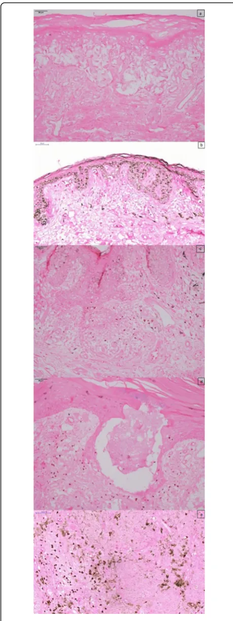
All melanomas diagnosed between the years of 2003 and 2011 totaled 195 cases. After applying excluding criteria, 112 patients have submitted to IMP-3 and FOX P3 im-munohistochemistry staining (Figs. 1 and 2). All metas-tasis, local or distant were considered as the same denouement event.
In this sample, 47.3% of the patients were females, which was not associated with metastasis (p= 0.495) (Table1). Age after 61.5 years old (established by a ROC curve application) was associated with metastasis, com-paring to the younger group (p= 0.032).
Subtypes frequency was similar to the previously de-scribed in literature [2.3]: 64.3% of SS, 15.2% of Ac, 10.7% of Nd and 9.8% of LM. Our sample had thin Breslow (0.7, ranging from 0 to 25 mm), low frequency of mitosis (median of 0, varying from 0 to 50), ulceration (30.4%) and progression to metastasis (24.1%) what rep-resents that most cancers were diagnosed early.
Nodular subtype was the one which had more associ-ation with metastasis (OR = 9.00), but all subtypes had a higher risk in comparison with superficial spreading.
Breslow, ulceration and mitosis were associated with metastasis. The OR for 1 to 4 mm of Breslow was 8.33, and for Breslow > 4 mm the OR was 14.44 (p< 0.001). (Table 1) When there were more than five mitoses, the OR was 15.65 against 13.44 when there were fewer mi-toses. Ulceration was related to metastasis with an OR of 5.44,p< 0.001.
Near half of the patients showed histological regres-sion (49.1%). Regresregres-sion was inversely associated with metastasis (OR = 0.36,p= 0.021).
The majority of cases had 25% or less positivity of both IMP3 and FOXP3. Their positivity could not be re-lated to metastasis (p= 0.947 andp= 0.936, respectively). It was not possible to analyze the event“death” because its number was very low.
Discussion
The main finding of that study was the uselessness of IMP3 an FOXP3 to predict melanoma metastasis in all subtypes, in the studied sample.
There are few studies of the use of IMP3 immunostain-ing in melanoma. However, it is considered positive if present in at least 10% of melanocytes by direct observa-tion (Pryor et al. 2008; Sheen et al.2015). Some demon-strated that it was negative in common and dysplastic nevi, slightly positive in thin melanomas and Spitz nevi and strongly positive in metastatic melanomas (Pryor et al. 2008). Others have suggested a worse prognosis in IMP3 positive cancers (Sheen et al. 2015). Nevertheless, Chokoeva et al. (2015) have shown positivity in only 40% of melanomas and 20% of dysplastic nevi, advocating that it is a weak prognostic marker.
In this study, approximately 10% of the melanomas had more than 25% positive cells. Five instances of superficial spreading melanoma, four instances of acral melanoma, one instance of nodular melanoma and one instance of lentigo maligna melanoma showed positivity of 2+. The positivity rate would be near 20%, if 2+ group had been considered positive too. Our study did not show a correlation between IMP3 and metastasis (p= 0.947), and resetting the cut off value did not ren-der the analysis significant (data not available in tables). Only three out of the metastatic melanomas, which
means only 11% of metastatic melanomas (3/27), had more than 25% of positive cells.
Several studies in animals indicated positive correlation between CD4 + CD25 + FOXP3+ cells presence and tumor progression. Murine melanomas had a delay in time to a visible lesion, decrease in tumor weight and increase in survival time when FOXP3 was inhibited (Franco-Molina et al.2016). On the other hand, Ladányi et al. (2010) and Hillen et al. (2008) did not associated FOXP3 presence with a worse prognosis. This study also did not show its correlation with metastasis (p= 0.936). Most of the posi-tive cases were superficial spreading subtype (7 cases), followed by lentigo maligna (3 cases), nodular (2 cases) and acral subtype (1 case). Our study analyzed the primary lesion, in agreement with other studies (Ladányi et al.
2010; Hillen et al.2008). FOXP3 could be useful in analyz-ing metastasis instead of primary lesions.
The low frequency of denouement events, mainly death, was the main limitation of the study. Therefore, the study collected all possible patients in 9 years, with the minimum follow-up of 4 years and a maximum of 12 years which represents an excellent portrait of our population.
Table 1Metastasis’risk related to clinical and histopathological variables
Variables Without metastasis With metastasis OR (CI95%) p
Gender Female 42(79%) 11(21%) 1.00
Male 43(73%) 16(27%) 1.30 (0.60–2.81) 0.495
Age* ≤61.5 55(85%) 10(15%) 1.00
>61.5 30(64%) 17(36%) 2.35 (1.07–5.13) 0.032
Subtype* SS 66(92%) 6(8%) 1.00
Ac 9(53%) 8(47%) 5.65 (1.95–16.27) 0.001
Nd 3(25%) 9(75%) 9.00 (3.20–25.28) <0.001
LM 7(64%) 4(36%) 4.36 (1.23–15.46) 0.022
Breslow* [0–1] 62(95%) 3(5%) 1.00
(1–4] 16(62%) 10(38%) 8.33 (2.29–30.27) 0.001
>4 7(33%) 14(67%) 14.44 (4.15–50.26) <0.001
Ulceration* Absence 70(90%) 8(10%) 1.00
Presence 15(44%) 19(56%) 5.44 (2.38–12.44) <0.001
Mitosis* 0 58(97%) 2(3%) 1.00
[1–5] 16(55%) 13(45%) 13.44 (3.03–59.59) 0.001
>5 11(48%) 12(52%) 15.65 (3.50–69.93) 0.001
IMP3 ≤2+ 76(76%) 24(24%) 1.00
>2+ 9(75%) 3(25%) 1.04 (0.31–3.45) 0.947
FOXP3 ≤2+ 75(76%) 24(24%) 1.00
>2+ 10(77%) 3(23%) 0.95 (0.28–3.16) 0.936
Regression* Absence 37(65%) 20(35%) 1.00
Presence 48(87%) 7(12%) 0.36 (0.15–0.85) 0.021
Cox’s Regression model.p< 0,05*
Conclusions
In conclusion, there is no evidence of benefit in the use of IMP3 or FOXP3 as prognosis markers in primary melanomas in our population. Clinical and pathological features as mitoses, Breslow, ulceration and histological subtype were associated with metastasis in accordance with previous studies. Regression was inversely associ-ated with metastasis. More investigative studies are ne-cessary to reinforce those statements. Pathologists and dermatologists could be cautious before accomplishing new prognostic markers in their routine.
Acknowledgements
FAPESP and Hélio R.C.Nunes for statistical analysis.
Funding
Fapesp:16013-5
Availability of data and materials
Data and Materials avaliable for requests.
Authors’contributions
All the authors participated of data collecting, statistics analyses, writing and review. All authors read and approved the final manuscript.
Ethics approval and consent to participate
The study was approved by Institutional Review Board approval (Registration number: 33405714.1.0000.5411/2014).
Consent for publication
Not applicable.
Competing interests
The authors declare that they have no competing interests.
Publisher’s Note
Springer Nature remains neutral with regard to jurisdictional claims in published maps and institutional affiliations.
Author details
1Department of Dermatology and Radiotherapy, Botucatu Medical School
-Paulista State University (UNESP), Rubião Junior District s/n, Botucatu, SP ZIP code 18618-970, Brazil.2Private Clinic, Araçatuba, SP, Brazil.3Pathology
Department, Paulista State University (UNESP), Rubião Junior District s/n, Botucatu, SP ZIP code 18618-970, Brazil.
Received: 11 January 2018 Accepted: 26 February 2018
References
Albreski D, Sloan SB (2009) Melanoma of the feet: misdiagnosed and misunderstood. Clin Dermatol 27:556–563
Chokoeva AA, Ananiev J, Wollina U et al (2015) IMP-3 expression in benign melanocytic nevi, dysplastic nevi and malignant melanoma: preliminary findings in Bulgarian patients. J Biol Regul Homeost Agents 29:695–699 de Vries E, Bray F, Coebergh JW et al (2006) Melanocytic tumours. In: LeBoit PE, Burg
G, Weedon D, Sarasin A (eds) Skin tumours. IARC Press, Lyon, pp 49–120 Franco-Molina MA, Miranda-Hernández DF, Mendoza-Gamboa E et al (2016)
Silencing of Foxp3 delays the growth of murine melanomas and modifies the tumor immunosuppressive environment. Onco Targets Ther 9:243–253 Hillen F, Baeten CI, van de Winkel A et al (2008) Leukocyte infiltration and tumor
cell plasticity are parameters of aggressiveness in primary cutaneous melanoma. Cancer Immunol Immunother 57:97–106
Ladányi A, Mohos A, Somlai B et al (2010) FOXP3+ cell density in primary tumor has no prognostic impact in patients with cutaneous malignant melanoma. Pathol Oncol Res 16:303–309
Niu J, Jiang C, Li C et al (2011) Foxp3 expression in melanoma cells as a possible mechanism of resistance to immune destruction. Cancer Immunol Immunother 60:1109–1118
Pryor JG, Bourne PA, Yang Q et al (2008) IMP-3 is a novel progression marker in malignant melanoma. Mod Pathol 21:431–437
SEER Cancer Statistics Review, 1975–2012. [database online] Bethesda: National Cancer Institute. Avaliable at:http://seer.cancer.gov/csr/1975_2012/. Accessed 2 July 2017