Aspiration Biopsy and Brushing Cytology*
WILLIAM
J.
FRABLE, M.D.
Professor of Surgical Pathology and Director of Cytopathology,
Medical College of Virginia, Health Sciences Division of
Virginia Commonwealth University, Richmond
Since its introduction some twenty years ago the Papanicolaou technique for cervical-vaginal cytology has had a tremendous impact on the de-tection of precancerous lesions and in situ cancer in the female genital tract. It has forced the clinician and pathologist to study the "early lesions" of cancer, particularly in the female genital tract and has brought about a better understanding of their biology. Cytology has over the years been extended to other areas, particularly the respiratory tract. Two recent areas of emphasis are the thin-needle aspiration biopsy and direct brushing of mucosal surfaces through the various fiber optic scopes now available, particularly for the respiratory and gastrointestinal tract.
Thin-Needle Aspiration Biopsy. This is an old technique having been employed at Memorial Center for Cancer and Allied Diseases for over thirty years. It has never become popular in the United States, but in the last ten or fifteen years has become widely used in Europe, particularly in the Scandinavian countries. At the Karolinska Institute, approximately 12,000 such aspirations are done per year, mainly for mass lesions of the breast, prostate, lung, thyroid and lymph nodes. The technique ren-ders a rapid diagnosis, particularly on benign and malignant tumors, and is usually done on an out-patient basis. In the setting described, a saving of time and expense to the patient is obvious.
The technique of thin-needle aspiration is done with a narrow gauge 22 or 23 needle of various lengths, percutaneously, without anesthesia. Because * Presented by Dr. Frable at the 44th Annual McGuire Lecture Series, March 23, 1973, at the Medical College of Virginia, Richmond.
310
of the nature of the needle it is nontraumatic and less painful than a venipuncture. Important is the use of a syringe such as the Franzen type, which may be operated with one hand (11). The other hand is used to fix the tumor mass and thereby control the needle point. The type of syringe holder that will employ disposable 20 ml plastic syringes is illus-trated in figure 1.1
The needle attached to the syringe is introduced into the mass to be aspirated without the application of vacuum. The vacuum is applied and the needle is moved back and forth in the mass several times. The operator should watch the barrel of the syringe and avoid getting material into the barrel. It is not necessary to obtain a core of tissue, but if tissue fragments are obtained that is an added bonus. The idea is to obtain cells and to keep the material within the needle.
If a cyst is aspirated this may be evacuated by filling the syringe. If the fluid is clear it is unreward-ing to process it either as a smear from the cell button of the centrifuge specimen or by one of the filter techniques, either nucleopore or millipore. If the fluid is cloudy or hemorrhagic it is advisable to process it as described, either by filter techniques or smears from the cell button. Cell blocks are not use-ful but any fragments of tissue may be processed as a biopsy. ·
Once the needle has been moved back and forth in the mass several times the pressure in the syringe is allowed to equalize before the needle is removed from the mass. This is extremely important. 1 The Cameco Syringe Pistol® purchased from Preci-sion Dynamics Corporation, 3031 Thornton Ave., Burbank,
California 91504.
Fig. I-Syringe pistol for thin-needle aspiration biopsy.
It prevents a chance to deposit the cells along the needle tract. The syringe is then withdrawn and the needle is held over plain glass slides while the syringe is removed. Air is then introduced into the syringe, the needle reattached and a small drop or two of material is expressed onto the slides. There is usually enough material to make two or three slides. Smears are prepared much in the manner of making a blood smear, but the entire slide need not be covered and the material may be confined in a smaller area. Smears to be stained by the Papani-colaou technique should be immediately fixed while still wet in methyl alcohol. Smears for May-Grun-wald Giemsa stain or Wrights-Giemsa or hema-toxylin and eosin are allowed to air dry. In addition, on air dried smears we have used a metachrome B stain with success. This is the same stain that we employ on frozen section material. Smears prepared in this way may be made permanent by simply wash-ing the metachrome B stained slide with water, al-lowing this to evaporate and then coverslipping with Permount®.
European investigators have favored the hema-tology stains while pathologists in the United States have favored the hematoxylin and eosin or Papani-colaou stain. I have found the metachrome B stain combined with the Papanicolaou stain to be ex-tremely useful.
In the aspiration biopsy technique using the thin-needle, the clinical evaluation is extreme,ly im-portant. Since the material is small in amount and of a cellular type rather than a tissue fragment, clinical evaluation of the mass and its orientation becomes of utmost importance. The greatest success with this technique has been when the pathologist who is reading the aspiration has also taken the ma-terial. He has palpated the tumor mass and has a first hand knowledge of the clinical situation. If clinicians wish to adopt this technique and send their material to be read by a pathologist, then they must supply extremely detailed clinical information to avoid errors, especially of the false positive type. The technique has been used particularly to aspirate lymph nodes with metastatic tumors, for breast tu-mors, thyroid lesions, salivary gland tutu-mors, lung tumors through the transthoracic route, prostate and kidney tumors and soft part sarcomas. There has been limited use in bone tumors. Prostate aspirations are done transrectally with a metal guide which fits over the examining finger and is so positioned that the tip of the needle will pass into the mass palpated by the tip of the examining finger.
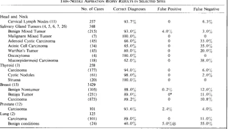
Results. Results from several selected sites in a large series are summarized in Table 1. One of the most useful areas has been cervical lymph nodes, particularly for metastatic tumors. It can be seen that in a large series of cases there were no false positive reports and only a small percentage of false negative reports. Primary sites of these metastatic tumors were equally divided between those above the clavicle and those below (11 ) . In 3 5 % of these cases the aspiration was the first indication of can-cer (11). Zajicek, et al. (27), obtained similar results in 1,200 consecutive aspirations of cervical nodes.
Two difficult areas are congenital cysts of the neck and carotid body tumors. Engzell and Zajicek ( 8) reported the aspiration of 100 consecutive con-genital cysts of the neck and compared these with
TABLE 1
THIN-NEEDLE ASPIRATION BIOPSY RESULTS IN SELECTED SITES
No. of Cases Correct Diagnoses False Positive False Negative
Head and Neck
Cervical Lymph Nodes ( 11) 257 93.7% 0 6.3% Salivary Gland Tumors (4, 5, 6, 7, 26) 368
Benign Mixed Tumor (215) 93.0% 4.0% 3.0%
Malignant Mixed Tumor (7) 100.0% 0 0
Adenoid Cystic Carcinoma (45) 66.0% 0 33.0%
Acinic Cell Carcinoma (34) 65.0% 0 35.0%
Warthin's Tumor (45) 80.0% 0 20.0%
Oncocytoma (4) 100.0% 0 0
Mucoepidermoid Carcinoma (18) 62.0% 0 38.0%
Thyroid (3) 258
Carcinoma (177) 94.0% 0 6.0%
Cystic Nodules (61) 98.0% 0 2.0%
Struma (20) 100.0% 0 0
Breast (15) 1429
Benign Nontumor (305) 88.0% 0.2% 12.0%
Benign Tumor (251) 89.0% 0* 11.0%
Carcinoma (873) 89.2% 0 10.8%
Prostate (12)
Carcinoma IOI 93.6% 2.4% 4.0%
Lung (2) 125
Carcinoma (101) 89.0% 0 11.0%
Benign conditions (24) 40.0% 5.0%@ 55.0%
* 5.3% of cases suspected as carcinoma were all histologically proven fibroadenomas @ one case
location of the cyst, being only 5 5 % in medial cysts and 92 % in lateral cysts. They found benign looking squamous cells in 6% of the smears from metastatic carcinoma.
Engzell, et al. ( 10), have reported their ex-perience with 13 cases of aspiration of carotid body tumors. In one case, there were no cells and in two, metastasis was suspected from an unknown primary. Three cases were diagnosed as neurofibroma or neurofibrosarcoma and in seven cases a correct diag-nosis of carotid body tumor was made. The authors indicate some caution in aspirating this tumor when it is suspected, as one of their cases became he mo-plegic following the procedure.
Aspiration of salivary gland tumors has gen-erally been quite successful by this technique as in-dicated in Table 1 ( 4, 5, 6, 7). Adenoid cystic carcinoma, acinic cell carcinoma and mucoepide r-moid carcinoma carry fairly high false negative rates
(26). Follow-up on benign mixed tumors indicates no increase in recurrence rate due to the aspira-tion technique. In seven cases following resection, the needle tract was serially sectioned and no tumor
cells were found ( 5). Important in the aspiration smears in the diagnosis of adenoid cystic carcinoma are mucinous globules. These were present in 22 of 45 patients ( 6). While the false negative rate for adenoid cystic carcinoma over the 24-year period of the original study is high, it has improved over the last 11 years, being only one out of 23 aspira-tions or 4 % ( 6). This is true also for 34 cases of acinic cell carcinoma in which aspirations of the last 11 years have resulted in a false negative rate of two in 16 or 12% (7).
Other salivary gland carcinomas are too infr e-quent to document the reliability of the technique. The most difficult has been mucoepidermoid tumors of low grade malignancy. In the report by Zajicek and Eneroth ( 26), 18 cases of mucoepidermoid carcinoma yielded only cystic fluid, cell detritus and lymphocytes. Poorly differentiated mucoepidermoid carcinoma as well as the unclassified adenocarcino-mas were easily identified by the aspiration technique.
no false positives in carcinoma, cystic nodules or struma, and a small percentage of false negatives in carcinoma and cystic nodules ( 3).
The major area of interest in the United States has been needle aspiration of the breast. Franzen and Zajicek ( 15) have attained a high degree of accuracy with this technique, not only with carci-noma, but with the benign disease. A conservative attitude is required in dealing with breast lesions since radiation or surgical treatment is frequently done on the basis of aspiration diagnosis. False negative aspirations are generally on the basis of tumors less than 1 cm, very fibrotic, poorly cel-lular carcinomas or failure to aspirate the residual mass after evacuating a cyst. With increased ex-perience in the recent series, the false negative rate has been cut to 5 % .
It can also be seen from Table 1 that the aspira-tion technique is highly accurate in prostate car-cinoma. Since the approach is transrectal, there have been a few cases, less than 2 % , of gram negative septicemia attributable to the aspiration ( 12).
The lung has been the most recent area where the thin-needle technique has been used. Lesions of the lung yield grudgingly to diagnostic tech-niques of exfoliative cytology or conventional bron-choscopy and biopsy. Prior to the thin-needle technique, a large bore-type needle was used with a frequent tendency to hemorrhage, pneumothorax or air embolism. Dahlgren (2) has used the thin-needle technique under fluoroscopic control with only topi-cal skin anesthesia. In 101 malignant tumor cases, there have been no false positives and in the 24 benign conditions, only one false positive. In the total series of 3,000 thin-needle aspirations, there have been no complications (2). Sputum cytology in the same material has yielded only a 40%
diag-nostic rate. Nasiell (22) correctly diagnosed from 83 lung cancer cases, 72 % by the thin-needle tech-nique. About half of the false negative cases had inadequate material. The results were better than conventional sputum cytology. The technique is thought by that author to be particularly good for peripheral and small tumors or for clinically inop-erable cases. The author reported no complications. Sanders, et al. (25), also reported a large series of 164 patients with an accuracy of 84 % in malignant neoplasms. The authors did not find the technique accurate in nonmalignant localized parenchymal dis-ease and not particularly good in chest wail or dif-fuse parenchymal disease. They did find it helpful
in the mediastinal mass lesions, particularly peri-cardia!, bronchogenic or thymic cysts. They listed the contraindications as: 1 ) hemorrhagic diathesis, 2) a patient on anticoagulants, 3) severe pulmonary hy-. pertension, 4) pulmonary hydatid cyst, 5) uncon-trolled cough, 6) advanced emphysema, 7) patient with suspected arteriovenous malformation.
These authors used the large bore needle (2.1-3.0 ml) and had a 30% complication rate of small pneumothoraces. This needle is twice as large as that recommended in the thin-needle technique from the European workers (25). Most of these pneu-mothoraces were asymptomatic. Five percent in this series, however, required tube drainage. In the follow-up of this series, there were no verified cases of tumors spread by the needle technique employed. Janower and Land (20) also used the thicker needle of the Vin-Silverman type. As expected, they had a 10-15% minimal complication rate of hemop-tysis and pneumothorax. These larger needles can-not be recommended any longer, therefore, in this technique.
The most frequent criticism leveled at the thin-needle technique is the implantation of tumor cells along the needle tract. Berg and Robbins ( 1), in a 15-year follow-up of 370 cases of breast cancer that were aspirated and 370 matched controls, found that the long term prognosis slightly favored the aspirated cases. This would seem to indicate no un-toward effects from the thin-needle aspiration tech-nique. Engzell, et al. (9), have also studied aspira-tion experimentally in the popliteal lymph node in rabbits seeded with the Vx2 carcinoma cells. The efferent lymphatics and efferent vein were cannulated and material was collected during the aspiration with massage of the lymph node. In one case out of 16, tumor cells were found in the blood and lymph after massage and thin-needle aspiration.
The authors (9) also reported the clinical follow-up of 124 cases of benign mixed tumors after ten years and found three local recurrences at 4, 5 and 9 years. No tumor was found in the needle tract on s~rial sectioning. In addition, they studied 242 patients with prostate carcinoma who had been followed for five years after thin-needle aspiration. One patient of this group developed transrectal growth three years after needle aspiration. It would seem from these studies that this technique is safe from the criticism of implantation of tumor cells in
the needle tract.
cyto-logic investigation has been brushing cytology us-ing the various fiber optic scopes, particularly in the respiratory and gastrointestinal tract. In the res-piratory tract, this is the outgrowth of the selective catheterization technique of Nordenstrom and Car-lens (23) and the disposable bronchial brush de-signed by Fennessey (13, 14).
We prefer the bronchial brushing under direct vision using the fiber optic bronchoscope. This al-lows visualization of any lesion in the major and
most of the secondary bronchi as well as the res-piratory excursions. Any lesion seen may be sam-pled directly and the brush may be pushed into the parenchyma of most of the bronchial segments for peripheral sampling. Our experience to date, involves 39 cases with cytologic abnormalities, including 16 carcinomas mostly of the squamous type and 18 cases with various atypias of reserve cell hyperplasia, squamous metaplasia or bronchial hyperplasia. We have had four unusual cases as follows: cytomegalic inclusion disease in a transplantation case, pneumo-cystis carinii in a patient with myeloma, adenovirus infection and the identification of ferruginous bodies along with atypical alveolar pneumonocytes.
We have had the best success in smear prepara-tion by having a technologist available in the room where the brushing is being done to make the smears. Two or more smears are made using totally frosted Dakins slides2 which have been premoistened with ethyl alcohol. These are immediately fixed in 95% ethyl alcohol. Just because the slides are damp, cel-lular material is not lost and drying artifacts which occur very rapidly with bronchial epithelial cells are avoided. This is extremely important. Failure to fol-low this simple technique will result in poor quality smears and will discourage the physicians from using this very valuable technique. Following the making of the smears, we shake the brush in a small amount, 10 ml, or physiologic saline or Polysal®3; from this material filters are prepared, either nucleopore or millipore. The material is all stained by the Papani-colaou technique. If fragments of tissue ar.e obtained, as they occasionally are, they may be processed as a biopsy.
The results from several series are reported in Table 2 (16, 17, 18). A false positive rate has been obtained in these series of less than 1 % or one
2 Trident Microscope Slides-Rectangular Dakin, Aloe Scientific, St. Louis, Missouri 63103.
3 Polysal® Balanced Electrolyte Injectable. Cutter Laboratories Inc., Berkeley, California 94710.
case. The best results are reported by Hattori et al. ( 18), who attained an accuracy of 91.6 % in 12 cases of lung cancer. In this series there were no false positive cases. Even in the community hospital where there might not be special interest in this tech-nique, Funkhouser and Meininger ( 17) reported 70% accuracy. Combining this with bronchial and sputum exfoliative cytology, an overall accuracy of 83.8% was obtained in the diagnosis of lung cancer.
Contraindications to the technique as outlined by Janower and Land (20) are patients who are in poor general condition or who cannot tolerate any bronchial irritation. The advantages to the tech-nique, in our opinion, are that the quality of the material is greatly improved from the cytologic point of view. Secondly, a more specific diagnosis can be rendered, particularly in inflammatory and infectious conditions. Thirdly, secondary segmental bronchi and peripheral lesions can be reached and bronchi can be selectively sampled, particularly in "early" lung cancer cases.
The technique is also very valuable in lesions of the esophagus and stomach. The Japanese have pioneered the technique through their use of fiber optics. In their hands, with the early diagnosis of gastric cancer by this method, they have reported a survival in 365 patients of 92.5 % for five years ( 19). While this technique has not generally been used in the United States, the results reported by Prolla et al. (24), summarized in Table 2, are quite impressive. The false positive rate is very low and is better than conventional lavage cytology. The false negative rate is much lower than the conven-tional lavage cytology. The advantages are the simul-taneous brushing with endoscopy. The cytology is selected from the lesion seen. The cytologic pro-cedure takes only a short time in contrast to the collection, preservation and centrifugation for con-ventional lavage cytology. The preservation and cel-lularity of the material is much better and there are no complications (21). Our own experience with this technique in a limited series to date would con-firm all of these findings. Particularly to be stressed is that the quality of the material cytologically is much improved.
TABLE 2
BRUSHING CYTOLOGY RESULTS IN SELECTED SITES
Bronchi and Lung* (16, 17, 18) Stomach and Esophagus (24)
Carcinoma and Lymphoma Benign Conditions
* Combination of three series
No. of Cases
114
269 (54) (215)
t No false negatives among malignant tumors
@ One case
the lesion, particularly in the brushing cytology; the quality of the material is much improved over con-ventional cytologic techniques and the complications to the patient and the expense of the procedures are minimal.
Author's note: Grateful acknowledgment is given
to Mrs. Harriet Kent who prepared the manuscript.
REFERENCES
1. BERG, J. w. AND ROBBINS, G. F. A late look at the
safety of aspiration biopsy. Cancer 15: 826, 1962.
2. DAHLGREN, S. E. AND LIND, B. Comparison between
diagnostic results obtained by transthoracic needle
bi-opsy and by sputum cytology. Acta Cytol. 16:53, 1972.
3. EINHORN, J. AND FRANZEN,
s.
Thin-needle biopsy in thediagnosis of thyroid disease. Acta Radial. 58:321, 1962.
4. ENEROTH, C. M. AND ZAJICEK, J. Aspiration biopsy of
salivary gland tumors. II. Morphologic studies on smears
and histologic sections from oncocytic tumors ( 45
cases of papillary cystadenoma lymphomatosum and
4 cases of oncocytoma). Acta Cytol. 9:355, 1965.
5. ENEROTH,
c
.
M. AND ZAJICEK, J. Aspiration biopsy ofsalivary gland tumors. III. Morphologic studies on
smears and histologic sections from 368 mixed tumors.
Acta Cytol. 10:440, 1966.
6. ENEROTH,
c.
M. AND ZAJICEK, J. Aspiration biopsy ofsalivary gland tumors. IV. Morphologic studies on
smears and histologic sections from 45 cases of adenoid
cystic carcinoma. Acta Cytol. 13:59, 1969.
7. ENEROTH, C. M., JAKOBSSON, P. AND ZAJICEK, J.
Aspira-tion biopsy of salivary gland tumors. V. Morphologic
investigation on smears and histologic sections of acinic
cell carcinoma. Acta Radio/. Suppl. 310:85, 1971.
Correct Diagnoses
75.0%
90.7%
98.6%
False Positive
<1.0%@
0
1.4%
False Negative
9.3%
0
8. ENGZELL, U. AND ZAJICEK, J. Aspiration biopsy of
tumors of the neck. I. Aspiration biopsy and cytologic
findings in 100 cases of congenital cysts. Acta Cytol.
14:51, 1970.
9. ENGZELL, U., ESPOSTI, P. L., RUBIO, C., SIGURDSON, A.
AND ZAJICEK, J. Investigation on tumor spread in
con-nection with aspiration biopsy. Acta Radial. 10: 385,
1971.
10. ENGZELL, U., FRANZEN, S. AND ZAJICEK, J. Aspiration
biopsy of tumors of the neck. II. Cytologic findings in
13 cases of carotid body tumor. Acta Cytol. 15:25,
1971.
11. ENGZELL, U., JAKOBSSON, P. A., SIGURDSON, A. AND
ZAJICEK, J. Aspiration biopsy of metastatic carcinoma
in lymph nodes of the neck. A review of 1,101
con-secutive cases. Acta Otolaryng. 72: 138, 1971.
12. EsPOSTI, P. L. Cytologic diagnosis of prostatic tumors
with the aid of transrectal aspiration biopsy. A critical
review of 1,110 cases and a report of morphologic and
cytochemical studies. Acta Cytol. 10: 182, 1966.
13. FENNESSEY, J. J. A technique for selective catheteriza
-tion of segmental bronchi using arterial catheters. Am.
J. Roentgen. 96:936, 1966.
14. FENNESSEY, J. J. Bronchial brushing in the diagnosis
of peripheral lung lesions. A preliminary report. Am.
J. Roentgen. 98:474, 1967.
15. FRANZEN, S. AND ZAJICEK, J. Aspiration biopsy in
diag-nosis of palpable lesions of the breast. Critical review
of 3,479 consecutive biopsies. Acta Radial. 7:241, 1968.
16. FRY, w. A. AND MANALO-ESTRELLA, P. Bronchial
brush-ing. Surg. Gyneco/. Obstet. 130:67, 1970.
17. FUNKHOUSER, J. w. AND MEININGER, D. E. Cytologic
aspects of bronchial brushing in a community hospital.
18. HATTORI, S., MATSUDA, M., SUGIYAMA, T. AND MATSUDA,
H. Cytologic diagnosis of early lung cancer: Brushing
method under x-ray television fluoroscopy. Dis. Chest 45:129, 1964.
19. HAYASHIDA, T. AND KIDOKORO, T. End results of early
gastric cancer collected from 22 institutions. Stomach
a11d /11testi11e (Japanese) 4: 1077, 1969.
20. JANOWER. M. L. AND LAND, R. E. Lung biopsy.
Bron-chial brushing and percutaneous puncture. Radio/. Cli11.
N. Am. 9:73, 1971.
21. KOBAYASHI, S., PROLLA, J. C. AND KIRSNER, J. B.
Brushing cytology of the esophagus and stomach under
direct vision by fiberscopes. Acta Cytol. 14:219, 1970.
22. NASIELL, M. Diagnosis of lung cancer by aspiration
biopsy and a comparison between this method and
exfoliative cytology. Acta Cytol. 11: 114, 1967.
23. NORDENSTROM, B. AND CARLENS, E. Bronchial biopsy
in connection with bronchography. A eta Radio/.
(Diagn.) 3:37, 1963.
24. PROLLA, J. C., YOSHII, Y., ROGERIO, G. X. AND KJRSNER,
J. B. Further experience with direct vision brushing
cytology of malignant tumors of upper gastrointestinal
tract. Histopathologic correlation with biopsy. A eta Cyto/. 15:375, 1971.
25. SANDERS, D. E., THOMPSON, D. W. AND PUDDEN, B. J.E.
Percutaneous aspiration lung biopsy. Can. Med. Assoc. 104: 139, 1971.
26. ZAJICEK, J. AND ENEROTH, C. M. Cytological diagnosis
of salivary-gland carcinomata from aspiration biopsy smears. Acta Oto/aryng. 263: 183, 1970.
27. ZAJICEK, J., ENGZELL, U. AND FRANZEN,
s.
Aspirationbiopsy of lymph nodes in diagnosis and research. In: