Cysts of the Jaws in Pediatric Population:
A 12-Year Institutional Study
1SK Padmakumar, 2VT Beena, 3Devu Aloka, 4Rupali Lav, 5R Sivakumar
ABSTRACT
Aim: The aim of the study was to find the clinicopathological correlation of the cysts of the jaws in the pediatric population from the data obtained from a major tertiary dental care insti tution in the state of Kerala.
Materials and methods: The cases dated between 1st January 2001 and 31st December 2012 were retrieved from the archives of the Department of Oral Pathology and Microbiology, Government Dental College, Thiruvananthapuram, India. The data were analyzed for age, gender, site of the cyst and histo pathologic type.
Results: Out of a total of 5894 biopsies received, cysts of the jaws accounted for 1396 (23.68%) cases. Among the cystic lesions, pediatric cysts were found to be 187 (13.39%) in number. Of these, 125 were intraosseous cystic lesions of the jaws of which, 81 (64.8%) were developmental in origin, 42 (33.6%) were inflammatory in origin.
Conclusion: From the data, it is possible to conclude that among all types of cysts of the jaws, developmental cysts are the most commonly encountered in the pediatric population. The lesions show a male predominance with anterior maxilla being the most common site.
Keywords:Odontogenic cyst, Pediatric population, Develop mental cyst, Inflammatory cyst.
How to cite this article: Padmakumar SK, Beena VT, Aloka D, Lav R, Sivakumar R. Cysts of the Jaws in Pediatric Population: A 12Year Institutional Study. Oral Maxillofac Pathol J 2015; 6(1):532536.
Source of support: Nil Conflict of interest: None
InTRoDuCTIon
Kramer (1974) has defined a cyst as ‘a pathological cavity having fluid, semifluid or gaseous contents and which is not created by the accumulation of pus’.1
Epithelium-lined cysts in bone are seen only in the jaws, with rare exceptions.2 Cysts of odontogenic origin are among the
commonest lesions encountered in dental practice and
Original research
1,5Assistant Professor, 2Professor and Head, 3Junior Resident 4Senior Resident
15Department of Oral Pathology and Microbiology, Government
Dental College, Thiruvananthapuram, Kerala, India
Corresponding Author: Devu Aloka, Junior Resident Department of Oral Pathology and Microbiology, Government Dental College, Thiruvananthapuram, Kerala, India, Phone: 09496734344, email: [email protected]
hence, it is imperative for oral and maxillofacial patholo-gists to get well-acquainted with these pathologies.
Cysts of the jaws are known to occur across different age and ethnic groups. Although existing literature on the incidence and clinicopathological features of cysts of the jaw is extensive, little has been discussed about these cysts occurring in the pediatric population. To the best of our knowledge, there is limited reported literature on jaw cysts in the pediatric age group within the South Indian population.
The present study was aimed at clinicopathological correlation of cysts of the jaws in the pediatric popula-tion reporting to a tertiary government dental healthcare educational institution located in the state of Kerala, in the southern most part of India, with a predominantly Dravidian population.
MATeRIALS AnD MeThoDS
The past 12-year records of the Department of Oral Pathology and Microbiology, Government Dental Col-lege, Thiruvananthapuram, India, were retrieved from the archives dated between 1st January 2001 and 31st December 2012. A total of 5894 biopsies were obtained, out of which, cysts of the jaws accounted for 1396 cases. Since our study was aimed at estimating the frequency of jaw cysts in the pediatric population, only cases belong-ing to the age group 0 to 14 years were included in the sample population.
Cysts of the Jaws in Pediatric Population: A 12-Year Institutional Study
OMPJ
The data were collected and entered into a Microsoft Excel spread sheet and descriptive statistical results were drawn using the SPSS version 16.0 statistical software package.
ReSuLTS
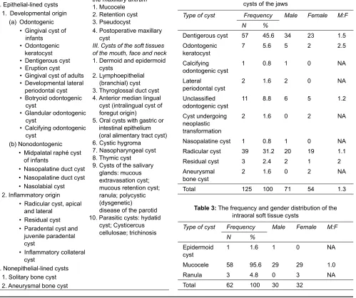
Out of a total of 5894 biopsies received at our department during the 12-year-period, cysts of the jaws accounted for 1396 (23.68%) cases. Among the cystic lesions, pediatric cysts were found to be 187 (13.39%) in number. Of these, 125 were intraosseous cystic lesions of the jaws of which, 81 (64.8%) were developmental in origin, 42 (33.6%) were inflammatory in origin and the rest of the cases included aneurysmal bone cysts, epidermoid cyst and nasopalatine cyst. Table 1 shows the 2005 WHO classification of cysts of the head and neck region which was the basis for our case segregation.
Among the developmental cysts, dentigerous cysts (45.6%) were found to be the most common followed by
odontogenic keratocysts (5.6%) and among the cysts of inflammatory origin, radicular cysts (31.2%) were found to be the predominant type. In our study, we also noted that mucoceles accounted for 95.08% of the total number of intraoral soft tissue cysts.
Among the 125 cysts, 71 (56.8%) were in males and 54 (43.2%) were in females. This male predominance was however not significant statistically (p > 0.05). The overall male:female ratio was found to be 1.3:1. The overall fre-quency and gender distribution of various cysts is shown in Tables 2 and 3. Graph 1 shows distribution of various cysts of the jaws and Graph 2 shows gender distribution among predominant cysts.
A recurrence was noted in five cases, two each of odontogenic keratocyst and radicular cyst and one of calcifying odontogenic cyst type I-C. In addition to this, two neoplastic transformations of dentigerous cysts into ameloblastoma were encountered, both in females aged 13 years. Five syndromic cases were also recorded.
Table 1: WHO classification of the cysts of the head and neck region (2005)
I. Cysts of the jaws II. Cysts associated with
the maxillary antrum
1. Mucocele 2. Retention cyst 3. Pseudocyst A. Epitheliallined cysts
1. Developmental origin (a) Odontogenic
• Gingival cyst of infants
• Odontogenic keratocyst • Dentigerous cyst • Eruption cyst • Gingival cyst of adults • Developmental lateral periodontal cyst • Botryoid odontogenic cyst
• Glandular odontogenic cyst
• Calcifying odontogenic cyst
4. Postoperative maxillary cyst
III. Cysts of the soft tissues of the mouth, face and neck
1. Dermoid and epidermoid cysts
2. Lymphoepithelial (branchial) cyst 3. Thyroglossal duct cyst 4. Anterior median lingual cyst (intralingual cyst of foregut origin)
5. Oral cysts with gastric or intestinal epithelium (oral alimentary tract cyst) 6. Cystic hygroma
7. Nasopharyngeal cyst 8. Thymic cyst
9. Cysts of the salivary glands: mucous extra vasation cyst; mucous retention cyst; ranula; polycystic (dysgenetic) disease of the parotid 10. Parasitic cysts: hydatid
cyst; Cysticercus cellulosae; trichinosis (b) Nonodontogenic
• Midpalatal raphé cyst of infants
• Nasopalatine duct cyst • Nasopalatine duct cyst • Nasolabial cyst 2. Inflammatory origin
• Radicular cyst, apical and lateral
• Residual cyst • Paradental cyst and juvenile paradental cyst
• Inflammatory collateral cyst
B. Nonepitheliallined cysts 1. Solitary bone cyst 2. Aneurysmal bone cyst
Table 2: The frequency and gender distribution of cysts of the jaws
Type of cyst Frequency Male Female M:F
N %
Dentigerous cyst 57 45.6 34 23 1.5 Odontogenic
keratocyst 7 5.6 5 2 2.5
Calcifying
odontogenic cyst 1 0.8 1 0 NA Lateral
periodontal cyst 2 1.6 2 0 NA Unclassified
odontogenic cyst 11 8.8 6 5 1.2 Cyst undergoing
neoplastic transformation
2 1.6 0 2 NA
Nasopalatine cyst 1 0.8 1 0 NA Radicular cyst 39 31.2 20 19 1.1
Residual cyst 3 2.4 2 1 2
Aneurysmal
bone cyst 2 1.6 0 2 NA
Total 125 100 71 54 1.3
Table 3: The frequency and gender distribution of the intraoral soft tissue cysts
Type of cyst Frequency Male Female M:F
N %
Epidermoid
cyst 1 1.6 1 0 NA
Mucocele 58 95.6 29 29 1.0
Ranula 3 4.8 0 3 NA
Graph 1: Frequency of types of cysts Graph 2: Gender distribution of dentigerous cyst, odontogenic keratocyst, radicular cyst and residual cyst
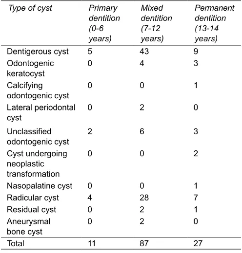
Table 4: The age distribution of cysts of the jaws
Type of cyst Primary
dentition (0-6 years)
Mixed dentition (7-12 years)
Permanent dentition (13-14 years)
Dentigerous cyst 5 43 9
Odontogenic
keratocyst 0 4 3
Calcifying
odontogenic cyst 0 0 1
Lateral periodontal
cyst 0 2 0
Unclassified
odontogenic cyst 2 6 3
Cyst undergoing neoplastic transformation
0 0 2
Nasopalatine cyst 0 0 1
Radicular cyst 4 28 7
Residual cyst 0 2 1
Aneurysmal
bone cyst 0 2 0
Total 11 87 27
Table 5: The mean age distribution of developmental and inflammatory cysts
Type of cyst Mean age
(years) Standard deviation Minimum Maximum
Developmental 10.14 2.5 3.5 14.0 Inflammatory 9.98 2.4 5.0 14.0
The highest frequency of jaw cysts of the three age intervals which we had considered in the study was noted in the mixed dentition period. 68.75% of odontogenic cysts and 71.42% of the cysts of inflammatory origin were encountered in the mixed dentition cases. The distribu-tion of cysts according to the age of occurrence is shown in Table 4. The mean age of occurrence of developmental cysts and inflammatory cysts were found to be 10.14 and 9.98 years respectively, as shown in Table 5.
Most of the cysts of developmental origin were seen in the maxillary anterior region (28.39%), with a maxilla to mandible ratio of 1.2:1. In the case of inflammatory cysts, mandibular posterior region was the most frequently encountered site with 45.23% of cases, followed by maxil-lary anterior region (38.09%). However, the overall distri-bution of cysts was found to be more or less uniform with
a maxilla to mandible ratio of 1.03:1. The site distribution of various cysts is shown in Tables 6 and 7.
DISCuSSIon
Literature concerning the epidemiology of cysts in the jaws, within the pediatric population is relatively scarce. Although several such analyses have been reported by various groups in Europe3 and America, we found a gross
scarcity in the reported literature among Asians and in particular, the Dravidians. In our retrospective analysis spanning over 12 years, out of the 5894 cases, only 125 accounted for the cysts of the jaws in the pediatric popu-lation. These figures have been reported from a premier tertiary government dental healthcare institution in the southern most part of India, where we receive patient referrals from several surrounding districts as well. This part of the country is populated by a relatively homo-genous population of Dravidian origin. We thus feel that figures projected in our study would be representative of this population.
The occurrence rate of cysts in pediatric age group is relatively low. In the present study, we found the incidence of pediatric jaw cysts to be predominated by cysts of developmental origin (63.2%) while those of inflammatory origin accounted only for 33.6%. Similar findings were reported by Bodner et al4 in 2012 where
in developmental cysts were 44% and inflammatory cysts were 17% in the pediatric population. da Cruz et al5 reported 66.7% developmental cysts in their 21-year
Cysts of the Jaws in Pediatric Population: A 12-Year Institutional Study
OMPJ
Table 6: The site distribution of cysts of the jaws
Type of cyst Maxilla Mandible Not
recorded Max:Mand
Class 1 Class 2 Class 3 Class 1 Class 2 Class 3
Dentigerous cyst 20 10 6 4 14 2 1 1.8
Odontogenic keratocyst 0 0 1 3 1 1 1 0.2
Calcifying odontogenic cyst 0 0 0 0 0 1 0 NA
Lateral periodontal cyst 2 0 0 0 0 0 0 NA
Unclassified odontogenic cyst 0 2 0 2 6 0 1 0.25
Cyst undergoing neoplatic
transformation 0 0 0 2 0 0 0 NA
Nasopalatine cyst 1 0 0 0 0 0 0 NA
Radicular cyst 16 3 0 1 17 1 1 1
Residual cyst 0 0 0 0 2 0 1 NA
Aneurysmal bone cyst 0 0 0 1 0 1 0 NA
Total 39 15 7 13 40 6
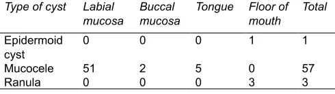
Table 7: The site distribution of intraoral soft tissue cysts
Type of cyst Labial
mucosa Buccal mucosa Tongue Floor of mouth Total
Epidermoid
cyst 0 0 0 1 1
Mucocele 51 2 5 0 57
Ranula 0 0 0 3 3
accordance with the distribution of cysts in the general population in Southern India as reported by Donoghue et al6 which showed a predominance of inflammatory
cysts. Also Telang et al7 had reported 43.2% of radicular
cysts and 20.2% dentigerous cysts in their 19-year retro-spective study of cystic lesions of pediatric and adolescent population.
As suggested in previous literature this difference in the distribution of developmental and inflammatory cysts may probably be attributed to the state of dynamism of dentoalveolar complex.4 This may be a result of interplay
of several factors including the development and erup-tion of the succedaneous dentierup-tion and the simultaneous skeletal growth of the maxilla and mandible in this age group. In addition, we also suggest that the incidence of jaw cysts in the pediatric population is probably under reported owing to the fact that exfoliation/loss of primary teeth may result in the resolution of certain cystic lesions that are limited in size and are asymptomatic particularly when they do not involve the underlying tooth follicles of permanent teeth. The exfoliation of primary teeth may also limit the development and expansion of these cystic lesions which again would help in widening the disparity in reported incidence of developmental and inflamma-tory cysts in the pediatric population when compared to that of the adult population.
The increased number of developmental cysts also suggests a probable role of genetic factors in its formation where as inflammatory cysts have obviously more of an environmental etiology.4
The odontogenic cysts and inflammatory cysts showed a male predominance with an overall male to female ratio of 1.3:1. This is in accordance with the previ-ous studies done by da Cruz et al5 who found 66.7% cases
in males. Nineteen-year institutional study by Telang et al7 reported a male to female ratio of 1.52:1. In the
general population too, similar findings were reported
by Bodner et al,4 Ramachandra et al,8 Donoghue et al,6
Varinauskas et al,9 Ackigoz et al,10 Ansari et al,11
Men-ingaud et al12 and Tortoici et al13 Relatively poor oral
hygiene and increased susceptibility to trauma among boys might be the reason for male predominance.12
Anterior maxilla (28.39%) was found to be the pre-dominant site of cyst occurance in the case of develop-mental cysts. Similar results were reported by Donoghue et al,6 Varinauskas et al,9 Ackigoz et al,10 Ansari et al,11
Tortoici et al13 Gehani et al14 and Prockt et al15 in their
studies on the general population. But, mandibular predominance was reported in other studies done by Meningaud et al12 and Avelar et al.16
Out of all the cystic lesions, we encountered five synd-romic cases, two of which were associated with multiple dentigerous cysts and three cases presented with multiple odontogenic keratocysts. The associated syndromes were one case each of Sotto syndrome, Carpenters syndrome and cleidocranial dysplasia and two cases of nevoid basal cell carcinoma syndrome (NBCCS).
The most common presenting complaint was swell-ing and/or delayed or noneruption of permanent teeth, a finding that is consistent with previously reported literature.13 Imaging modalities like intraoral periapical
radiograph, panoramic radiograph and occlusal radio graph were employed in the evaluation of these pathologic jaw cysts.12 Extraoral radiographs, computed
transformation.
Marsupialization is the most commonly performed surgical treatment modality for pediatric jaw cysts in our institution, in order to salvage the involved/underlying permanent tooth.17 Enucleation is done only in a few
cases. This is in sharp contrast to the treatment usually rendered in case of the jaw cysts in adults where enuclea-tion is the most commonly employed technique in the management of jaw cysts.12
ConCLuSIon
From the data, it is possible to conclude that among all types of cysts of the jaws, developmental cysts are the most commonly encountered in the pediatric population. The lesions show a male predominance with anterior maxilla being the most common site. Marsupialization was the most preferred surgical treatment modality. Fur-ther studies across different age groups are warranted in order to better establish the variability in the frequency of jaw cysts in individuals of various racial and ethnic backgrounds.
ACKnowLeDgMenT
We thank Dr Sreelal T, MD Community Medicine and Dr P Pamagal Kavithai, Junior Resident, Department of Community Medicine, Government Medical College, Thiruvananthapuram for lending their statistical exper-tise in the completion of this study.
RefeRenCeS
1. Shear M, Speight P. Cysts of the oral and maxillofacial regions. 4th ed. Oxford: Blackwell Munksgaard; 2007. Classifi cation and frequency of cysts of the oral and maxillofacial tissues; 1-2. 2. Neville BW, Damm DD, Allen CM, Bouquot JE. Odontogenic
cysts and tumors. Oral and maxillo facial pathology. 2nd ed. Philadelphia, PA: WB Saunders CO; 2002. p. 590-610. 3. Jones AV, Craig GT, Franklin CD. Range and demographics
of odontogenic cysts diagnosed in a UK population over a 30-year period. J Oral Pathol Med 2006;35(8):500-507.
5. Serra VG, Conde DM, Marques RVCF, de Freitas CVS, Lopes FF, da Cruz MCFN. Odontogenic cysts in children and adole - s cents: a 21-year retrospective study. Braz J Oral Sci 11(2):81-83. 6. Selvamani M, Donoghue M, Basandi PS. Analysis of 153
cases of odontogenic cysts in a South Indian sample popula-tion: a retrospective study over a decade. Braz Oral Res 2012 Jul-Aug;26(4):330-334.
7. Telang A, Lahari K, Pushparaj S. Odontogenic cysts in chil-dren: A 19-year institutional review. Lat Am J Orthod Ped Dent [cited 2011 June]. Available at: http://www.ortodoncia. ws/publicaciones/2011/art13.asp
8. Ramachandra P, Maligi P, Raghuveer HP. A cumulative analysis of odontogenic cysts from major dental institutions of Bangalore city: a study of 252 cases. J Oral Maxillofac Pathol 2011;15(1):1-5.
9. Varinauskas V, Gervickas A, Kavoliu-niene O. Analysis of odontogenic cysts of the jaws. Medicina (Kaunas) 2006;42(3): 201-207.
10. Açikgöz A, Uzun-Bulut E, Özden B, Gündüz K. Prevalence and distribution of odontogenic and nonodontogenic cysts in a Turkish population. Med Oral Patol Oral Cir Bucal 2012 Jan;17(1):108-115.
11. Ansari S, U Rehman A, Rehman B. Frequency and demo-graphy of commonly occurring odontogenic cysts in Khyber Pakhtunkhwa (Pakistan). Pak Oral Dent J 2010;30(1):41-46 12. Meningaud JP, Oprean N, Pitak-Arnnop P, Bertrand JC.
Odontogenic cysts: a clinical study of 695 cases. J Oral Sci 2006;48(2):59-62.
13. Tortorici S, Amodio E, Massenti MF, Buzzanca ML, Burruano F, Vitale F. Prevalence and distribution of odontogenic cysts in Sicily: 1986-2005. J Oral Sci 2008;50(1):15-18.
14. Gehani REI, Krishnan B, Orafi H. The prevalence of inflam-matory and developmental odontogenic cysts in a Libyan population. Libyan J Med 2008;3(2):11-15.
15. Prockt AP, Schebela CR, Maito FDM , Sant’Ana-Filho M , Rados PV. Odontogenic cysts: analysis of 680 cases in Brazil. Head Neck Pathol 2008;2(3):150-156.
16. Avelar RL, Antunes AA, Carvalho RW, Bezerra PG, Neto PJ, Andrade ES. Odontogenic cysts: a clinicopathological study of 507 cases. J Oral Sci 2009;51(4):581-586.