Journal Homepage: vrf.iranjournals.ir
Anatomical and cytohistological study of the pituitary gland in adult turkey
Ramin Jahangirfard, Ali Shalizar-Jalali*, Rasoul Shahrooz, Gholamreza Najafi, Aram MinasDepartment of Basic Sciences, Faculty of Veterinary Medicine, Urmia University, Urmia, Iran.
Article Info Abstract
Article history:
Received: 28 January 2018 Accepted: 22 May 2018 Available online: 15 June 2019
In order to conduct this study, eight adult turkey heads were obtained. Pituitary glands were harvested following cranial bones removal and examined morphologically and anatomically as well as topographically. Then, tissue sections were prepared and stained using Hematoxylin and Eosin, Alcian blue, orange G and periodic acid-Schiff staining techniques. The results showed that turkey pituitary gland as a pea-sized structure is located in the ventral part of the cerebrum and composed of adenohypophysis and neurohypophysis parts. Moreover, histological analyses revealed that sinusoids are well-developed at the distal part of the adenohypophysis and irregular masses of endocrine cells exist among them. Distributions of basophilic cells in the distal part of adenohypophysis were significantly higher than those of other endocrine cells, while the acidophilic cells had the lowest distribution. Lower and higher numbers of chromophobe cells were also found compared to those of basophilic and acidophilic cells, respectively. These findings were mostly similar to the other birds’ pituitary gland anatomical and histological features, but there were also differences in cellular elements distributions along with infundibular cavity topography.
© 2019 Urmia University. All rights reserved. Key words:
Anatomy Histology Pituitary gland Turkey
غلاب نوملقوب رد زیفوپیه هدغ یکیژولوتسیهوتیس و یکیموتانآ هعلاطم
هدیکچ
ماجنا روظنم هب هعلاطم نیا
، نوملقوب رس ددع تشه غلاب
تشادرب بقاعتم زیفوپیه ددغ .دیدرگ هیهت ناوختسا
تخیر رظن زا و دندشادج رس هساک یاه ناکم زین و یموتانآ ،یسانش
یبایزرا دروم یراگن
نتفرگ رارق .د شور طسوت و هیهت یتفاب عطاقم ،سپس یاه
نیلیسکوتامه -،ولب نایشلآ ،نیزوئا دیسا کیدویرپ و یج جنروا
-گنر فیش .دندش یزیمآ تروص هب نوملقوب زیفوپیه هدغ هک دنداد ناشن جیاتن
شخب لماش و تسا هدش عقاو خم یمکش تمسق رد دوخن هزادنا هب یراتخاس فوپیهورون و زیفوپیهوندآ یاه
یم زی یبایزرا ،نینچمه .دشاب تفاب یاه
هک دنتخاس راکشآ یسانش دیئوزونیس
اه شخب رد ییاهتنا
هعسوت زیفوپیهوندآ یم هتفای
هدوت و دنشاب لولس مظنمان یاه نآ لصاف دح رد نیرکودنا یاه
اه یگدنکارپ .دنراد دوجو لولس یاه
یلیفوزاب یاه شخب رد
ییاهتنا م لباق لکش هب زیفوپیهوندآ هظحلا
رتشیب یا
یگدنکارپ زا لولس ریاس یاه یلاح رد ،دندوب نیرکودنا یاه
هک لولس یلیفودیسا یاه یگدنکارپ نازیم نیرتمک
دندوب اراد ار لولس اب هسیاقم رد . و یلیفوزاب یاه
لولس ،یلیفودیسا گنر یاه
هب زین زیرگ
.دندوب یرتشیب و رتمک یناوارف یاراد بیترت هتفای نیا
هب اه وط تفاب و یکیموتانآ یاه یگژیو هباشم هدمع ر یگدنکارپ رد زین یتافلاتخا یلو دوب ناگدنرپ ریاس زیفوپیه هدغ یسانش
هارمه یلولس یازجا یاه
یفیق هرفح یناکم تیعقوم اب .تشاد دوجو لکش
:یدیلک یاه هژاو
تفاب ،یموتانآ زیفوپیه هدغ ،نوملقوب ،یسانش
*Correspondence:
Ali Shalizar-Jalali. DVM, PhD
Department of Basic Sciences, Faculty of Veterinary Medicine, Urmia University, Urmia, Iran
E-mail: a.shalizar@urmia.ac.ir
Forum
Introduction
The pituitary gland, hypophysis cerebri, as a major endocrine gland is associated with hypothalamus through infundibular stalk and plays critical roles in other endo-crine glands activities via several hormones secretion.1 This small gland is located in sella turcica of the basi-sphenoid bone body and composed of adenohypophysis and neurohypophysis.2 In mammals, the adenohypophysis is composed of pars distalis, pars intermedia and pars tuberalis; however, pars intermedia does not exist in birds and adenohypophysis is mostly made of pars distalis.3 The avian pars distalis is divided into cephalic and caudal regions containing acidophilic, basophilic and chromophobe cells as well as extracellular colloid and fibrous materials.4-6 Reportedly, presence of folliculo-stellate agranular cells containing immuno-reactive S-100 protein has also been described in avian pars distalis. It has been suggested that these cells may play roles in autocrine-paracrine regulation of hormone secretion along with immune-endocrine interactions.7 The neurohypophysis includes infundibular stalk, median eminence and pars nervosa consisting of neurosecretory terminals.1
Recently, turkey has been suggested as an animal model to mimic human diseases and to promote pharmacotherapies.8 Further, it has been suggested that poultry productions will increase remarkably than those of ruminant meat in the coming three decades9 and efficient veterinary services can lead to poultry diseases control and their economic losses reduction.10,11
Based on this concept, the present study was carried out to disclose anatomical and cytohistological features of the pituitary gland in adult turkeys and to provide a clear understanding of the avian endocrinology resulting in veterinary diagnostics promotions.
Materials and Methods
In this study, eight adult turkey heads were provided randomly from slaughterhouses of Urmia, West Azerbaijan province, Iran. Pituitary glands were harvested following cranial bones removal, examined anatomically using stereomicroscope (SZX-ILLB200; Olympus Co., Tokyo, Japan) and fixed in 10% neutral buffered formalin for 72 hr. Then, the samples were embedded in paraffin, serially cut using a rotary microtome (Leitz Wetzlar GmbH, Wetzlar, Germany), stained using Hematoxylin and Eosin (H & E; Merck, Darmstadt, Germany), Alcian blue (AB; Merck), orange G (OG; Merck) and periodic acid-Schiff (PAS; Asian Chemical Company, Amol, Iran) staining techniques and analyzed via light microscopy (Eclipse E200-LED; Nikon, Tokyo, Japan). Cell distribution per unit area was determined using the unbiased counting frame12 and photomicrographs were taken using SONY onboard camera (Zeiss, Tokyo, Japan).
Statistical analysis. All quantitative data were expressed as mean ± SEM. Statistical analyses were conducted using SPSS (version 20.0, IBM Company, Chicago, USA) and one-way analysis of variance (ANOVA) followed by Tukey multiple comparison. The p values less than 0.05 were considered as statistically significant.
Results
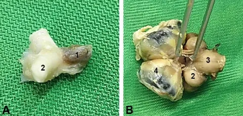
Anatomical findings. Anatomical examinations revealed that turkey pituitary gland as a pea-sized structure is located in the ventral part of cerebrum and associated with an optic nerve as a second pair of the cerebral nerve in the frontal part of cerebrum as well as hypothalamus via infundibular stalk (Fig. 1A). The gland was observed in sella turcica fossa of the basisphenoid bone body and attached to the periosteum via meninges. The large and dark anterior part of the gland, adenohypophysis, was separated fromthe pale and small dorsal part of the gland, neurohypophysis, by a narrow and dark line known as Rathke’s cleft.
Furthermore, the optic lobes were found at lateral sides of the gland and pons and medulla oblongata were observed dorsally (Fig. 1B).
Fig. 1. A) Dorso-lateral view of the pituitary gland in adult turkey. 1: Pituitary gland; 2: Optic nerve. B) Ventral view of the pituitary gland in adult turkey. 1: Pituitary gland; 2: Optic lobes; 3: Medulla oblongata; 4: Eyeball.
Histological findings. The present study revealed that turkey has a well-developed adenohypophysis and the neural part is located at the vicinity of third ventricle near diencephalon. The distal part was composed of cephalic and caudal lobes. Although these two lobes were not different from each other, distinct different cell diversity was found. Pituicytes with round to oval nuclei and pale cytoplasms were observed in neural part. The unmyelinated axons terminal regions with Herring bodies were also located in neural part.
The connective tissue was evident along the capsule surrounding the pituitary gland and the Rathke’s cleft was seen between the distal part and connective tissue. Numerous blood vessels were also observed in the connective tissue around adenohypophysis and neuro-hypophysis. Additionally, the neural part contained infundibular stalk and median eminence with similar histoarchitectures.
Fig. 2. Photomicrograph of the pituitary gland in adult turkey. PD: Pars distalis; PN: Pars nervosa; CT: Connective tissue; IC: Infundibular cavity; APC: Acidophilic cells; BPC: Basophilic cells; CPB: Chromophobe cells; S: Sinusoid (Orange G staining; A: 100× and B: 400×).
Fig. 3. Photomicrograph of the pituitary gland in adult turkey. PD: Pars distalis; PN: Pars nervosa; CT: Connective tissue; IC: Infundibular cavity; BPC: Basophilic cells; CPB: Chromophobe cells(Periodic acid-Schiff staining; A: 100× and B: 400×).
Moreover, histological analyses via OG and PAS stainings indicated that sinusoids are well-developed at the distal part of the adenohypophysis and irregular masses of endocrine cells exist among them. The growth hormone-secreting acidophilic cells (APCs), somatotrophs, with cytoplasm filled with acidophilic secretory granules and poorly stained chromophobe cells (CPCs) were also evident in pars distalis following OG and PAS stainings (Figs. 2B and 3B). The AB staining exhibited that basophilic cells (BPCs) with basophilic secretory granules are a major cellular constituent of turkey pars ditalis (Fig. 4).
Stereological analyses following H&E staining revealed that distribution of BPCs with deep purple-colored cytoplasm in the distal part of adenohypophysis is significantly higher than that of other endocrine cells, while APCs have the lowest distribution. Lower and higher numbers of CPCs with pale-colored cytoplasms were also found compared to those of BPCs and APCs, respectively (Fig. 5).
Fig. 4. Photomicrograph of the pituitary gland in adult turkey. PD: Pars distalis; PN: Pars nervosa; CT: Connective tissue; IC: Infundibular cavity; BPC: Basophilic cells (Alcian blue staining; A: 100× and B: 400×).
Fig. 5. A) Photomicrograph of the pituitary gland in adult turkey. BPC: Basophilic cells; CPB: Chromophobe cells (Hematoxylin and Eosin staining; 400×). B) Distribution of acidophilic, basophilic and chromophobe cells in pars distalis of adult turkey pituitary gland. CPCs: Chromophobe cells; BPCs: Basophilic cells; APCs: Acidophilic cells. abc Different letters indicate significant
differences (p < 0.05).
Discussion
Although birds and mammals share general biological characteristics, they also bear major differences due to different metabolic demands and genetic features as well as biochemical adaptations.13,14 Moreover, it has been shown that ecophysiological factors and food habits can lead to various anatomical, cytohistochemical and histophysiological characteristics of the same tissue among bird species.15,16
Since thorough understanding of tissues normal histo-anatomy is pivotal in a diagnostic pathology,17 the current study was undertaken to shed further lights on morphological and cytohistological appearances of the pituitary gland in adult turkey.
Confirming previous reports, anatomical findings in this study showed that pea-sizedpituitary gland in turkey is located in the ventral part of cerebrum and observed in sella turcica fossa of the basisphenoid bone body.18 Its well-developed adenohypophysis was also separated from small neurohypophysis via narrow Rathke’s cleft as described previously.2,4
absence of pars intermedia in the pituitary gland has been reported in birds and cetaceans including bottlenose dolphin (Tursiops truncatus), sperm whale
(Physeter macrocephalus) and stripeddolphin (Stenella
coeruleoalba).3,19,20 Accordingly, it was found that
adenohypophysis and neurohypophysis are separated by a thick layer of connective tissue in a bottlenose dolphin19 and a thin fibrous membrane in melon-headed whale
(Peponocephala electra), spinner dolphin (Stenella
longirostris), striped dolphin (Stenella coeruleoalba),
pantropical spotted dolphin (Stenella attenuata), pygmy sperm whale (Kogia breviceps), dwarf sperm whale
(Kogia sima), Risso's dolphin (Grampus griseus), short
-finned pilot whale (Globicephala macrorhyncha), false killer whale (Pseudorca crassidens), Fraser's dolphin
(Lagenorhynchus hosei), rough-tooth dolphin (Steno
bredanensis), Gervais's beaked whale (Mesoplodon
europaeus) and infant sperm whale (Physeter catodon).21
Conversely, it has been demonstrated that teleosts adenohypophysis is divided into pars distalis and pars intermediaregions.22 Additionally, histological analysis in this study indicated that no follicle was found in the pars distalis of turkey adenohypophysis, which stands in contrast to previous findings.19,21
Stereological evidence in current study exhibited that distribution of BPCs in pars distalis of turkey adenohypophysis is noticeably higher than that of other endocrine cells, while APCs have the lowest distribution. Contrarily, it has been reported that APCs are the most predominant cell type within the rat23 and Egyptian insectivorous bat (Rhinopomahardwickei) adenohypophysis.24
On the whole, anatomical and cytohistological characteristics of turkey pituitary gland were mostly similar to those of other birds, but there were also differences in cellular components distribution in adenohypophysis providing novel cytological insights into the avian endocrinology.
Conflict of interest
The authors declare that there is no conflict of interest regarding the publication of this article.
References
1. Whittow GC. Sturkie's avian physiology. 5th ed. San Diego, USA: Academic Press 1999; 437-460.
2. Eurell JA, Frappier BL. Dellmann's textbook of veterinary histology. 6th ed. Iowa, USA: Wiley-Blackwell 2013; 300-306.
3. Ritchie M. Neuroanatomy and physiology of the avian hypothalamic/pituitary axis: Clinical aspects. Vet Clin North Am Exot Anim Pract 2014; 17(1): 13-22.
4. Bacha WJ, Bacha LM. Color atlas of veterinary
histology. 2nd ed. West Sussex, USA: Wiley-Blackwell 2000; 191-193.
5. Mohanty B. Extracellular accumulations in the avian pituitary gland: Histochemical analysis in two species of Indian wild birds. Cells Tissues Organs 2006; 183 (2): 99-106.
6. Sasaki F, Doshita A, Matsumoto Y, et al. Embryonic development of the pituitary gland in the chick. Cells Tissues Organs 2003; 173(2): 65-74.
7. Harrisson F. Primary cilia associated with striated rootlets in granulated and folliculo-stellate cells of the avian adenohypophysis. Anat Embryol (Berl) 1989; 180(6): 543-547.
8. Yoo YS, Park HS, Choi GH, et al. Recent advances in the development of experimental animal models mimicking human aortic aneurysms. Vasc Specialist Int 2015; 31(1): 1-10.
9. LexB, Klaas van der H, Gerard van D, et al. World livestock and crop production systems, land use and environment between 1970 and 2030. In: Brouwer F, McCarl BA (Eds). Agriculture and Climate Beyond 2015: A new perspective on future land use patterns. Netherlands, Dordrecht: Springer Science & Business Media 2006; 77-80.
10.Pavade G, Awada L, Hamilton K, et al. The influence of economic indicators, poultry density and the performance of veterinary services on the control of high-pathogenicity avian influenza in poultry. Rev Sci Tech 2011; 30(3): 661-671.
11.Knight-Jonesa TJD, RushtonbJ. The economic impacts of foot and mouth disease – What are they, how big are they and where do they occur? Prev Vet Med 2013; 112(3-4): 161-173.
12.Jalali AS, Hasanzadeh S, Malekinejad H. Achillea
millefolium inflorescence aqueous extract ameliorates
cyclophosphamide-induced toxicity in rat testis: Stereological evidences. Chin J Nat Med 2012; 10(4): 247-254.
13.Tieleman BI, Versteegh MA, Fries A, et al. Genetic modulation of energy metabolism in birds through mitochondrial function. Proc Biol Sci 2009; 276(1662): 1685-1693.
14.Rioux P, Blier PU. Energetic metabolism and biochemical adaptation: A bird flight muscle model. Biochem Mol Biol Educ 2006; 34(2): 125-128.
15.Chiale MC, Fernández PE, Gimeno EJ, et al. Morphology and histology of the uropygial gland in Antarctic birds: Relationship with their contact with the aquatic environment? Aust J Zool 2014; 62(2) 157-165. 16.Klasing KC. Avian gastrointestinal anatomy and
physiology. Semin Avian Exot Pet Med 1999; 8(2): 42-50.
18.Dyce KM, Sack WO, Wensing CJG. Textbook of veterinary anatomy. 4th ed. St. Louis, USA: Elsevier Health Sciences 2009; 216-217.
19.Vuković S, Lucić H, Duras Gomercić M, et al. Anatomical and histological characteristics of the pituitary gland in the bottlenose dolphin (Tursiops truncatus) from the Adriatic Sea. Vet Arhiv 2011; 81(1): 143-151.
20.Oelschläger HH, Kemp B. Ontogenesis of the sperm whale brain. J Comp Neurol 1998; 399(2): 210-228. 21.Cowan DF, Haubold EM, Tajima Y. Histological,
immuno-histochemical and pathological features of the pituitary gland of odontocete cetaceans from the Western gulf of
Mexico. J Comp Pathol 2008; 139(2-3): 67-80.
22.Ekici A, Timur M. An anatomical and histochemical examination of the pituitary gland of carp (Cyprinus
carpio). Turk J Vet Anim Sci 2013; 37: 399-403.
23.Poole MC, Kornegay WD. Cellular distribution within the rat adenohypophysis: A morphometric study. Anat Rec 1982; 204(1): 45-53.
24.Mohammed SA. Differentiation between the anterior pituitary cells of the Egyptian insectivorous bats
Rhinopoma hardwickei using transmission electron