REVIEW ARTICLE
Vestibular contribution to memory processing
Abdollah Moossavi1, Meymaneh Jafari2,3*
1
- Department of Otolaryngology, School of Medicine, Iran University of Medical Sciences, Tehran, Iran
2- Department of Audiology, Faculty of Rehabilitation, Isfahan University of Medical Sciences, Isfahan, Iran 3- Department of Audiology, University of Social Welfare and Rehabilitation Sciences, Tehran, Iran
Received: 5 Aug 2018, Revised: 17 Sep 2018, Accepted: 30 Sep 2018, Published: 15 Apr 2019
Abstract
Background and Aim: The vestibular system contributes in the stabilization of the head and body, orientation, and gazing through the pro-cessing of sensory inputs. A wealth of evidence supports the involvement of vestibular informa-tion in higher funcinforma-tions, too.
Methods: In this paper, we reviewed the pre-vious studies on the effect of the vestibular sys-tem on memory as one of the cognitive func-tions.
Results: Clinical and laboratory findings indi-cate the association of vestibular inputs (besides postural control and oculomotor) with a variety of higher functions, especially memory func-tion. Because part of the memory function is determined by other cognitive processes i.e. att-ention capacity, emotional disturbances, and executive functions, the study of the effect of vestibular inputs on these functions provides a more accurate view of how the vestibular inputs affect memory performance.
Conclusion: Although our current knowledge on vestibular-memory interaction is increasing, the exact involvement of vestibular signals in memory representations is still unclear and needs further studies to determine the theore-tical basis of vestibular involvement in memory
processing.
Keywords: Vestibular system; vestibular cor-tex; cognitive function; memory
Citation: Moossavi A, Jafari M. Vestibular contribution to memory processing. Aud Vestib Res. 2019;28(2):62-74.
Introduction
The vestibular system is one of the oldest sen-sory systems [1]. This system is enclosed deeply in the inner ear and surrounded by thick tem-poral bones. This small but complex structure provides sensory information about acceleration and head movement in space [2,3]. Vestibular signals are important in vertebrates, because they provide information of self-movement and the implementation of these moves in any envi-ronment [2]. These signals are also important in human embryonic life so that vestibular inputs tend to orient the embryo during delivery [4]. In everyday life, vestibular receptors continu-ously monitor the movement of the head to help maintain its balance and orientation toward the target. These signals are also important for some autonomic reflexes, which tend to sta-bilize the gaze (oculomotor) and the posture (muscular) [5]. However, vestibular afferent sig-nals continuously work (even when the person is in a stable position). Usually, the final signal by the vestibular system is ignored unless it receives an unusual stimulus (e.g. a jerky boat) * Corresponding author: Department of Audiology,
Faculty of Rehabilitation, Isfahan University of Medical Sciences, Hezarjerib Ave., Isfahan, 8174656831, Iran. Tel: 0098313-6693089,
or gets damaged. Then, the significant vesti-bular responses such as vertigo (illusion of rot-ation or swaying), visual disturbances or insta-bility occur and the vital role of the vestibular system becomes clear [6].
After vestibular dysfunction, besides severe abnormal symptoms of dizziness and imbalance, disabling mental outcomes such as cognitive, psychological, and autonomic impairments occ-ur [7]. These effects are not noticeable in other sensory disturbances (such as vision, olfactory), so it may be a reflection of the wide network of vestibular projection which extends into multi-ple cortical and subcortical regions such as brain stem, basal ganglia, hippocampus, cerebellum and cerebral cortex –all related to higher func-tions [2]. This broad network shows multimodal vestibular processing compared to other sensory modalities. Unlike other senses that send their information to a specific area in the cerebral cortex, information of the vestibular system int-egrates with other sensory inputs (i.e. vision, joints, skin and muscles) throughout the central nervous system (CNS). As a result, there is no central vestibular area (similar to other primary sensory cortex areas), which only responds to vestibular inputs [8].
Considering what was discussed, the unique role of the vestibular system in everyday functions cannot be ignored. Although, the role of the ves-tibular system in cognitive functions has been previously overlooked, several studies have rec-ently examined the involvement of the vesti-bular system in motor, cognitive, affective and perceptive functions [9]. Clinical and laboratory evidence indicates the association of vestibular inputs (besides postural control and oculomotor) with a variety of higher functions, especially memory, as one of the cognitive functions [10]. Various studies have investigated the relation-ship between vestibular system and memory-related processes in different vestibular distur-bances; some of these studies have used arti-ficial stimulation of the vestibular system, too. Since part of the memory function is determined by other cognitive processes (i.e. attention, per-ception, motivation), the study of vestibular inputs on these functions provides a more
accurate view of how the vestibular system affect memory performance. In the continuation, after a brief overview of the vestibulo-cortical maps, the involved pathways are explained. Then, we will review the studies of various vestibular disorders as well as those studies that used artificial stimulation of the vestibular sys-tem. Finally, the effect of other cognitive func-tions on the relafunc-tionship between the vestibular system and the memory processing will be dis-cussed.
The vestibulo-cortical maps
Sending projections from vestibular signals to cortical and subcortical regions shows how the information of the vestibular system integrates in the central nervous system and participates in a series of reflexive and higher functions [11,12]. By artificial stimulation of the vesti-bular system (reducing visual and propriocep-tive inputs), several important cortical regions activate which represents the neuroanatomical basis for the involvement of vestibular signals in a variety of cognitive processes. These areas include the parietal cortex, the temporo-parieto-insular and retro-temporo-parieto-insular cortex, the cingulate cortex, the frontal cortex and numerous subcor-tical structures, including thalamus, basal gan-glia, and cerebellum [13].
modalities, is weak [14].
The current knowledge about the target areas of vestibulo-cortical projections has increased after using various research methods (neuroche-mical, neuroimaging, brain stimulation, lesion examination). Through this new methods and information, we can show the potential role of vestibular inputs in functions beyond main-taining balance [11,13,17,18]. Most vestibular signals in the cortex are reflected in three main pathways: 1) to the brain stem nuclei (raphe nucleus and locus coeruleus), 2) to subcortical structures or cortical regions of motion and vis-ion (i.e. the cerebellum and occipital lobe) and 3) to the cortical regions related to cognitive processing, autonomic functions, and mental health through several direct, such as parabra-chial nucleus (PBN), and indirect, such as the hippocampus, prefrontal cortex, mainly through thalamus [19].
Neuroanatomical information plays an impor-tant role in providing evidence for vestibular involvement in cognition [7]. One of the str-ongest anatomical evidence for cognitive ves-tibular interactions is vesves-tibular projections to memory cortical regions [20]. In the next sec-tion, we will review these pathways.
Vestibular-memory pathways
Vestibular pathways that reach (indirectly) the hippocampus, memory processing area, are imp-ortant to create anatomical substrate for the par-ticipation of vestibular signals in cognition, esp-ecially spatial memory [21]. There are four hyp-othetical main pathways for sending vestibular signals to hippocampus. Fig. 1 shows an outline of these pathways, including the (a) vestibulo-thalamo-cortical pathway, (b) theta pathway, (c) head direction pathway and (d) the vestibulo-cerebello-cortical pathway [18].
Pathway (a) sends the spatial and self-move-ment information through the parietal, entor-hinal, and perirhinal cortices to the hippocam-pus. Pathway (b) sends information to the hipp-ocampus via the pontine reticular formation, supramammillary nucleus and medial septum and is responsible for memory processing. Path-way (c) is related to the head orientation and
before the hippocampus, it sends information through the dorsal tegmental nucleus to the lateral mammillary nucleus and anterodorsal thalamic nucleus. Finally, information on spatial learning is probably transmitted through path-way (d), which passes through the cerebellum and ventral lateral nucleus of thalamus [13,18,22]. In addition to hippocampus, basal ganglia is also a key area in spatial recognition. There is probably a fifth hypothetical path that goes through the striatum and plays a role in spatial learning and spatial memory [13].
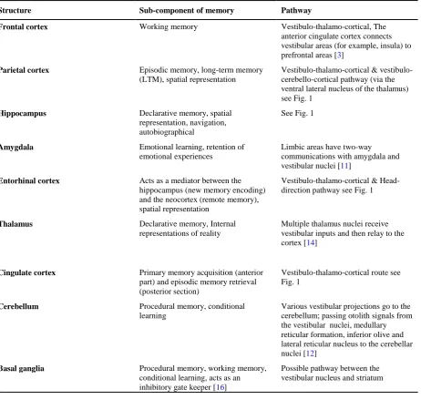
By considering the pathways and nerve net-works that are activated by vestibular signals (including the above paths), it is clear that there are several regions that receive vestibular sig-nals and also play a role in memory processing [23,24]. Table 1 presents the neuroanatomical overlap between main memory centers and vestibulo-cortical pathways.
The temporal region of the brain (especially the hippocampus) is responsible for guiding various aspects of memory, especially spatial memory and navigation and is involved in the vestibular cortical network [23]. In these anatomical mod-els, vestibular signals are not sent directly to memory cortical regions and effect of these signals on memory functions is not shown, but the overlap between different networks indicates the potential association of vestibular signals and memory functions [24].
The effect of vestibular signals on memory is related to cognitive maps formed in the region of hippocampus and parahippocampus (i.e. ent-orhinal, perirhinal, and postrhinal cortices) [13]. The activity of the place cells (activity in certain places), the border cells (indicating the boun-daries of the environment), the head direction cells (activity when the head is directed in a certain direction) and the grid cells (creating information about the variable position of the person in space) combine to represent the inner impression of the environment [25]. These maps represent the spatial characteristics of an event and are probably the first step in the formation of spatial memory [13].
is. According to them, since the movement is vital for the continuation of life [26], the ves-tibular system is designed specifically to detect changes in the individual's movement (i.e. is the head upright, is it fixed, what speed/direction do I go?) [27], so probably the vestibular signals somehow show themselves in memory repre-sentations [1]. Although some vestibular inputs are expanded in the cortex and converge with other sensory inputs and motor signals, they are not fully isolated in memory functions [8,13]. By providing basic information about the indi-vidual's movements (as a reference for other
sensory inputs), these inputs provide an unpara-lleled contribution to memory processing and allow accurate and simultaneous cognitive and motor functions [26]. In addition, the vestibular disorder causes damage to spatial memory and navigation, so the patients with vestibular dis-order show spatial memory impairment regard-less of having normal cognitive function. Thus, the strong link between vestibular performance and spatial-cognitive-vision skills is confirmed. The role of the vestibular system in spatial per-ception and spatial memory has been inves-tigated in numerous studies [1]. In this regard,
Table 1. Neuroanatomical overlap between vestibulo-cortical pathways and main memory centers
Structure Sub-component of memory Pathway
Frontal cortex Working memory Vestibulo-thalamo-cortical, The anterior cingulate cortex connects vestibular areas (for example, insula) to prefrontal areas [3]
Parietal cortex Episodic memory, long-term memory (LTM), spatial representation
Vestibulo-thalamo-cortical & vestibulo-cerebello-cortical pathway (via the ventral lateral nucleus of the thalamus) see Fig. 1
Hippocampus Declarative memory, spatial representation, navigation, autobiographical
See Fig. 1
Amygdala Emotional learning, retention of emotional experiences
Limbic areas have two-way communications with amygdala and vestibular nuclei [11]
Entorhinal cortex Acts as a mediator between the hippocampus (new memory encoding) and the neocortex (remote memory), spatial representation
Vestibulo-thalamo-cortical & Head-direction pathway see Fig. 1
Thalamus Declarative memory, Internal representations of reality
Multiple thalamus nuclei receive vestibular inputs and then relay to the cortex [14]
Cingulate cortex Primary memory acquisition (anterior part) and episodic memory retrieval (posterior section)
Vestibulo-thalamo-cortical route see Fig. 1
Cerebellum Procedural memory, conditional learning
Various vestibular projections go to the cerebellum; passing otolith signals from the vestibular nuclei, medullary reticular formation, inferior olive and lateral reticular nucleus to the cerebellar nuclei [12]
Basal ganglia Procedural memory, working memory, conditional learning, acts as an inhibitory gate keeper [16]
after the vestibular disorder, damage to the short-term memory capacity, in addition to spatial memory, are discussed [28].
Effect of vestibular dysfunction on memory
In the 1960s, it was first shown that, animals for navigation and remembering paths, in add-ition to the visual signs, use other idiothetic and non-visual signs such as atrial info and deep sense [26,29]. The first clinical evidence of the impact of vestibular disorders on spatial mem-ory impairment was presented in 1989 by Grimm et al. [30]. In the following decades, various studies have shown that abnormalities in one or both vestibular labyrinths lead to non-orientation of animals in memory exercises (searching or navigating paths learned) and there is no possibility of learning new spatial locations for them [31]. The obtained data from individuals with vestibular disorders such as perilymph fistula syndrome [30], bilateral vesti-bular neuroectomy [22,32], bilateral partial ves-tibular disorder [33] and unilateral compensated labyrinthine impairment (non-vertigo) [34] also confirm these findings. Of course, it should be
noted that perilymph fistula syndrome also causes hearing impairment, which can be a reason for other cognitive impairments [30]. However, a new epidemiological study on the impact of vestibular disorder on age-related cognitive functions (with control of auditory and visual impairments) has been shown that declining vestibular function is associated with worsening of cognitive function in visuospatial tests (including visual memory), but does not affect executive function or verbal memory [35]. In these studies, after the vestibular dis-order, damage to spatial memory, delay in spa-tial learning and navigating and damage to spatial short-term memory capabilities have been seen [22,30,32-34] which does not corr-elate with the normal cognitive, perceptive, and movement capacities (including non-spatial memory) or other standard assessments [36]. With regard to this information, if there is a disturbance in the vestibular system, at least some of memory functions are damaged. Thus, normal vestibular function is essential for some memory processes, especially for visuospatial information [10,26].
A B C D
Vestibular nucleus Medial geniculate and
ventral posterior inferior thalamic
Nucleus Parietal cortex Perirhinal cortex Entorhinal cortex Hippocampus
Vestibular nucleus Dorsal tegmental nucleus Lateral mammillary nucleus Anterodorsal thalamic nucleus
Retrosplenial cortex Postsubiculum Entorhinal cortex
Hippocampus
Vestibular nucleus Pedunculopontine tegmental
nucleus, pontine reticular formation Flocculus, paraflocculus
Thalamus Temporal, parietal,
frontal cortex
Dentate nucleus Nucleus post interpositus
Hippocampus
Vestibular nucleus Pedunculopontine tegmental
nucleus, pontine reticular formation Posterior hypothalamic region
Supramammillary region Medial septum Hippocampus
Fig. 1. Neuroanatomical model of vestibular-hippocampal routes: A) Thalamo-cortical route, B) Head-direction route, C) Vestibulo-cerebello-cortical pathway, D) Theta-generating route
Hippocampus plays a key role in the study of how to store and represent spatial information; therefore, following a vestibular dysfunction, neuroanatomical effects are likely to be exp-ected in addition to behavioral changes [7]. Damage to spatial memory that occurs follo-wing vestibular impairment is probably related to dysfunction of hippocampus, because both the place cell responses [37] and theta rhythm [38-40] become abnormal. In contrast, the field potentials of CA1 and dentate gyrus and long-term potentiation (LTP) are not significantly affected by bilateral vestibular impairment [41]. Lack of vestibular inputs changes the functions of the hippocampus [22]. Along with this theory, studies on the hippocampal morphology following bilateral vestibular impairment have shown numerous and different results, such as bilateral hippocampal atrophy (approximately 17%) about 5 to 10 years after surgery [22] to no significant changes after a shorter period [42
-44]. In patients with unilateral vestibular neu-rectomy, no results of hippocampal atrophy has been observed [18]. In patients with unilateral Meniere disorder, bilateral hippocampal atrophy has been reported with a further reduction on the left side [45]. It has also been observed that bilateral vestibular impairment leads to a bilateral reduction in the gray matter of the CA3 region of the hippocampus, and the rate of reduction is consistent with the degree of clinical vestibular impairment [46]. In addition, bilateral vestibular disorders leads to morpholo-gical changes in the hippocampus, in the form of decrease in the number of basal dendrites (not apical) of CA1 [47] and N-Methyl-D-Aspartate (NMDA) receptors in the hippocam-pus [43]. There is a hypothesis that vestibular dysfunction also contributes in the development of the Alzheimer disease by affecting the hipp-ocampus and other regions of the medial tem-poral lobe [48]. Such changes in the hippo-campal atrophy have not been observed in rats with bilateral vestibular disorders [43,44]. One possible explanation for this observation is that in rats with bilateral vestibular disorders, loco-motor hyperactivity is observed, which may neutralize the effects of vestibular impairment
on hippocampus [37,43,49].
spatial coding and navigating [57].
Effect of artificial vestibular stimulation on memory
Information from the vestibular system for self-movement is essential for the growth of normal spatial memory. For this purpose, various stu-dies have examined the effect of changing or enhancing the signals of the vestibular system on memory [26]. Astronauts' findings in space have created a unique opportunity for temporary changes in vestibular inputs, because in weight-lessness status, the participation of the otolithic sense in gravity is eliminated [10]. Although microgravity cognitive effects (conditions whe-re no otolithic inputs exist) varies, the impair-ment of visuospatial functions (for example, referring to memorized places with closed eyes) has been considered in various studies [10,58,59]. The results of these studies, besides confirming previous findings, suggest that temporary reduction in memory performance in healthy subjects is similar to those who have recently had an experience of vestibular disorder (without any specific symptoms, such as hearing loss or prolonged dizziness). Of course, in healthy people, non-vestibular factors such as anxiety and sleep deprivation can also lead to cognitive impairment [58].
Laboratory methods are used to study the effect of increased vestibular stimulation. These prac-tices, besides clinical applications, can be used in research because of their low cost, non-inv-asiveness, safety and easy to use implementa-tion [60-62]. In some studies, the effect of ves-tibular stimulation on memory has been studied. Extra-threshold electrical currents in galvanic vestibular stimulation (GVS) induce a similar effect to mild vestibular impairment [63]. GVS activates vestibular nerve by inducing a small flow through the mastoid prominent skin to the vestibular system. GVS activates the discharge zone of primary vestibular afferents and by producing irregular firings of vestibular affe-rents, leading to depolarization (cathodic stimu-lation) or hyperpolarization (anodic stimustimu-lation) of vestibular nerve [64,65]. Functional imaging studies in humans show that using galvanic
atrium stimulation, a network of multimodal cortical regions (including insular cortex, supe-rior temporal gyrus, temporo-parietal cortex, basal ganglia and anterior cingulate gyrus [66], PIVC, temporo-parietal joint [67] and hippo-campus [64] are activated. Therefore, GVS may have a positive effect on the activation of memory-related brain regions [68]. Stimulation with more GVS intensity can sometimes lead to disturbances in the vestibular system and will have a positive effect when less stimulation occurs [26,31]. This stimulation method has led to a shift in the behavior of normal individuals in the practice of short-term visuospatial memory [63]. Therefore, there is probably a close connection between GVS, vestibular sys-tem, the memory [68]. However, the reason for memory impairment with extra-threshold GVS is unknown, this may be due to indirect relation to visual impairment (i.e. oscillopsia or nystag-mus), or direct relation to cognitive resource conflicts due to inappropriate inputs of vesti-bular, visual and proprioceptive systems [10]. To avoid these limitations, Wilkinson et al. used small sub-sensory GVS [69]. GVS leads to improved visual memory recall, speed and accuracy [68,69]. Although several studies have shown improvement in cognitive performance after a session of using noisy or stochastic GVS in healthy people, there is little information on the efficacy of repeated GVS in pathologic conditions. In a study with presentation of low frequency noisy GVS presentation during learning and recall stages, spatial memory fun-ction improved in rats with cognitive impair-ment. This effect may be related to the effective and frequent activity of vestibular neurons and the hippocampal regions of the vestibular system. The study results have shown the pot-ential benefits of this approach in encountering the cognitive decline caused by sporadic AD [64].
water, increased spatial memory for visual places is observed and by stimulation of the right ear, increased verbal memory in visually presented words is observed. Thus, as a result of CVS, facilitating cerebral blood flow in the opposite brain structures, which is needed for verbal and spatial cognitive processing, leads to improved visual memory [70]. Of course, dur-ing interpretdur-ing the results, attention should be paid to the complications of CVS, such as nau-sea and visual disturbances (nystagmus), as it may lead to impaired vestibular influences on cognitive functions [72]. Considering the results of various studies, memory functions are affec-ted by increased activity of the vestibule after artificial stimulation and this effect is likely to be positive and beneficial. These methods stimulate different parts of the vestibular system and have different applications; therefore, bef-ore drawing any conclusions regarding the sta-bility and mechanism of the effect, more exp-lanations and studies are needed [10].
Related vestibular-cognitive effects
Evidence from various studies suggests that other cognitive processes may also be affected by changes in vestibular inputs. Therefore, in order to investigate the relationship between vestibular signals and memory, their effects on cognitive functions should be considered regar-dless of reflexive functions [73].
Since part of the memory function is determined by other cognitive processes (attention capaci-ties, emotional disturbances, executive func-tion), the study of the effect of vestibular inputs on these functions provides a more accurate view of how the vestibular inputs affect memory performance [10]. In this section, we will rev-iew the available evidence for various func-tional interactions.
Visuospatial abilities
The results of various studies indicate that the vestibular system is likely to have a specific and significant contribution to the organization of the representation of two or three-dimensional spaces. Therefore, the stimulation of the vesti-bular system or its impairment affects cognitive
processes such as spatial memory and naviga-ting (which are used to determine the position of an individual or an external goal in a three dimensional space and rely on these mental representations) [10]. Because there are several brain regions that respond both to vestibular inputs and to visuospatial processing (e.g. the hippocampus, insula, superior temporal gyrus, inferior parietal lobe, temporo-parietal joint, ventral intraparietal cortex and posterior parietal cortex), these effects can be interpreted by con-sidering cortical networks activated by stimu-lation of the vestibular system [10,62,74,75]. Therefore, when the vestibular system is imp-aired, a cascade of neurological changes (thro-ugh the vestibulo-cortical network) is likely to occur, which will damage the cognitive aspects of spatial vision, such as navigation, mental imagery, and memory [26].
Attention capacities
Studies in normal people have shown that cog-nitive functions decrease with increasing need for preservation of the posture [10,29]. Thus, maintaining the balance is a complex process and extends beyond a simple reflex perfor-mance. It uses different cognitive resources when needed and weakens processes such as memory activities. This effect is especially noti-ceable in the elderly, because they are at risk of vestibular dysfunction and cognitive impairment [76].
causes: limited overall capacity (i.e. reduction of attention resources due to postural instability and widespread cognitive damage after that), or competition in the use of specific spatial processing resources (disturbance in spatial reference detection after vestibular disorder or direct effect of postural disturbance on specific spatial cognitive exercises) [77]. The results of various studies indicate that postural function is associated with limited capacity and increased attention needs in equilibrium exercises. If this relationship exists, vestibular involvement in the process of memory processing may indicate a widespread effect of vestibular stimulation or the general discharge of attentional resources due to a vestibular dysfunction that extends to multiple cognitive functions, for example, att-ention and information processing, and it is not just limited to the functions of spatial memory [20].
Executive function
Sensory stimulation during physical exercises is effective on the proliferation and neurogenesis of the cells in the hippocampus. Therefore, ves-tibular stimulation during physical exercises may cause neurogenesis of the cell in the den-tate gyrus [78]. Equilibrium exercises increase memory performance and spatial recognition in people with a healthy vestibular system [79]. Moderate physical exercises for at least 20−30 minutes, two or three times a week, improve cognitive performance in the elderly [80] and reduces the risk of developing AD [81]. This effect has been seen even with the control of vascular disorders, locomotor disorders and other risk factors for AD, which can be related to exercise. The beneficial effects of exercises on delaying and preventing AD are parallel to increasing blood flow in vestibular nucleus and higher vestibular centers, but such an effect was not observed in cochlear nuclei and other hearing centers [82]. Physical exercise results in improved vestibulospinal and vestibulo-ocular reflexes [83]. Physical exercise affects the volume of the anterior hippocampus (the entry of vestibular inputs) in AD but has no effect on the caudate core or volume of thalamus [84].
Psychiatric disorders
In addition to the vestibular-cognitive com-munication described above, clinical reports show a clear relationship between vestibular dysfunction and psychiatric symptoms like panic, anxiety, and depression. Patients with vestibular dysfunction suffer from emotional disturbances and depression [11,58,86] and people with psychiatric conditions often com-plain of feelings of dizziness and instability [87]. The emotional state of individuals is a fac-tor in changing learning and memory abilities [88]. Animals with high levels of anxiety usu-ally have poor learning and memory functions [89]. Anxious people tend to scan the environ-ment more in comparison with relaxed people (regardless of the existence of intimidating sti-muli or threats), with more head movements and different orientations [90]. Such a tedious scan causes conflicts in their capacity with environ-mental events and reflects the general inability to keep their attention focused on targeted stimuli, which, in turn, affects the information needed to learn and memory [89].
When vestibular involvement is considered in memory function, this two-way communication should also be taken into account; because mental problems can lead to cognitive impair-ment (such as damage to attention and working memory in schizophrenia or excessive attention to threatening triggers in panic disorder) [91]. In fact, PBN creates a direct link between the vestibular system and the limbic system (inclu-ding amygdala, locus coeruleus, hypothalamus and prefrontal cortex) that are involved in emo-tional processing and mental disorders [11]. Vestibulo-hippocampal interactions can also cause psychological symptoms, as this area is involved in both spatial and emotional proce-ssing [29]. In other words, any cognitive disorder can potentially have a psychological and or vestibular origin [7,21,29].
Conclusion
especially memory. Ascending vestibular path-ways send vast projections to areas involved in spatial memory and navigation, such as hippo-campus, entorhinal cortex, and thalamus. There-fore, damage to the vestibular system causes spatial memory disturbance and results in struc-tural changes in the hippocampus. In addition, various studies in healthy adults indicate that microgravity conditions lead to memory errors, while artificial stimulation of the vestibular sys-tem with sys-temperature or electrical flow facili-tates memory function.
Although, the two systems are connected and our current knowledge about vestibule-memory interactions is increasing, the involvement of atrial signals in memory representations are still unclear. It seems that balance is a complex pro-cess based on cognitive resources and factors. Therefore, further studies are needed to diag-nose the relationship between vestibular and memory systems and to determine the theore-tical basis of vestibular involvement in memory processing.
Conflict of interest
The authors declared no conflicts of interest.
References
1. Smith PF, Darlington CL, Zheng Y. Move it or lose it--is stimulation of the vestibular system necessary for normal spatial memory? Hippocampus. 2010;20(1):36-43. doi: 10.1002/hipo.2058
2. Highstein SM. Anatomy and physiology of the central and peripheral vestibular system: overview. In: Highstein SM, Fay RR, Popper AN, editors. Springer handbook of auditory research. New York: Springer; 2004. p. 1-10.
3. Preuss N, Hasler G, Mast FW. Caloric vestibular stimulation modulates affective control and mood. Brain Stimul. 2014;7(1):133-40. doi:
10.1016/j.brs.2013.09.003
4. Eliot L. What's going on in there? How the brain and mind develop in the first five years of life. 1st ed. New York: Bantam; 1999.
5. Holstein GR, Friedrich VL Jr, Martinelli GP, Ogo-rodnikov D, Yakushin SB, Cohen B. Fos expression in neurons of the rat vestibulo-autonomic pathway acti-vated by sinusoidal galvanic vestibular stimulation. Front Neurol. 2012;3:4. doi: 10.3389/fneur.2012.00004
6. Khan S, Chang R. Anatomy of the vestibular system: A review. NeuroRehabilitation. 2013;32(3):437-43. doi:
10.3233/NRE-130866
7. Smith PF. The vestibular system and cognition. Current opinion in neurology. 2017;30(1):84-9. doi:
10.1097/WCO.0000000000000403
8. Angelaki DE, Cullen KE. Vestibular system: the many facets of a multimodal sense. Annu Rev Neurosci. 2008;31:125-50. doi:
10.1146/annurev.neuro.31.060407.125555
9. Grabherr L, Macauda G, Lenggenhager B. The moving history of vestibular stimulation as a therapeutic intervention. Multisens Res. 2015;28(5-6):653-87. 10. Bigelow RT, Agrawal Y. Vestibular involvement in
cognition: Visuospatial ability, attention, executive function, and memory. J Vestib Res. 2015;25(2):73-89. doi: 10.3233/VES-150544
11. Gurvich C, Maller JJ, Lithgow B, Haghgooie S, Kulkarni J. Vestibular insights into cognition and psychiatry. Brain Res. 2013;1537:244-59. doi:
10.1016/j.brainres.2013.08.058
12. Büttner-Ennever JA. A review of otolith pathways to brainstem and cerebellum. Ann N Y Acad Sci. 1999;871:51-64.
13. Hitier M, Besnard S, Smith PF. Vestibular pathways involved in cognition. Front Integr Neurosci. 2014;8:59. doi: 10.3389/fnint.2014.00059
14. Lopez C, Blanke O. The thalamocortical vestibular system in animals and humans. Brain Res Rev.
2011;67(1-2):119-46. doi:
10.1016/j.brainresrev.2010.12.002
15. Mazzola L, Lopez C, Faillenot I, Chouchou F, Mauguière F, Isnard J. Vestibular responses to direct stimulation of the human insular cortex. Ann Neurol. 2014;76(4):609-19. doi: 10.1002/ana.24252
16. Baier B, Karnath HO, Dieterich M, Birklein F, Heinze C. Keeping memory clear and stable--the contribution of human basal ganglia and prefrontal cortex to working memory. J Neurosci. 2010;30(29):9788-92. doi:
10.1523/JNEUROSCI.1513-10.2010
17. Shinder ME, Taube JS. Differentiating ascending vestibular pathways to the cortex involved in spatial cognition. J Vestib Res. 2010;20(1):3-23. doi:
10.3233/VES-2010-0344
18. Hüfner K, Hamilton DA, Kalla R, Stephan T, Glasauer S, Ma J, et al. Spatial memory and hippocampal volume in humans with unilateral vestibular deafferentation. Hippocampus. 2007;17(6):471-85. doi:
10.1002/hipo.20283
19. Lopez C. A neuroscientific account of how vestibular disorders impair bodily self-consciousness. Front Integr Neurosci. 2013;7:91. doi: 10.3389/fnint.2013.00091
20. Besnard S, Lopez C, Brandt T, Denise P, Smith PF. Editorial: the vestibular system in cognitive and memory processes in mammalians. Front Integr Neurosci. 2015;9:55. doi: 10.3389/fnint.2015.00055
21. Hanes DA, McCollum G. Cognitive-vestibular interactions: a review of patient difficulties and possible mechanisms. J Vestib Res. 2006;16(3):75-91.
22. Brandt T, Schautzer F, Hamilton DA, Brüning R, Markowitsch HJ, Kalla R, et al. Vestibular loss causes hippocampal atrophy and impaired spatial memory in humans. Brain. 2005;128(Pt 11):2732-41. doi:
10.1093/brain/awh617
23. Glikmann-Johnston Y, Saling MM, Reutens DC, Stout JC. Hippocampal 5-HT1A receptor and spatial learning and memory. Front Pharmacol. 2015;6:289. doi:
10.3389/fphar.2015.00289
10.1523/JNEUROSCI.3575-09.2009
25. Moser EI, Kropff E, Moser MB. Place cells, grid cells, and the brain's spatial representation system. Annu Rev Neurosci. 2008;31:69-89. doi:
10.1146/annurev.neuro.31.061307.090723
26. Smith PF, Geddes LH, Baek JH, Darlington CL, Zheng Y. Modulation of memory by vestibular lesions and galvanic vestibular stimulation. Front Neurol. 2010;1:141. doi: 10.3389/fneur.2010.00141
27. Wilkinson D, Morris R, Milberg W, Sakel M. Caloric vestibular stimulation in aphasic syndrome. Front Integr Neurosci. 2013;7:99. doi: 10.3389/fnint.2013.00099
28. Popp P, Wulff M, Finke K, Rühl M, Brandt T, Dieterich M. Cognitive deficits in patients with a chronic vestibular failure. J Neurol. 2017;264(3):554-563. doi:
10.1007/s00415-016-8386-7
29. Smith PF, Zheng Y. From ear to uncertainty: vestibular contributions to cognitive function. Front Integr Neurosci. 2013;7:84. doi: 10.3389/fnint.2013.00084
30. Grimm RJ, Hemenway WG, Lebray PR, Black FO. The perilymph fistula syndrome defined in mild head trauma. Acta Otolaryngol Suppl. 1989;464:1-40.
31. Ghaheri F, Adel Ghahraman M, Jarollahi F, Jalaie S. [Visuo-spatial memory enhancement by galvanic vestibular stimulation: A preliminary report]. Audiol. 2014;23(1):50-61. Persian.
32. Schautzer F, Hamilton D, Kalla R, Strupp M, Brandt T. Spatial memory deficits in patients with chronic bilateral vestibular failure. Ann N Y Acad Sci. 2003;1004:316-24.
33. Kremmyda O, Hüfner K, Flanagin VL, Hamilton DA, Linn J, Strupp M, et al. Beyond dizziness: virtual navigation, spatial anxiety and hippocampal volume in bilateral vestibulopathy. Front Hum Neurosci. 2016;10:139. doi: 10.3389/fnhum.2016.00139
34. Guidetti G, Monzani D, Trebbi M, Rovatti V. Impaired navigation skills in patients with psychological distress and chronic peripheral vestibular hypofunction without vertigo. Acta Otorhinolaryngol Ital. 2008;28(1):21-5. 35. Bigelow RT, Semenov YR, Trevino C, Ferrucci L,
Resnick SM, Simonsick EM, et al. Association between visuospatial ability and vestibular function in the baltimore longitudinal study of aging. J Am Geriatr Soc. 2015;63(9):1837-44. doi: 10.1111/jgs.13609
36. Wiener-Vacher SR, Hamilton DA, Wiener SI. Vestibular activity and cognitive development in children: perspectives. Front Integr Neurosci. 2013;7:92. doi: 10.3389/fnint.2013.00092
37. Russell NA, Horii A, Smith PF, Darlington CL, Bilkey DK. Long-term effects of permanent vestibular lesions on hippocampal spatial firing. J Neurosci. 2003;23(16):6490-8.
38. Russell NA, Horii A, Smith PF, Darlington CL, Bilkey DK. Lesions of the vestibular system disrupt hippocampal theta rhythm in the rat. J Neurophysiol. 2006;96(1):4-14. doi: 10.1152/jn.00953.2005
39. Tai SK, Ma J, Ossenkopp KP, Leung LS. Activation of immobility-related hippocampal theta by cholinergic septohippocampal neurons during vestibular stimulation. Hippocampus. 2012;22(4):914-25. doi:
10.1002/hipo.20955
40. Neo P, Carter D, Zheng Y, Smith P, Darlington C, McNaughton N. Septal elicitation of hippocampal theta rhythm did not repair cognitive and emotional deficits
resulting from vestibular lesions. Hippocampus. 2012;22(5):1176-87. doi: 10.1002/hipo.20963
41. Zheng Y, Mason-Parker SE, Logan B, Darlington CL, Smith PF, Abraham WC. Hippocampal synaptic transmission and LTP in vivo are intact following bilateral vestibular deafferentation in the rat. Hippocampus. 2010;20(4):461-8. doi:
10.1002/hipo.20645
42. Cutfield NJ, Scott G, Waldman AD, Sharp DJ, Bronstein AM. Visual and proprioceptive interaction in patients with bilateral vestibular loss. Neuroimage Clin. 2014;4:274-82. doi: 10.1016/j.nicl.2013.12.013
43. Besnard S, Machado ML, Vignaux G, Boulouard M, Coquerel A, Bouet V, et al. Influence of vestibular input on spatial and nonspatial memory and on hippocampal NMDA receptors. Hippocampus. 2012;22(4):814-26. doi: 10.1002/hipo.20942
44. Zheng Y, Balabhadrapatruni S, Baek JH, Chung P, Gliddon C, Zhang M, et al. The effects of bilateral vestibular loss on hippocampal volume, neuronal number, and cell proliferation in rats. Front Neurol. 2012;3:20. doi: 10.3389/fneur.2012.00020
45. Seo YJ, Kim J, Kim SH. The change of hippocampal volume and its relevance with inner ear function in Meniere's disease patients. Auris Nasus Larynx. 2016;43(6):620-5. doi: 10.1016/j.anl.2016.01.006
46. Göttlich M, Jandl NM, Sprenger A, Wojak JF, Münte TF, Krämer UM, et al. Hippocampal gray matter volume in bilateral vestibular failure. Hum Brain Mapp. 2016;37(5):1998-2006. doi: 10.1002/hbm.23152
47. Balabhadrapatruni S, Zheng Y, Napper R, Smith PF. Basal dendritic length is reduced in the rat hippocampus following bilateral vestibular deafferentation. Neurobiol Learn Mem. 2016;131:56-60. doi: 10.1016/j.nlm.2016.03.009
48. Previc FH. Vestibular loss as a contributor to Alzheimer's disease. Med Hypotheses. 2013;80(4):360-7. doi: 10.1016/j.mehy.2012.12.023
49. Aitken P, Zheng Y, Smith PF. Ethovision™ analysis of open field behaviour in rats following bilateral vestibular loss. J Vestib Res. 2017;27(2-3):89-101. doi:
10.3233/VES-170612
50. zu Eulenburg P, Stoeter P, Dieterich M. Voxel-based morphometry depicts central compensation after vestibular neuritis. Ann Neurol. 2010;68(2):241-9. doi:
10.1002/ana.22063
51. Brandt T, Zwergal A, Glasauer S. 3-D spatial memory and navigation: functions and disorders. Curr Opin Neurol. 2017;30(1):90-97. doi:
10.1097/WCO.0000000000000415
52. Helmchen C, Ye Z, Sprenger A, Münte TF. Changes in resting-state fMRI in vestibular neuritis. Brain Struct Funct. 2014;219(6):1889-900. doi: 10.1007/s00429-013-0608-5
53. Hüfner K, Binetti C, Hamilton DA, Stephan T, Flanagin VL, Linn J, et al. Structural and functional plasticity of the hippocampal formation in professional dancers and slackliners. Hippocampus. 2011;21(8):855-65. doi:
10.1002/hipo.20801
54. Stackman RW, Clark AS, Taube JS. Hippocampal spatial representations require vestibular input. Hippocampus. 2002;12(3):291-303. doi:
10.1002/hipo.1112
path integration and landmark navigation, episodic and semantic memory. Hippocampus. 2005;15(7):827-40. doi: 10.1002/hipo.20113
56. Yoder RM, Taube JS. Head direction cell activity in mice: robust directional signal depends on intact otolith organs. J Neurosci. 2009;29(4):1061-76. doi:
10.1523/JNEUROSCI.1679-08.2009
57. Jacob PY, Poucet B, Liberge M, Save E, Sargolini F. Vestibular control of entorhinal cortex activity in spatial navigation. Front Integr Neurosci. 2014;8:38. doi:
10.3389/fnint.2014.00038
58. Grabherr L, Mast FW. Effects of microgravity on cognition: The case of mental imagery. J Vestib Res. 2010;20(1-2):53-60. doi: 10.3233/VES-2010-0364
59. Oman C. Spatial orientation and navigation in microgravity. In: Mast FW, Jäncke L, editors. Spatial processing in navigation, imagery and perception. New York: Springer; 2007. p. 209-48.
60. Fitzpatrick RC, Day BL. Probing the human vestibular system with galvanic stimulation. J Appl Physiol. 2004;96(6):2301-16. doi:
10.1152/japplphysiol.00008.2004
61. Palla A, Lenggenhager B. Ways to investigate vestibular contributions to cognitive processes. Front Integr Neurosci. 2014;8:40. doi: 10.3389/fnint.2014.00040
62. Lopez C, Blanke O, Mast FW. The human vestibular cortex revealed by coordinate-based activation likelihood estimation meta-analysis. Neuroscience.
2012;212:159-79. doi:
10.1016/j.neuroscience.2012.03.028
63. Dilda V, MacDougall HG, Curthoys IS, Moore ST. Effects of galvanic vestibular stimulation on cognitive function. Exp Brain Res. 2012;216(2):275-85. doi:
10.1007/s00221-011-2929-z
64. Adel Ghahraman M, Zahmatkesh M, Pourbakht A, Seifi B, Jalaie S, Adeli S, et al. Noisy galvanic vestibular stimulation enhances spatial memory in cognitive impairment-induced by intracerebroventricular-streptozotocin administration. Physiol Behav. 2016;157:217-24. doi: 10.1016/j.physbeh.2016.02.021
65. Cheng YY, Kuo CH, Hsieh WL, Lee SD, Lee WJ, Chen LK, et al. Anxiety, depression and quality of life (QoL) in patients with chronic dizziness. Arch Gerontol Geriatr. 2012;54(1):131-5. doi:
10.1016/j.archger.2011.04.007
66. Utz KS, Dimova V, Oppenländer K, Kerkhoff G. Electrified minds: transcranial direct current stimulation (tDCS) and galvanic vestibular stimulation (GVS) as methods of non-invasive brain stimulation in neuropsychology--a review of current data and future implications. Neuropsychologia. 2010;48(10):2789-810. doi: 10.1016/j.neuropsychologia.2010.06.002
67. Fink GR, Marshall JC, Weiss PH, Stephan T, Grefkes C, Shah NJ, et al. Performing allocentric visuospatial judgments with induced distortion of the egocentric reference frame: an fMRI study with clinical implications. Neuroimage. 2003;20(3):1505-17. doi:
10.1016/j.neuroimage.2003.07.006
68. Lee JW, Lee GE, An JH, Yoon SW, Heo M, Kim HY. Effects of galvanic vestibular stimulation on visual memory recall and EEG. J Phys Ther Sci. 2014;26(9):1333-6. doi: 10.1589/jpts.26.1333
69. Wilkinson D, Nicholls S, Pattenden C, Kilduff P, Milberg W. Galvanic vestibular stimulation speeds
visual memory recall. Exp Brain Res. 2008;189(2):243-8. doi: 10.1007/s00221-008-1463-0
70. Bächtold D, Baumann T, Sándor PS, Kritos M, Regard M, Brugger P. Spatial- and verbal-memory improvement by cold-water caloric stimulation in healthy subjects. Exp Brain Res. 2001;136(1):128-32.
71. Miller SM, Ngo TT. Studies of caloric vestibular stimulation: implications for the cognitive neuro-sciences, the clinical neurosciences and neurophi-losophy. Acta Neuropsychiatr. 2007;19(3):183-203. doi:
10.1111/j.1601-5215.2007.00208.x
72. Gopinath A, Archana R, Sailesh KS, Mukkadan JK. Effect of caloric vestibular stimulation on memory. Int J Pharm Bio Sci. 2015;6(3):(B)453-59.
73. Deroualle D, Lopez C. Toward a vestibular contribution to social cognition. Front Integr Neurosci. 2014;8:16. doi: 10.3389/fnint.2014.00016
74. Blanke O, Mohr C, Michel CM, Pascual-Leone A, Brugger P, Seeck M, et al. Linking out-of-body experience and self processing to mental own-body imagery at the temporoparietal junction. J Neurosci. 2005;25(3):550-7. doi: 10.1523/JNEUROSCI.2612-04.2005
75. Smith PF. Dyscalculia and vestibular function. Med Hypotheses. 2012;79(4):493-6. doi: 10.1016/j.mehy.2012.06.032
76. Redfern MS, Müller ML, Jennings JR, Furman JM. Attentional dynamics in postural control during perturbations in young and older adults. J Gerontol A Biol Sci Med Sci. 2002;57(8):B298-303.
77. Yardley L, Papo D, Bronstein A, Gresty M, Gardner M, Lavie N, et al. Attentional demands of continuously monitoring orientation using vestibular information. Neuropsychologia. 2002;40(4):373-83. doi:
10.1016/S0028-3932(01)00113-0
78. Smith PF. Is hippocampal neurogenesis modulated by the sensation of self-motion encoded by the vestibular system? Neurosci Biobehav Rev. 2017;83:489-495. doi:
10.1016/j.neubiorev.2017.09.013
79. Rogge AK, Röder B, Zech A, Nagel V, Hollander K, Braumann KM, et al. Balance training improves memory and spatial cognition in healthy adults. Sci Rep. 2017;7(1):5661. doi: 10.1038/s41598-017-06071-9
80. Hindin SB, Zelinski EM. Extended practice and aerobic exercise interventions benefit untrained cognitive outcomes in older adults: a meta-analysis. J Am Geriatr Soc. 2012;60(1):136-41. doi: 10.1111/j.1532-5415.2011.03761.x
81. Rovio S, Kåreholt I, Helkala EL, Viitanen M, Winblad B, Tuomilehto J, et al. Leisure-time physical activity at midlife and the risk of dementia and Alzheimer's disease. Lancet Neurol. 2005;4(11):705-11. doi:
10.1016/S1474-4422(05)70198-8
82. Delp MD, Armstrong RB, Godfrey DA, Laughlin MH, Ross CD, Wilkerson MK. Exercise increases blood flow to locomotor, vestibular, cardiorespiratory and visual regions of the brain in miniature swine. J Physiol. 2001;533(Pt 3):849-59.
83. Gauchard GC, Gangloff P, Jeandel C, Perrin PP. Physical activity improves gaze and posture control in the elderly. Neurosci Res. 2003;45(4):409-17. doi:
10.1016/S0168-0102(03)00008-7
hippocampus and improves memory. Proceedings of the National Academy of Sciences. 2011;108(7):3017-22. doi: 10.1073/pnas.1015950108
85. Lahmann C, Henningsen P, Brandt T, Strupp M, Jahn K, Dieterich M, et al. Psychiatric comorbidity and psychosocial impairment among patients with vertigo and dizziness. J Neurol Neurosurg Psychiatry. 2015;86(3):302-8. doi: 10.1136/jnnp-2014-307601
86. Balaban CD, Thayer JF. Neurological bases for balance-anxiety links. Journal of balance-anxiety disorders. 2001;15(1-2):53-79. doi: 10.1016/S0887-6185(00)00042-6
87. Eagger S, Luxon LM, Davies RA, Coelho A, Ron MA. Psychiatric morbidity in patients with peripheral vestibular disorder: a clinical and neuro-otological study. J Neurol Neurosurg Psychiatry. 1992;55(5):383-7. 88. Hariri AR, Goldberg TE, Mattay VS, Kolachana BS, Callicott JH, Egan MF, et al. Brain-derived neurotrophic
factor val66met polymorphism affects human memory-related hippocampal activity and predicts memory performance. J Neurosci. 2003;23(17):6690-4.
89. Herrero AI, Sandi C, Venero C. Individual differences in anxiety trait are related to spatial learning abilities and hippocampal expression of mineralocorticoid receptors. Neurobiol Learn Mem. 2006;86(2):150-9. doi:
10.1016/j.nlm.2006.02.001
90. Mathews A, May J, Mogg K, Eysenck M. Attentional bias in anxiety: selective search or defective filtering? J Abnorm Psychol. 1990;99(2):166-73. doi:
10.1037/0021-843X.99.2.166