Original Article
Effect of Small Intestine Strangulation Obstruction on Clinical and
Histopathological Parameters
An Experimental Study in Donkeys
Haroun Ali Youssef 1 Magda M. Ali 1* Heba Mohamed M. Kuraa1
Department of veterinary Surgery, Faculty of Veterinary Medicine, Assiut University, Assiut, Egypt
Received: 06 May 2011, Accepted: 10 June 2011
Abstract
To study clinical and histopathological changes occur within the first 12 hours of strangulating obstruction of the small intestine in equine, twenty five adult donkeys were used in an experimental study. Strangulation obstruction of the small intestine was performed for 3, 6, 9 and 12 hours, respectively. Clinical examination was done before surgery and at 3 hours intervals postoperatively. After euthanasia, histopathological examination was made 10 cm, 1, 2 and 3 meters proximal to the strangulated part. Three hours postoperatively, the animals began to show signs of abdominal pain, they were looking around, stamping the hind feet, falling down suddenly. Nine hours postoperatively, animals showed signs of depression with intermittent nervous movements in the form of circle movement. After 12 hours, the animals were lying down; There were a significant reduction in the body temperature, respiratory rate, pulse rate, heart rate with significant increase in capillary refill time. Macroscopic changes of the strangulated part were congestion, edema, and dark red discoloration of the intestinal wall and mesentery. Distension of the intestine proximal to the strangulation extended more with increase the period of strangulation. Microscopic examination showed showed severe congestion, dark brown to blackish discoloration with fibrous shreds on the strangulated segment. Peticheal hemorrhages were observed in the intestinal wall and its mesentery for a distance up to 3 meters. The severity of signs varies according to the duration of obstruction which could give a remarkable justification of the prognosis of the patient and the availability of treatment.
Key words: Strangulation, Small intestine, Intestinal obstruction, Equine, Colic
*Corresponding author:
Magda M. Ali, Ph.D.
Introduction
Strangulating intestinal obstruction is one of the leading causes of death in horses. 1 Various reasons have been identified to account for the relatively high morbidity and mortality following surgical treatment of horses with strangulated small intestine. 2 In one study 7 of 10 horses (70 %) that recovered from anesthesia after small intestinal resection and anastomosis, required another celiotomy for resection of small intestine that was considered viable at the first surgery.3 In another study, 11 of 53 (21 %) horses that underwent second celiotomy for treatment of acute gastrointestinal disease were found to have intestinal necrosis due to unresectable or unrecognized ischemic bowel at the time of the primary celiotomy.4 Other contributory causes of mortality in horses with strangulating obstruction of the small intestine include post operative ileus5 and adhesions.6 Horses with intestinal obstruction commonly have luminal distension of the small intestine proximal to the primary obstruction. A relation between the degree of intraluminal distension and survival was observed in a series of horses that had surgery for small intestinal obstructions.7 In clinical cases, small intestinal distension was associated with mucosal lesions proximal to the primary lesion that was similar to those seen in horses with experimentally induced strangulating obstructions of the small intestine.8 Otherwise equine jejunum increases in mural thickness when subjected to venous strangulation. Obstruction under experimental conditions resembles the clinical lesions.9 Surgical treatment of the strangulation obstruction of the small intestine in horses may be complicated by failure to determine the extent and the severity of the intestinal ischemia and therefore it is possible that the ischemic-injured intestine that is not visible grossly can remain unresected in horses with strangulation intestinal
obstruction.4 Although the surgeon may succeed in removing all devitalized regions in small intestine, horses often develop complications in the postoperative period because of consequences of an ischemia-reperfusion injury. Therefore, the surgically treated cases revealed high mortality rate 10. The adhesions develop after small intestinal distension indicates that preoperative damage might account for some postoperative complications after correction of small intestinal obstructions, these adhesions could be bowel-to-bowel or bowel-to-mesentry 11. Complications after small intestinal resection cause malabsorption syndrome.12 Although tremendous studies have been done on the different forms of intestinal obstruction, determination of the affected parts which have to be resected presents a great challenge to the equine surgeons. Resection of > 60 % of the small intestine can cause malabsorption, diarrhea, weight loss and liver damage.13 However, the length of the small intestine that could be resected in comparison with the extension of damage in the intestine following strangulation has not reported.
The objectives of this study were to
determine the clinical and
Materials and Methods
Twenty five apparently healthy adult donkeys of both sexes, weighting 150 to 200 kg were studied. The animals were divided into five groups (each including five donkeys) according to the duration of an experimentally induced small intestinal obstruction. In the first four groups (I, II, III, IV), the animals were exposed to strangulation periods of (3, 6, 9 and 12 hours respectively). In group V (control group), the animals were exposed to laparotomy and the distal part of the jejunum was manipulated without strangulation.
Before anesthesia and surgery, physical examination, complete cell blood count, serum electrolytes and biochemical analysis were performed to confirm that donkeys were healthy. Food, but not water, was withheld for 12 hours before each experiment. Clinical findings, including heart rate (HR), respiratory rate (RR), pulse rate (P), capillary refilling time (CRT) and body temperature (BT) were recorded pre-operatively for each animal.
Anesthesia was induced with
chlorpromazine HCla (0.4 mg kg-1 IM) as premedication and Thiopental sodiumb 5 % (5.5 mg kg-1 IV) for induction and (1 mg kg-1 IV) for maintenance of anesthesia. The animals were positioned on right lateral recumbency and the operation site at the left flank region was aseptically prepared in a routine manner. The abdomen of each donkey was opened through a vertical 20-cm long left flank incision (midway between the tuber coxae and the last rib). The small intestine strangulation obstruction was induced by applying a closure ligature at each end of a thirty centimeters long segment of the distal part of the jejunum using an umbilical tape ligature with ligature of its draining veins after dissected from the mesentery using size 1 silk ligature. The arcuate veins were ligated massively along with the arcuate arteries close to the intestinal ligature. The abdominal wall was closed in layers in a routine manner. All animals were
euthanized at the end of the experiment while under general anesthesia by an overdose of thiopental sodium (88 mg kg-1 IV). The animals were exposed for the following procedures:
Clinical examination. The clinical findings were recorded for all groups at 3, 6, 9 and 12 hours postoperatively, the examination include signs of abdominal pain, HR, RR, P, CRT and BT.
Macroscopic changes. Changes in the color and size of the strangulated part, its blood vessels and the surrounding mesentery were recorded for each animal soon after strangulation before closure of the abdominal wound and after euthanasia in the postmortem examination.
Histopathological examination. Specimens were taken from the euthanized animals post operatively from all groups. The specimens were taken from the strangulated part, 10 cm, 1 m, 2 m and 3 m long proximal to the strangulated part and were fixed in 10 % neutral buffered formalin, dehydrated in a graded alcohol series, cleared in methyl benzoate and embedded in paraffin wax. Five-micron thick sections were cut and stained with hematoxylin and eosin.14
Results
Clinical findings. The clinical
examinations revealed marked
and pulse rate, respiratory rate and capillary refilling time were observed postoperatively in all groups. Results of clinical examinations of the studied animals before and after surgical operation are shown in Table 1. Examination of the animals of group V revealed no detectable changes after surgery.
Macroscopic changes. Soon after applying of the strangulation to the small intestine, the blood vessels of the strangulated part of the intestine appeared
congested (Fig. 1). After three hours, the strangulated part was dark red in color, mildly edematous, congested, had scattered pitechia on the serosa and mesentery with intraluminal accumulation of the blood- tinged contents. The mesentery was congested and hemorrhagic (Fig. 2). These changes increased dramatically with the increase of the strangulation period for 6, 9, 12 hours (groups: II, III, IV) (Figs. 3, 4, 5).
Table1. Clinical variations in experimental studied animals: Before and 3, 6, 9 and 12 hours post - operation. P a ra met er s Before operation
Post - operation
3 hrs (group I) 6 hrs (group II) 9 hrs (group III) 12 hrs (groupIV) Mean ± SE Min. Max. Mean ± SE Min. Max. Mean ± SE Min. Max. Mean ± SE Min. Max. Mean ± SE Min. Max.
BT (ºC) 38.14 ± 0.17 37.8 -38.8 38.28 ± 0.24 37.6 – 38.8 38.14 ± 0.20 37.8 – 38.9 37.12** ± 0.26 36.9 – 37.3 36.34*** ± 0.28 36.1 – 36.6 RR 12.80 ± 0.37 12.0– 14.0 14.00* ± 0.31 13.0 – 15.0 21.20** ± 1.85 18.0 – 28.0 26.80** * ± 0.73 25.0 – 29.0 30.40*** ± 1.50 26.0 – 35.0 PR 51.40 ± 1.20 48.0– 55.0 60.40*** ± 0.92 58.0 – 63.0 71.60** * ± 1.86 68.0 – 77.0 77.00** * ± 1.58 72.0 – 81.0 87.80*** ± 0.86 85.0 – 90.0 HR 52.40 ± 1.56 49.0 – 58.0 63.40*** ± 2.11 58.0 – 69.0 73.40** * ± 2.54 66.0 – 79.0 77.20** * ± 1.85 72.0 – 83.0 86.00*** ± 2.00 80.0 – 92.0 CRT
(sec.) 3.40 ± 0.24 3.0 – 4.0 3.60 ± 0.24 3.0 – 4.0 5.60** ± 0.40 5.0 – 7.0 11.20** * ± 1.15 8.0 – 15.0 16.00*** ± 1.50 14.0 – 18.0 L a pa ro to my ( g ro up V) BT (ºC) 38.14 ± 0.17 37.8 -38.8 37.76 ± 0.22 37.2 – 38.5 37.76 ± 0.22 37.2 – 38.5 37.98
± 0.19 37.7 – 38.2 37.86 ± 0.29 36.9 – 38.5 RR 12.80 ± 0.37 12.0 – 14.0 ±14.20*
0.37 13.0 – 15.0
14.60*
± 0.74 12.0 – 16.0 14.40* ± 0.81 12.0 – 16.0 15.60*
± 0.87 13.0 – 18.0 PR 51.40 ± 1.20 48.0 – 55.0 66.80***
± 2.26 59.0 – 72.0 64.60** * ± 1.99 60.0 – 70.0 57.8** ± 1.35 54.0 – 62.0 56.40*
± 0.92 54.0 – 59.0 HR 52.40 ± 1.56 49.0 – 58.0 69.00***
± 1.92 63.0 – 75.0 67.20** * ± 1.93 62.0 – 73.0 62.4** ± 1.50 58.0 – 66.0 59.80*
± 0.66 58.0 – 62.0 CRT sec. 3.40 ± 0.24 3.0 – 4.0 3.20 ± 0.20 3.0 – 4.0 3.60 ± 0.24 3.0 – 4.0 4.00 ± 0.31 3.0 – 5.0 3.60
The strangulated part in group II showed extensive congestion, edema, and an increase in the diameter with diffuse ecchymoses and patches of hemorrhage in the bowel wall and mesentery (Fig. 3). The intensity of bowel wall discoloration was worse in groups III and VI after surgery. The strangulated part in these two groups showed dark brown to blackish discoloration with severe congestion and fibrinous shreds on the intestinal wall. No detectable changes were observed in the intestine proximal to the strangulated part in group I; except in two animals whose intestine proximal to strangulated part showed slight edema, distention and slight congestion of the arcuate blood vessels. In group II, the intestine proximal to the strangulated segment appeared slightly distended and edematous with yellow colored mucoid material inside the lumen (Fig. 3). On the other hand in group III and ІV, the intestinal segment proximal to the seat of strangulation was severely distended, with thinning of the wall and accumulation of large amount of fluid inside the lumen (Figs. 4 and 5). Congestion and hemorrhages were not observed in group I, but were observed slightly in group II and were clearly obvious in groups III and ІV. The peticheal hemorrhages were observed for a distance of three meters proximal to the site of obstruction in group III and ІV (Fig. 6). Examination of the intestine in animals of group V revealed no detectable changes.
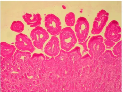
Histopathological examination. The strangulated part of the small intestine showed necrosis and sloughing of epithelium, congestion, edema and hemorrhage in the lamina propria as common features in all groups. Specimens that have been taken for a distance of 10 cm long proximal to the strangulated part showed necrosis and sloughing of epithelium, blunting of some villi, congestion and edema in the lamina propria. These lesions were marked in segments removed in group I and were worse in segments removed in groups II
and III. Thickening and blunting of the intestinal villi appeared at 6 and 9 hours, while blunting of the villi was observed at 12 hours after obstruction (Figs. 7- 14). Microscopic examination of the intestine for a distance of 1 m long proximal to the strangulated part of the small intestine revealed necrosis of epithelium, thickening of intestinal villi, congestion and edema in the lamina propria. These lesions were marked in segments removed in group I and were worse in other groups. Blunting of the intestinal villi was observed in group IV. Microscopic examination of the intestine for a distance of 2 m long proximal to the strangulated part showed blunting of the villi and edema in the lamina propria. These lesions were marked in the segments removed in group I and were worse in segments removed other groups. Necrosis of the epithelium appeared in groups II, III, and IV hours after surgery. Microscopic examination of the intestine for a distance of 3 m proximal to the strangulated part showed edema in the lamina propria. This lesion was marked in segments removed in group I and was more prominent in segments removed from other groups. Necrosis of epithelium appeared in groups II, III and IV. Thickening and blunting of intestinal villi and congestion in the lamina propria were observed in groups II and III. Adhesion of the epithelial villi was observed only in group IV. None of these changes were observed in the group V.
Fig 2. Three hours strangulation obstruction, A. Strangulated part, B. proximal to the strangulated part, C. distal to the strangulated part, D. Mesentery
Fig 3. Six hours strangulation obstruction, A. Strangulated part, B. proximal to the strangulated part, C. distal to the strangulated part, D. Mesentery
Fig 4. Nine hours strangulation obstruction, A. strangulated part, B. proximal to the strangulated part, C. distal to the strangulated part, D. Mesentery
Fig 5. Twelve hours strangulation obstruction, A. Strangulated part, B. proximal to the strangulated part, C. distal to the strangulated part, D. Mesentery
Fig 6. The intestinal segment cranial to strangulated part of small intestine subjected to 12 hours of VSO showing petecheal hemorrhages for great distance up to 3 meters proximal to the site of obstruction. Black arrows
Fig 7. The strangulated part of intestine, 3 hrs post-operation, showing necrosis and sloughing of epithelium (asterisk) and congestion and hemorrhages in the lamina propria (arrows). H & E (400×)
A
B
C D
A
B
C D
B
C
D A
B
C D
A
Fig 8. The strangulated part of intestine, 6 hrs post-operation, shows extensive necrosis of epithelium (asterisk), hemorrhages into intestinal villi (arrows) and congestion and edema in the lamina propria. H & E (400×)
Fig 9. The strangulated part of the intestine, 9 hrs post-operation, shows extensive necrosis of epithelium, hemorrhage into intestinal villi and severe congestion and hemorrhage in the lamina propria. H & E (400×)
Fig 10. The strangulated part of the intestine, 12 hrs post-operation, shows massive necrosis of epithelium and absence of villi and severe congestion in the lamina propria. H & E (400×)
Fig 11. The intestine 3 hrs post-operation, 10 cm proximal to strangulation, shows necrosis and sloughing of epithelium (asterisk), blunting of some villi (arrow) and congestion and edema in the lamina propria (thick arrows) . H & E (400×)
Fig 12. The intestine 6 hrs post-operation, 10 cm proximal to strangulation, shows necrosis of epithelium, thickening and blunting of intestinal villi and edema in the lamina propria. H & E (400×).
Fig 13. The intestine 9 hrs post-operation, 10 cm proximal to strangulation, shows necrosis of epithelium, thickening and blunting of intestinal villi and congestion and edema in the lamina propria. H & E (400×)
Fig 14. The intestine 12 hrs post-operation, 10 cm proximal to strangulation, shows necrosis of epithelium, blunting of intestinal villi. H & E (400×)
Discussion
Intestinal displacements and strangulations are the common causes of colic in horses.15 The clinical picture of strangulation obstruction in equine small intestine depends on the site of the obstruction and the degree of the strangulation.16 The results obtained from this study indicated that marked disturbances in the form of abdominal pain with changes in the animal behavior and clinical parameters increased dramatically with the increase of the strangulation period. These changes could be used to justify the prognosis of surgery for correction of the small intestine strangulation and to determine the methods for stabilizing the patient before anesthesia. The decrease in the level of abdominal pain after the elapse of nine hours was explained due to necrosis of the intestinal mucosa, and rapidly development of endotoxemia at this stage.17-19 The cause of the visceral pain was the interruption of the mesenteric circulation by strangulation or intestinal displacement causing ischemia of the intestine.20-22 Duration of colic signs, heart rate, intestinal sounds, skin tenting, level of pain and appearance of peritoneal fluid are significant predictors of survival in cases of strangulation obstruction.15 The significant reduction in the body
quickly develop and the segment becomes black red in color. 28 The extravascular pressure compromises the venous return without effect on the arterial blood flow to the involved segment resulting in intestinal ischemia. 29,30 Horses often develop postoperative complications that result from intraluminal distension proximal to the site of obstruction and consequences of ischemia, adhesions, and mucosal necrosis. These adhesions indicate that the preoperative damage might account for postoperative complications. 31 In the present study the distension, edema and hemorrhagic patches of the bowel wall and mesentery were obvious after 6 hours of venous strangulation obstruction. The intensity of bowel wall discoloration and distension was more dramatic at 9 and 12 hours after strangulation. Jubb et al (1985) attributed the intraluminal distension to the fluid and gas content of the intestinal lumen. In our study, the lumen distension proximal to the strangulated segment occurred as a sequel to ischemia which induced flaccidity of the smooth muscle of the intestinal wall. 29
As the intestine distends, its function is further compromised, leading to increased secretion, reduced absorption, and poor propulsion along its length. 17,32 The histopathological examination of the strangulated part of the small intestine in the present study were similar to those described by Freeman and Kilgallon (2001) who found mucosal epithelial damage of the bowel wall and the mesentery subjected to venous strangulation obstruction. The reason for the severe mucosal damage in case of strangulation obstruction is not definitely understood, but the severity of the lesions may be correlated with the severity of the condition in the affected horses. 33 Three overlapping mechanisms (absorption of endotoxin, hypoxia and reperfusion injury) have been suggested as contributing factors to the development of mucosal lesions remote from the site of the strangulation obstruction. In the horses
with strangulating obstruction have the same or less similar results such as sub epithelial lifting at the villus tip, with occasional necrolytic vacuolization of mucosal cells, large focal sloughs of mucosal epithelium and development of prominent sub epithelial spaces. In the more severe cases, epithelium necrosis extended further down the villus, extensive necrosis of the lamina propria and continued sloughing of the mucosal epithelium to the crypt area. 7 Gangrene of the affected intestinal wall in case of strangulation obstruction leads to disruption of the mucosal epithelial barrier, allowing trans mural movement of endotoxin into the peritoneal cavity, from which it is absorbed. Because endotoxin may damage capillary endothelium and increase capillary permeability, it has been considered as a cause of vascular and mucosal damage in the remainder of the small intestine. 34,35 In this study, the histopathological examination of the intestine for a distance of 10 cm long proximal to the strangulated part was the same noticed by Dabareiner et al (1993). While the Histopathological changes occurred 3 m proximal to the strangulated part were similar to the study of Freeman and Kilgallon (2001) and Allen et al (1986). False interpretation of intestinal viability after correcting intestinal strangulating obstruction results in continued shock, ileus, peritonitis and an eventual death (usually 2-3 days postoperatively). Therefore, the surgeon should choose to resect some normal bowel rather than the risk of leaving any compromised bowel in place. 36 According to clinical experience, horses can tolerate loss of 10 m and 10.7 m of small intestine.
3,37,38
Resection of long segments does not increase postoperative mortality compared with short segment. 3
justification of the prognosis of the patient and the availability of treatment. The extent of intestinal wall damage to a long distance proximal to the seat of obstruction indicated the need of resection of a considerable distance of the normal intestine to avoid the need for second operation and decrease the postoperative complications. In addition, the decision to perform laparotomy should not be postponed until the condition of the patient deteriorates to a great extent. This stresses the importance of an early decision to perform laparotomy. Further studies are required to determine the exact length of histopathological changes proximal to the strangulated part.
Manufacture addresses
Neurazine, Misr co., Saed sleem Str. 4 8734 Cairo, Egypt
Sandoz Pty Ltd, Level 4, Suite 7-19, 100 Harris Street, Pyrmont NSW 2009, Australia
Conflict of interest statement
None of the authors has any financial or personal relationships that could
inappropriately influence or bias the content of the paper.
This study was approved by the committee of animal care of the Faculty of Veterinary Medicine, Assiut University.
Acknowledgements
The authors thank Dr. Khaled Radad for his work in histopathological examination, and colleagues at the Department of Surgery, Faculty of Veterinary Medicine for assistance with the management of these cases.
References
1. White NA. Epidemiology and etiology of colic: The equine acute abdomen. White, NA. (ed), Lea & Febiger, Philadelphia. 1990; 50 – 64.
2. Gerard MP, Blikslager AT, Roberts MC, et al. The characteristics of intestinal injury peripheral to
strangulating obstruction lesions in the equine small intestine Equine vet. J. 1999; 31: 4: 331-335.
3. MacDonald MH, Pascoe JR, Stover SM. Survival after small intestine resection and anastomosis in horses. Vet. Surg. 1989; 18: 415 – 423.
4. Parker JE, Fubini SL, Todhunter RJ. Retrospective evaluation of repeat celiotomy in 53 horses with acute gastrointestinal disease. Vet. Surg. 1989; 18: 424-43 1.
5. Blikslager AT, Bowman KF, Levine JF, et al. Evaluation of factors associated with post-operative ileus in horses: 31 cases (1990-1992). J. Am. Vet. Med. Assoc. 1994; 205: 1748-1752.
6. Baxter GM. Abdominal adhesions after small intestinal surgery in the horse. Vet. Surg. 1989; 18: 409-414.
7. Allen DJ, White NA, Tyler DE. Factors for prognostic use in equine obstructive small intestinal disease. J. Am. Vet. Med. Assoc. 1986; 189:7: 777 – 780. 8. Meschter CL, Tyler DE, White NA, et
al. Histological findings in the gastrointestinal tract in horses with colic. Am. J. Vet. Res. 1986; 47: 598 – 606.
9. Freeman DE, Cimprich RE, Richardson DW. Early mucosal healing and chronic changes in pony jejunum after various types of strangulation obstruction. Am. J. Vet. Res. 1988; 49: 810-818.
10. Van Hoogmoed LM, Snyder J, Nieto JE. Use of an extracorporeal circuit to evaluate effects of ischemia and reperfusion of the equine large colon. Am. J. Vet. Res. 2000; 61: 1042 – 1051. 11. Lundin C, Sullins KE, White NA, et al. Induction of peritoneal adhesions with small intestinal ischaemia and distention in the foal. Equine vet. J. 1989; 21: 451-458.
small intestine in the pony. Am. J. Vet. Res. 1983; 44:1187-1191.
14. Bancroft JD, Stevens A. Theory and practice of Histologic Techniques, 3rd ed. Long Man Group Limited. 1993; 332-338
15. Van der Linden AM, Laffont CM, van Oldruitenborgh-Oosterbaan S. Prognosis in equine Medical and surgical colic J. Vet. Intern Med. 1993; 17:343-348
16. Datt OS, Usenik EA. Intestinal obstruction in the horse. Physical signs and blood chemistry. Cornell Vet. 1975; 152 – 159.
17. Youssef HA, Abd El-Salaam MN. A comparative study regarding of experimental strangulated obstruction of the small and large intestine in donkeys. In proceeding of the 5th Sci. Cong., Fac. Vet. Med. Assiut Univ. 1992; Nov. 8 – 10. Egypt.
18. Venugopalan A. Essentials of veterinary surgery, 7th ed. Oxford & IBH Publishing Co. PVT. LTD., New Delhi Bombay, Calcutta.1994; 325 – 338.
19. Mair T, Love S, Schumacher J. Equine medicine, surgery and reproduction, W. B. Saunders. Company, L.T. London, Philadelphia, Toronoto. 1998; 21 – 53. 20. Stashak TS. Clinical evaluation of the
equine colic patient. Vet. Clin. North Am. (Large Anim. Pract.); 1979; 1: 275 – 287.
21. Schall WB, Coffman JR. Defining gastrointestinal problems. In: Anderson NV. Ed. Veterinary gastroenterology, 1st ed. London: Lea & Febiger. 1980; 30 – 43.
22. Robertson JT. Conditions of the stomach and small intestine. Vet. Clin. North Am. (Large Anim. Pract.). 19824; 105 – 127.
23. Johnston JK, Morris DD. Comparison of duodenitis/ proximal jejunitis and small intestinal obstruction in horses: 68 cases (1977–1985) J. Am. Vet. Med. Assoc. 1987; 191:7: 849 – 854.
24. Freeman DE, Kilgallon EG. Effect of venous strangulation obstruction on length of equine jejunum and relevance to small-intesinal resection. Vet. Surg. 2001; 30: 218-222.
25. Dabareiner RM, Sullins KE, Snyder JR, et al. Evaluation of the microcirculation of the equine small intestine after intraluminal distention and subsequent decompression. Am. J. Vet. Res. 1993; 54:1673 – 1682.
26. Reeves MJ, Curtis CR, Salman MD. Development and validation of multivariable models to predict the need for surgery and prognosis in equine colic patients. Acta Vet Scand, 1988; 84 (Suppl.): 329–332.
27. Granger DN, Kvietys PR, Mortillaro, NA. Effects of luminal distention on intestinal transcapillary fluid exchange. Am. J. Physiol. 1980; 239:516 – 523. 28. Jubb KV, Kennedy PC. The lower
alimentary system. In: Pathology of domestic animals. 2nd ed., Academic Press Inc, New York. 1970; 89 – 98. 29. Jubb KF, Kennedy PC, Palmer N.
Pathology of domestic animals. 3rd ed., Academic Press Inc, New York. 1985; 59 – 64.
30. Sullins KE, Stashak TS, Mero KN. Pathologic changes associated with induced small intestinal strangulation obstruction and nonstrangulating infarction in horses. Am. J. Vet. Res. 1985; 46: 913 – 916.
31. Dabareiner RM, White NA, Donaldson LL. Effects of intraluminal distention and decompression on microvascular permeability and hemodynamics of the equine jejunum. Am. J. Vet. Res. 2001; 62: 2: 225 – 236.
32. Kohn CW. Preparative management of the equine patient with an abdominal crisis. Vet. Clin. North Am. Large Anim. Pract. 1979; 1: 289 – 311.
of the 1st Equine Colic Research Symposium, University of Georgia, Athens. 1982; 261 – 262.
34. McClure JJ. Endotoxic shock, Vet. Clin. North Proc. Soc. 1976; 6: 193 – 202.
35. Baris C, Guest WM, Frazer ME. Direct effects of endotoxin on the microcirculation. Adv. Shock Res. 1980; 4: 153 – 160.
36. Hickman J. Equine surgery and medicine. Academic Press. Harcourt Brace Jovanovich, London Orlando, San Diego, New ork, 1985; vol. 1 37. Collier MA, Trent AM. Jejunal
ntussusceptions associated with leiomyoma in an aged horse. J Am Vet Med Assoc. 1983; 182: 819-821. 38. Mair T, Divers T, Ducharme N.