R E S E A R C H
Open Access
Expression of neural cell adhesion molecule
and polysialic acid in human bone
marrow-derived mesenchymal stromal cells
Maria S. Skog
1*, Johanna Nystedt
2, Matti Korhonen
2, Heidi Anderson
1,3, Timo A. Lehti
1, Maria I. Pajunen
1,4†and Jukka Finne
1†Abstract
Background:In order to develop novel clinical applications and to gain insights into possible therapeutic mechanisms, detailed molecular characterization of human bone marrow-derived mesenchymal stromal cells (hBM-MSCs) is needed. Neural cell adhesion molecule (NCAM, CD56) is a transmembrane glycoprotein modulating cell–cell and cell–matrix interactions. An additional post-translational modification of NCAM is theα2,8-linked polysialic acid (polySia). Because of its background, NCAM is often considered a marker of neural lineage commitment. Generally, hBM-MSCs are considered to be devoid of NCAM expression, but more rigorous characterization is needed.
Methods:We have studied NCAM and polySia expression in five hBM-MSC lines at mRNA and protein levels. Cell surface localization was confirmed by immunofluorescence staining and expression frequency in the donor-specific lines by flow cytometry. For the detection of poorly immunogenic polySia, a fluorochrome-tagged catalytically defective enzyme was employed.
Results:All five known NCAM isoforms are expressed in these cells at mRNA level and the three main isoforms are present at protein level. Both polysialyltransferases, generally responsible for NCAM polysialylation, are expressed at mRNA level, but only very few cells express polySia at the cell surface.
Conclusions:Our results underline the need for a careful control of methods and conditions in the characterization of MSCs. This study shows that, against the generally held view, clinical-grade hBM-MSCs do express NCAM. In contrast, although both polysialyltransferase genes are transcribed in these cells, very few express polySia at the cell surface. NCAM and polySia represent new candidate molecules for influencing MSC interactions.
Keywords:Bone marrow, Mesenchymal stromal cell, Clinical grade, Neural cell adhesion molecule, Polysialic acid
Background
Human bone marrow-derived mesenchymal stromal cells (hBM-MSCs) are attractive candidates for cellular therapy, regenerative medicine, and tissue engineering. They are adult progenitor cells that hold potential for fast clonal expansion, secretion of trophic and immuno-modulatory factors, and multilineage differentiation. In addition, they are relatively easy to harvest, isolate, and
culture. However, it is widely acknowledged that follow-ing in-vitro culture, MSCs undergo replicative senes-cence and appear to lose their beneficial traits [1]. Furthermore, MSC cultures typically consist of a hetero-geneous mixture of cells at different stages of commit-ment and potential [1, 2]. In order to develop novel clinical applications and to gain more insight into the possible therapeutic mechanisms, detailed molecular characterization of hBM-MSCs is needed. This will ul-timately improve the safety and efficacy of cell therapy.
Neural cell adhesion molecule (NCAM, CD56) is a calcium-independent binding protein engaged in homophi-lic cell–cell and heterophilic cell–matrix interactions [3].
* Correspondence:maria.skog@helsinki.fi †Equal contributors
1Biochemistry and Biotechnology, Department of Biosciences, University of Helsinki, P.O. Box 56, FI-00014 Helsinki, Finland
Full list of author information is available at the end of the article
NCAM is transcribed from a single gene, but exists in multiple isoforms as a result of alternative splicing [4]. In humans, at least five distinct NCAM isoforms are known. A modification of NCAM results post-translationally from the addition of linear polymers of
N-acetylneuraminic acid units, polysialic acid (polySia) [5, 6]. Being a large negatively charged and highly hy-drated structure, polySia regulates NCAM activity by altering its biophysical properties [7]. PolySia has an important role in maintaining developmental plasticity and cell migration in tissues by influencing cellular inter-actions [8]. In addition, it is involved in various medical conditions such as tissue repair, neurodegenerative dis-eases, and progression of metastatic cancers [9, 10].
Expression of NCAM and polySia is strong and dy-namic during embryogenesis, decreases and focuses dur-ing development, and is limited to just few tissues and cell types in the adult [11, 12]. Because of its original discovery site, NCAM is often considered a marker of neural lineage commitment [13, 14]. However, it is known that expression of NCAM and polySia is wide-spread during organogenesis, particularly in undifferenti-ated mesenchymal cells [15]. Many in-vivo and in-vitro studies in animal models suggest that NCAM is an im-portant regulator of cell migration and condensation during skeletal development [16–18]. Hence, NCAM and polySia represent promising candidates for influen-cing MSC interactions.
It is generally held that hBM-MSCs do not express NCAM [19–25], while, for example, placental and um-bilical cord blood-derived MSCs are eminently NCAM positive [21, 24, 25]. Brooke et al. [26] have reported that
hBM-MSCs do express NCAM at the mRNA level, but
protein expression was not investigated. NCAM protein expression, which may indicate increased chondrogenic potential, has been reported in a small fraction of pri-mary bone marrow mononuclear cells (0.5–5.5 %), but expression diminished over time in culture [27, 28]. In con-trast, murine BM-MSCs predominantly express NCAM, which plays a crucial role, for example, in hematopoiesis [29]. Furthermore, experiments with NCAM knockout mice have shown reduced multilineage differentiation potential of BM-MSCs compared with wild-type controls [30, 31]. Thus, because of the role of NCAM and polySia in the control of cellular differentiation and interaction, it is important to reliably determine whether they are expressed in clinical-grade hBM-MSCs.
In this study, we have investigated the expression sta-tus of NCAM and polySia in clinical-grade hBM-MSCs using a variety of methods. We have concentrated par-ticularly on NCAM expression, because we observed a striking discrepancy between our findings and previous reports [19–25]. Furthermore, NCAM is the most stud-ied molecule of the immunoglobulin superfamily of cell
adhesion molecules (CAMs), but has been largely neglected in stem cell research despite its role as a de-velopmental regulator. This study clearly demonstrates the need for comprehensive analyses and careful control of methods in the characterization of MSCs. Gene and protein expression analyses show that these cells do, in fact, express NCAM. In contrast, although polysialyl-transferases are transcribed in these cells, very few ex-press polySia on the cell surface.
Methods
Cells
The culture protocol developed by Laitinen et al. [32] for clinical-grade MSCs based on platelet lysate was utilized in this study. Bone marrow was collected from five healthy volunteer donors (donor 067: female, age 24; donor 068: female, age 31; donor 069: female, age 30; donor 072: female, age 21; donor 073: female, age 21). Bone marrow was aspirated under local anesthesia from the posterior iliac crest and collected in heparinized tubes after signed informed consent according to the Declaration of Helsinki. The protocol was approved by the ethics committee of the Hospital District of Helsinki and Uusimaa (Finland). The isolation and characterization of hBM-MSCs has been described in detail previously [32]. The isolated cells were cultured in heparinized (LEO Pharma, Ballerup, Denmark) low-glucose Dulbecco’s modified Eagle’s medium (DMEM; Gibco, Life Technolo-gies, Paisley, UK), supplemented with 10 % platelet lysate (Finnish Red Cross Blood Service, Helsinki, Finland), and 100 U/ml penicillin and 100 μg/ml streptomycin (Gibco) according to Laitinen et al. [32]. The medium was chan-ged twice weekly and the cultures were passachan-ged when subconfluent (80 % confluency) and subcultured at 1000– 1500 cells/cm2. The hBM-MSCs used in this study were freshly analyzed (i.e., noncryopreserved) at passage 2 or 3.
was discarded and isolated cells were washed with PBS. Washing and magnetic separation was repeated 10 times. Lastly, the isolated cells were plated on a cell culture dish and cultured accordingly. The NCAM and polySia expres-sion status was analyzed with flow cytometry; the propor-tion of NCAM and polySia-expressing cells was 98.3 %.
All cells were cultured under a humidified atmosphere at 37 °C and with 5 % CO2.
Differentiation of hBM-MSCs
Adipogenic differentiation of hBM-MSCs was performed as described previously by Laitinen et al. [32]. The cells were cultured in adipogenic differentiation condition for 1–2 weeks. After differentiation the cells were fixed with 4 % paraformaldehyde in PBS for Sudan III staining.
Osteogenic differentiation of hBM-MSCs was carried out essentially as described previously by Laitinen et al. [32]. The cells were cultured in osteogenic medium for 3–4 weeks. After differentiation the cells were fixed with 4 % paraformaldehyde in PBS for von Kossa staining.
Chondrogenic differentiation was performed as de-scribed previously by Skog et al. [34]. The pellet cultures were maintained up to 4 weeks, changing the medium twice a week. After differentiation the cell pellets were fixed with 10 % formalin, embedded in paraffin, and cut into 5μm sections for Safranin O staining.
Qualitative reverse transcription PCR
NCAM isoform-specific sequences were obtained from NCBI Unigene (http://www.ncbi.nlm.nih.gov/unigene). Primers were designed using NCBI Primer Blast (http:// www.ncbi.nlm.nih.gov/tools/primer-blast) and optimized
by OligoEvaluator™ (http://www.sigmaaldrich.com/technical-documents/articles/biology/oligo-evaluator.html) or Tm Calculator (https://www.thermofisher.com/fi/en/home/ brands/thermo-scientific/molecular-biology/molecular-biology-learning-center/molecular-biology-resource-library/ thermo-scientific-web-tools/tm-calculator.html) PCR web tools. All commercial kits were used according to the manufacturer’s instructions. Total RNA was isolated using the High Pure RNA Isolation Kit (Roche Diag-nostics, Mannheim, Germany). Reverse transcription was performed with High Capacity RNA-to-cDNA Kit (Applied Biosystems, Foster City, CA, USA) with 1 μg of total RNA. A control reaction omitting the reverse transcriptase was prepared for each sample. PCRs were performed in 20 μl final volume with Phusion Hot Start II High-Fidelity DNA Polymerase (Thermo
Scientific, Waltham, MA, USA) for all NCAM
iso-forms, polysialyltransferases (ST8SIA2 and ST8SIA4), and controls, except that OneTaq Hot Start DNA Poly-merase (New England BioLabs, Ipswich, MA, USA) was used for isoformNCAM-120.
Human NCAM isoform and
polysialyltransferase-specific PCR primers were used for the amplification (Table 1). For Phusion Hot Start II High-Fidelity DNA Polymerase, the reaction mixture was composed of 0.02 U/μl of DNA polymerase, Phusion HF Buffer (with 1.5 mM MgCl2), 0.2 mM dNTPs, 0.5 μM primers, 3 %
DMSO, sterile water (Baxter Healthcare, Zürich,
Switzerland), and 2.0 μl of template. For OneTaq Hot Start DNA Polymerase, the reaction mixture was composed of 0.025 U/μl of DNA polymerase, OneTaq Standard Reaction Buffer (with 1.8 mM MgCl2), 0.2 mM
Table 1Primers used in the study
Primer UniGene Forward (5′➔3′) Reverse (5′➔3′) Amplicon size
(base pairs)
GAPDH Hs.544577 GAAGGTGAAGGTCGGAGTC GAAGATGGTGATGGGATTTC 225
NCAM-All Hs.503878 GGACTTCTACCCGGAACATCAG CGAGCTTAGGTGCACTGGG
NCAM-140 (Variant 1), NCAM-125 (Variant 4)
798
NCAM-180 (Variant 2) 828
NCAM-120 (Variant 3) 903
NCAM Variant 5 906
NCAM-180 NM_181351.4 GCTTCGTGGACTCGACCAG GCAGATGTACTCTCCGGCATC 126
NCAM-140 NM_000615.6 CGAAGAAAAGACTCTGGATGG TCATGCTTTGCTCTCGTTCT 1498
NCAM-125/140 NM_001242608.1 / NM_000615.6 CGAAGAAAAGACTCTGGATGG GTTCCCCTTGGACTGGC 755
NCAM-125 NM_001242608.1 CAGCCAGTCCAAGGGG TGTAGGATGCAGAATTGCCTC 346
NCAM120/125 NM_001076682.3 / NM_001242608.1 GAGTATGAGGTCTACGTGGTGGC GCAGAGAAAAGCAATGAGACAAAG 152
NCAM-120 NM_001076682.3 TCTGCTAGCTCGTCTACCCC CCAAAGGGGGCACTGATCTT 541
NCAM Variant 5 NM_001242607.1 GCTTCGTGGACTCGACCAG GCAGATGTACTCTCCGGCATC 204
ST8SIA2 Hs.302341 GGGCAACTCGGGGGTCTTGC AAGGCCCGCTGGATGACCGA 162
dNTPs, 0.2 μM primers, 1.5 M betaine (Sigma), sterile water (Baxter Healthcare), and 2μl of template. Amplifi-cation was performed for 35 cycles. The PCR products were analyzed by agarose gel electrophoresis. Glyceralde-hyde 3-phosphate dehydrogenase (GAPDH) was used as the reference gene.
Western blot
Whole-cell extracts were prepared by lysing the cells in radioimmunoprecipitation assay (RIPA) buffer pH 6.8 containing 50 mM Trizma® base, 150 mM sodium chlor-ide, 0.5 % sodium deoxycholate (all from Sigma), and 1 % Triton X-100 (Roche Life Sciences, Indianapolis, IN, USA), supplemented with EDTA-free protease inhibitor cocktail (Thermo Scientific, Rockford, IL, USA). For re-moval of polySia, 10 μg/ml of active endosialidase-GFP fusion protein [35] was added and the samples were incubated at 37 °C for 45 minutes. Samples were mixed with Laemmli sample buffer, loaded, and run
on 4–20 % Mini-PROTEAN® TGX™ Precast gradient
gel (Bio-Rad Laboratories, Hercules, CA, USA) or 7 % SDS-polyacrylamide gel. Proteins were electro-transferred from the gels to nitrocellulose membranes (GE Healthcare, Little Chalfont, UK) overnight at 4 °C. The membrane was blocked with 3 % skimmed milk powder in PBS with 0.1 % Tween-20 for 3 hours at 4 °C.
For NCAM detection, the membrane was incubated with a primary antibody mixture containing 0.33 μg/ml rabbit polyclonal anti-human NCAM antibody (AB5032; Millipore, Temecula, CA, USA) and 0.067 μg/ml mouse monoclonal anti-human NCAM antibody (123C3; Santa Cruz, Dallas, Texas, USA) in blocking solution overnight at 4 °C. Because anti-NCAM antibodies may detect dif-ferent isoforms with varying affinity in western blot, two primary antibodies were used simultaneously to ensure accurate detection of all isoforms [36]. After three washes, the membrane was incubated with a secondary antibody mixture containing HRP-conjugated anti-rabbit secondary antibody 1:3000 and anti-mouse secondary antibody 1:3000 (both from Cell Signaling, Danvers, MA, USA) in blocking solution for 2 hours at room temperature. The immunoblots were developed with SuperSignal® West Pico Chemiluminescent Substrate (Thermo Scientific). Secondary antibody control con-firmed the specificity of the labeling, andα-Tubulin (B-51-2; Sigma) served as a loading control.
Flow cytometry
Cells were double labeled with 10μg/ml of rabbit poly-clonal anti-human NCAM antibody (AB5032; Millipore) followed by AlexaFluor 647-labeled goat anti-rabbit sec-ondary antibody (Molecular Probes, Invitrogen, Eugene, OR, USA) and inactive endosialidase-GFP fusion protein [33] in PBS. Parallel samples were labeled with
AlexaFluor 647 mouse anti-human alkaline phosphatase antibody (B4-78; BD Biosciences, San Jose, CA, USA) and FITC mouse anti-human CD44 antibody (BD Biosciences) according to the manufacturer’s instruc-tions. Appropriate fluorescence minus one (FMO) con-trols were used for analysis. The cells were analyzed with FACS LSR II flow cytometer and FACSDiva 5.0.3 software (BD Biosciences). Cell debris and dead cells were excluded from the analysis based on physical pa-rameters and propidium iodide (PI) fluorescence probing for cell viability (proportion of positive cells 1.2–7.0 %, data not shown).
Immunocytochemistry
For immunocytochemical staining, cells were grown on glass coverslips and fixed with 4 % paraformaldehyde in PBS. Nonspecific binding was blocked with 1.5 % normal horse serum (Vector Laboratories, Burlingame, CA, USA) in PBS. Cells were labeled with 10 μg/ml of rabbit poly-clonal anti-human NCAM antibody (AB5032; Millipore) followed by AlexaFluor 647 conjugated goat anti-rabbit secondary antibody (Molecular Probes) and inactive endosialidase-GFP fusion protein [33], all in PBS. Cover slips were mounted with ProLong Mounting Medium with DAPI (Molecular Probes). The staining was visual-ized with an Olympus BX50F-3 microscope and imaged by a PCO CCD Imaging SensiCam color camera and Image-Pro Plus 4.0 software.
Results
Multilineage differentiation of hBM-MSCs
The multilineage differentiation assay was performed to verify the differentiation capacity of hBM-MSCs accord-ing to the International Society for Cellular Therapy (ISCT) minimal criteria for MSCs [37]. All hBM-MSC lines displayed typical MSC differentiation capacity along the adipogenic, osteogenic, and chondrogenic lineages (Fig. 1).
Expression of NCAM and polysialyltransferase mRNA in hBM-MSCs
To gain an overview ofNCAMmRNA expression, quali-tative reverse transcription PCR analysis was performed with isoform-specific primer pairs (Fig. 2a). The reverse transcription PCR analysis revealed distinct expression of all NCAM isoform mRNAs in the three analyzed hBM-MSC lines (Fig. 2b).
Surface expression of NCAM and polySia in hBM-MSCs
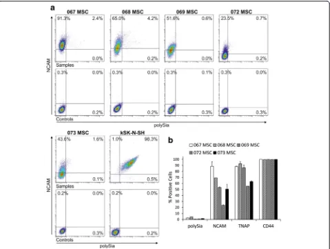
Using commercially available polyclonal human-specific NCAM antibody, we detected cell surface NCAM ex-pression on hBM-MSCs in flow cytometry (Fig. 4a), indi-cating that NCAMmRNA is translated into protein. All five hBM-MSC lines included in this study expressed NCAM on the cell surface. However, its occurrence was heterogeneous, because not all cells in the populations expressed NCAM at detectable levels. Differences be-tween donor-specific hBM-MSC lines were also ob-served, the proportion of positive cells ranging from 23.6 ± 0.8 % to 88.5 ± 7.4 % (Fig. 4b). Concurrent polySia detection was performed with inactive endosialidase-GFP fusion protein that binds to polySia. Surprisingly, only very few hBM-MSCs expressed polySia (Fig. 4a), the proportion of positive cells ranging from 0.5 ± 0.2 % to 4.4 % (Fig. 4b). All polySia-expressing cells were simultaneously labeled positively for NCAM, the main carrier of polySia.
For comparison, cell surface expression of CD44 and tissue nonspecific alkaline phosphatase (TNAP) was also analyzed from hBM-MSC lines. Flow cytometry data in-dicate that CD44, a common MSC marker, is fully expressed in all of the lines (>99 %, Fig. 4b). On the other hand, expression of the early osteogenic marker TNAP was more heterogeneous, ranging from 55.3 % to
92.8 ± 3.7 % (Fig. 4b). No correlation was found between NCAM and TNAP expression.
Expression of NCAM isoforms in hBM-MSCs
NCAM protein expression was further confirmed by western blot analysis (Fig. 5). Bands of approximately 180, 140, and 120 kDa in size were detected in all hBM-MSC lines analyzed, indicating the presence of the three main isoforms of NCAM. At the protein level, hBM-MSCs seem to express predominantly NCAM-180 and other isoforms to a lesser extent. However, distinction between NCAM-125 and NCAM-120 was not possible because the bands overlap. Some variation in the expres-sion of different isoforms was observed between the hBM-MSC lines, MSC 069 expressing different isoforms in more equal manner compared with other hBM-MSC lines. Because polySia may affect NCAM mobility in gel electrophoresis, endosialidase [35] was used to treat a set of parallel samples. In agreement with high polySia expression on kSK-N-SH cells (Fig. 5), endosialidase treatment revealed the presence of NCAM in these cells. In contrast, endosialidase treatment did not un-cover additional NCAM in hBM-MSCs. Altogether, the western blot results demonstrate that hBM-MSCs express various NCAM isoforms also at the protein level.
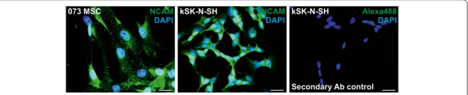
Cellular localization of NCAM and polySia
To determine the subcellular localization of NCAM and polySia, immunocytochemical detection was utilized. NCAM appears to be expressed in a clustered manner over the cell surface in hBM-MSCs (Fig. 6). In contrast, NCAM expression was even and smooth on the refer-ence neuroblastoma cell line kSK-N-SH. NCAM expres-sion in kSK-N-SH seems to be more intense around cell–cell contact sites, whereas this was not the case for hBM-MSCs.
Compared with NCAM, very little polySia expression was detected in hBM-MSCs (Fig. 7). Grainy polySia ex-pression was detected mainly on cellular extensions. This was very different from the polySia expression in kSK-N-SH cells, where polySia is distributed smoothly all over the cell surface. In kSK-N-SH cells, polySia ex-pression, together with NCAM, appears to be more
intense in cell–cell contacts; however, in hBM-MSCs such condensation was not detected. Altogether, the im-munocytochemistry results confirm that NCAM protein is expressed on the cell surface.
Discussion
migration, cytokine response, cell contact-dependent dif-ferentiation, and immune response modulation [41–44].
To date, definite information about NCAM and polySia expression in hBM-MSCs has been lacking. Because of the intrinsic heterogeneity of the MSC populations, donor variation, and diversity in culture conditions and analysis methods, biomolecular and cytometric characterization of MSCs from different laboratories is not easy to compare [2]. Furthermore, transcriptomic profiling has become a popular method for characterizing MSCs, but protein and mRNA expression levels do not always correlate. Analysis at the protein level is therefore needed before conclusions about the MSC phenotype can be drawn.
To gain more insight into the characteristics of xeno-free hBM-MSCs, we have utilized an established clinical-grade culture protocol based on human platelet lysate [32]. Platelet lysate has been approved as a safe and ef-fective supplement for MSC cultivation in vitro [45]. Conventional cell culture methods involve many animal-derived components; however, they are not desirable for clinical-grade cell production because of the increased risk of cross-contamination and host immune reactions [46]. In addition, xenogeneic additives, like bovine sera, may negatively alter the self-renewal and stemness of hBM-MSCs [47]. Because MSCs are very rare in the bone marrow, isolation and in-vitro expansion of the cells is usually required prior to their use. We analyzed noncryopreserved cells at passage 2 or 3, which is the time point of choice for most MSC applications because it offers the minimum required number of cells that still hold functional potency [48, 49]. NCAM and polySia ex-pression is possibly altered during in-vitro culture. How-ever, analysis of the properties of these cells directly
from the bone marrow would be challenging because re-liable markers for their identification are lacking. Culture of MSCs for at least two passages is commonly used to attain population purity [48].
Our findings regarding NCAM expression in hBM-MSCs differ from those reported previously [19–25], and show—in contrast to the generally held view—that the cells in fact do express NCAM. Furthermore, we conducted a more detailed analysis regarding NCAM gene and protein expression. Our data show that all five
known NCAM isoforms are transcribed in hBM-MSCs.
In particular, the main isoforms are detected at the pro-tein level. NCAM propro-tein is expressed throughout the cell surface in a clustered manner. However, flow cyto-metric analysis revealed quite broad donor-specific vari-ation in expression levels between the hBM-MSC lines. This is not unexpected even in our relatively homoge-neous donor population (healthy females, age 21–31 years), because it has been reported previously that hBM-MSC cultures are heterogeneous mixtures of cells, the properties and potency of which vary greatly be-tween individual donors independent of age or gender [50–52]. In this study, the donor population was not se-lected based on any specific characteristic, but samples were obtained from the volunteer donors in the order they came in based on the national guidelines for bone marrow donation eligibility. In Finland, as in most other western countries, the majority of bone marrow donors are female [53]. In general, gender has little effect on hBM-MSC features [50, 54, 55].
or serum free) and diverse procedures are employed in their management. Donor-dependent variation may also occur. Expression patterns may be regulated temporally and the cells may be in different stages and passages at the time of analysis, or have undergone replicative senes-cence. Also, the cellular phenotype may alter between fresh and cryopreserved cells. Technical variation may also be responsible; for example, the variable sensitivity and isoform specificity of anti-NCAM antibodies may give rise to misleadingly low or lacking protein expression. Fur-thermore, the expression of NCAM mRNA transcripts does not necessarily correlate with the expression of pro-tein on the cell surface.
For comparison, the hBM-MSC lines were also ana-lyzed by flow cytometry for the surface expression of CD44 and TNAP. CD44, a receptor for hyaluronic acid
and a common MSC marker, is involved in the contact between stem cells and the niche for stemness mainten-ance, as well as MSC homing [56, 57]. All five hBM-MSC lines expressed high levels of CD44 (>99 %), as ex-pected. TNAP is an early osteogenic marker that is expressed in a stage-specific manner during skeletal de-velopment [58]. Furthermore, TNAP deficiency causes bone hypomineralization, abnormalities in brain devel-opment, cortical malformations, as well as epileptic sei-zures [59]. Thus, TNAP and NCAM are developmentally involved in many of the same processes. However, no cor-relation between TNAP and NCAM expression was ob-served in the hBM-MSC lines. Also, it has been reported previously that TNAP expression may vary greatly be-tween individual donors [50, 60] and our results further support this finding.
To our knowledge, polySia expression in hBM-MSCs has not been reported previously. Our results show that both polysialyltransferases, ST8SIA2 and ST8SIA4, cata-lyzing polySia synthesis are transcribed in these cells. However, very few cells express polySia on the cell sur-face. On closer examination it was perceived that polySia is expressed mostly on the cell extensions, in accordance with its natural role as a promoter of cell projection out-growth and targeting [61]. Difference in polysialyltransfer-ase expression and polysialylation levels is an interesting finding, because traditionally it is thought that expression of polySia correlates with transcription of polysialyltrans-ferases [62, 63]. However, it has been previously shown that other, calcium-dependent, nontranscriptional regula-tory pathways also exist [64]. Such nontranscriptional regulation may be due to the spatiotemporal nature of
polySia, requiring specific cues for prompt and selective expression on the cell surface [65, 66].
Different cell types express different glycan signatures, a property which has also been utilized to identify and purify stem cells [67]. For example, the glycolipids SSEA-3 and SSEA-4 are amongst the most commonly used markers to identify embryonic stem cells; however, they are not necessary for the maintenance of pluripo-tency [68]. It is well known that expression of polysialy-lated NCAM decreases during postnatal development and mostly unpolysialylated NCAM is expressed in adult tissues, where it regulates cell interactions independent of polySia [69, 70]. In addition, a recent study shows that polysialylation is regulating human pluripotent stem cell differentiation into the three germ layers [63]. In the mesoderm, ST8SIA4 is the principle polysialyltransferase Fig. 5NCAM protein expression in hBM-MSCs. NCAM is expressed as transmembrane 180 and NCAM-140), membrane-anchored (NCAM-125 and NCAM-120), or secreted (any isoform) protein. Western blotting was performed to identify which NCAM isoforms are expressed at the protein level in hBM-MSCs. In gel electrophoresis, polySia may affect the mobility of NCAM. Parallel samples were thus treated with endosialidase
(+Endo) to remove polySia. NCAM was detected with a mixture of two primary antibodies. All of the main NCAM isoforms—180,
NCAM-140, and NCAM-120—were detected from the whole cell lysates of hBM-MSCs. The NCAM-120 and NCAM-125 isoforms could not be distinguished from one another. The NCAM-expressing neuroblastoma cell line kSK-N-SH served as a positive control. Secondary antibody control confirmed the specificity of the labeling andα-Tubulin served as a loading control.MSCmesenchymal stromal cell,NCAMneural cell adhesion molecule
under normal conditions, but this switches to ST8SIA2 when ST8SIA4 activity is eliminated [63]. The observed expression pattern of polysialyltransferases and restricted polysialylation may thus indicate that hBM-MSCs are dif-ferent from their prenatal pluripotent counterparts. How-ever, our differentiation results evidently demonstrate that these cells still possess multilineage differentiation cap-acity. Furthermore, the uncovered polySia and NCAM ex-pression may provide novel targets to modify MSC function [71].
Conclusions
Despite some promising clinical results related to refrac-tory graft-versus-host disease, the biological properties of MSCs remain largely unknown and clinical MSC ap-plications are lacking good markers, which reflect the clinical efficacy of the cells. Generally, NCAM expres-sion is considered to be absent from hBM-MSCs, but the results of our gene and protein expression analysis show that clinical-grade hBM-MSCs do, in fact, express NCAM as well as polySia. Hence, these results underline the need for comprehensive analyses and careful control of methods in the characterization of MSCs. NCAM and polySia represent promising new candidates to influence
MSC interactions. However, their functional and clinical significance needs to be explored in further studies.
Abbreviations
CAM, cell adhesion molecule; DAPI, 4′,6-diamidino-2-phenylindole; FBS, fetal bovine serum; FMO, fluorescence minus one; GFP, green fluorescent protein; hBM-MSC, human bone marrow-derived mesenchymal stromal cell; MSC, mesenchymal stromal cell; NCAM, neural cell adhesion molecule; PBS, phosphate-buffered saline; polySia, polysialic acid; ST8SIA2, ST8 alpha-N -acetyl-neuraminide alpha-2,8-sialyltransferase 2; ST8SIA4, ST8 alpha-N -acetyl-neuraminide alpha-2,8-sialyltransferase 4; TNAP, tissue nonspecific alkaline phosphatase
Acknowledgements
The authors thank Sirkka Hirschovits-Gerz and Susanna Räsänen (Finnish Red Cross Blood Service) for expert technical assistance. They also thank Dr Antti Rivinoja and Dr Reinhard Schwartz-Albiez (DKFZ (German Cancer Research Center), Translational Immunology, Heidelberg, Germany) for proposing isoform-specific PCR and providing most of the primer sequences. This study was supported by the Integrative Life Science Doctoral Program (ILS) of University of Helsinki, Finska Läkaresällskapet, the Academy of Finland (Grant 138365), and the Magnus Ehrnrooth Foundation.
Authors’contributions
JF and MIP conceived and supervised the study. MSS, JN, HA, TAL, MIP, and JF designed the experiments. MSS and JN performed the experiments. JN and MK provided ethical approval, cells, and reagents. JF provided tools and reagents. MSS wrote the manuscript. All authors made manuscript revisions. All authors read and approved the manuscript.
Competing interests
The authors declare that they have no competing interests.
Author details
1Biochemistry and Biotechnology, Department of Biosciences, University of Helsinki, P.O. Box 56, FI-00014 Helsinki, Finland.2Cell Therapy Services, Finnish Red Cross Blood Service, Kivihaantie 7, FI-00310 Helsinki, Finland.3Present Address: Genoscoper Laboratories Oy, P.O. Box 1040, FI-00251 Helsinki, Finland.4Present Address: Department of Bacteriology and Immunology, Medicum, Research Programs Unit, Immunobiology, University of Helsinki, P.O. Box 21, FI-00014 Helsinki, Finland.
Received: 21 December 2015 Revised: 28 June 2016 Accepted: 21 July 2016
References
1. Bara JJ, Richards RG, Alini M, Stoddart MJ. Concise review: Bone marrow-derived mesenchymal stem cells change phenotype followingin vitro culture: implications for basic research and the clinic. Stem Cells. 2014;32:1713–23.
2. Phinney DG. Biochemical heterogeneity of mesenchymal stem cell populations: clues to their therapeutic efficacy. Cell Cycle. 2007;6:2884–9. 3. Walmod PS, Kolkova K, Berezin V, Bock E. Zippers make signals:
NCAM-mediated molecular interactions and signal transduction. Neurochem Res. 2004;29:2015–35.
4. Cunningham BA, Hemperly JJ, Murray BA, Prediger EA, Brackenbury R, Edelman GM. Neural cell adhesion molecule: structure, immunoglobulin-like domains, cell surface modulation, and alternative RNA splicing. Science. 1987;236:799–806.
5. Finne J, Finne U, Deagostini-Bazin H, Goridis C. Occurrence of alpha 2-8 linked polysialosyl units in a neural cell adhesion molecule. Biochem Biophys Res Commun. 1983;112:482–7.
6. Nelson RW, Bates PA, Rutishauser U. Protein determinants for specific polysialylation of the neural cell adhesion molecule. J Biol Chem. 1995;270:17171–9.
7. Rutishauser U. Polysialic acid and the regulation of cell interactions. Curr Opin Cell Biol. 1996;8:679–84.
8. Rutishauser U. Polysialic acid in the plasticity of the developing and adult vertebrate nervous system. Nat Rev Neurosci. 2008;9:26–35.
9. Falconer RA, Errington RJ, Shnyder SD, Smith PJ, Patterson LH. Polysialyltransferase: a new target in metastatic cancer. Curr Cancer Drug Targets. 2012;12:925–39.
10. Schnaar RL, Gerardy-Schahn R, Hildebrandt H. Sialic acids in the brain: gangliosides and polysialic acid in nervous system development, stability, disease, and regeneration. Physiol Rev. 2014;94:461–518.
11. Thiery JP, Duband JL, Rutishauser U, Edelman GM. Cell adhesion molecules in early chicken embryogenesis. Proc Natl Acad Sci U S A. 1982;79:6737–41. 12. Rothbard JB, Brackenbury R, Cunningham BA, Edelman GM. Differences in
the carbohydrate structures of neural cell-adhesion molecules from adult and embryonic chicken brains. J Biol Chem. 1982;257:11064–9. 13. Pruszak J, Sonntag KC, Aung MH, Sanchez-Pernaute R, Isacson O. Markers
and methods for cell sorting of human embryonic stem cell-derived neural cell populations. Stem Cells. 2007;25:2257–68.
14. Hombach-Klonisch S, Panigrahi S, Rashedi I, Seifert A, Alberti E, Pocar P, et al. Adult stem cells and their trans-differentiation potential—perspectives and therapeutic applications. J Mol Med (Berl). 2008;86:1301–14.
15. Lackie PM, Zuber C, Roth J. Polysialic acid of the neural cell adhesion molecule (N-CAM) is widely expressed during organogenesis in mesodermal and endodermal derivatives. Differentiation. 1994;57:119–31. 16. Widelitz RB, Jiang TX, Murray BA, Chuong CM. Adhesion molecules in
skeletogenesis: II. Neural cell adhesion molecules mediate precartilaginous mesenchymal condensations and enhance chondrogenesis. J Cell Physiol. 1993;156:399–411.
17. Tavella S, Raffo P, Tacchetti C, Cancedda R, Castagnola P. N-CAM and N-cadherin expression duringin vitrochondrogenesis. Exp Cell Res. 1994;215:354–62.
18. Fang J, Hall BK. N-CAM is not required for initiation of secondary chondrogenesis: the role of N-CAM in skeletal condensation and differentiation. Int J Dev Biol. 1999;43:335–42.
19. De Ugarte DA, Alfonso Z, Zuk PA, Elbarbary A, Zhu M, Ashjian P, et al. Differential expression of stem cell mobilization-associated molecules on
multi-lineage cells from adipose tissue and bone marrow. Immunol Lett. 2003;89:267–70.
20. Vogel W, Grünebach F, Messam CA, Kanz L, Brugger W, Bühring HJ. Heterogeneity among human bone marrow-derived mesenchymal stem cells and neural progenitor cells. Haematologica. 2003;88:126–33. 21. Mariotti E, Mirabelli P, Abate G, Schiattarella M, Martinelli P, Fortunato G,
et al. Comparative characteristics of mesenchymal stem cells from human bone marrow and placenta: CD10, CD49d, and CD56 make a difference. Stem Cells Dev. 2008;17:1039–41.
22. Lecourt S, Marolleau JP, Fromigué O, Vauchez K, Andriamanalijaona R, Ternaux B, et al. Characterization of distinct mesenchymal-like cell populations from human skeletal musclein situandin vitro. Exp. Cell Res. 2010;316:2513–26.
23. Rozemuller H, Prins HJ, Naaijkens B, Staal J, Bühring HJ, Martens AC. Prospective isolation of mesenchymal stem cells from multiple mammalian species using cross-reacting anti-human monoclonal antibodies. Stem Cells Dev. 2010;19:1911–21.
24. Bosch J, Houben AP, Radke TF, Stapelkamp D, Bünemann E, Balan P, et al. Distinct differentiation potential of“MSC”derived from cord blood and umbilical cord: are cord-derived cells true mesenchymal stromal cells? Stem Cells Dev. 2012;21:1977–88.
25. Jaramillo-Ferrada PA, Wolvetang EJ, Cooper-White JJ. Differential mesengenic potential and expression of stem cell-fate modulators in mesenchymal stromal cells from human-term placenta and bone marrow. J Cell Physiol. 2012;227:3234–42.
26. Brooke G, Tong H, Levesque JP, Atkinson K. Molecular trafficking mechanisms of multipotent mesenchymal stem cells derived from human bone marrow and placenta. Stem Cells Dev. 2008;17:929–40.
27. Battula VL, Treml S, Bareiss PM, Gieseke F, Roelofs H, de Zwart P, et al. Isolation of functionally distinct mesenchymal stem cell subsets using antibodies against CD56, CD271, and mesenchymal stem cell antigen-1. Haematologica. 2009;94:173–84.
28. Harichandan A, Sivasubramaniyan K, Bühring HJ. Prospective isolation and characterization of human bone marrow-derived MSCs. Adv Biochem Eng Biotechnol. 2013;129:1–17.
29. Wang X, Hisha H, Mizokami T, Cui W, Cui Y, Shi A, et al. Mouse mesenchymal stem cells can support human hematopoiesis bothin vitro
andin vivo: the crucial role of neural cell adhesion molecule.
Haematologica. 2010;95:884–91.
30. Yang HJ, Xia YY, Wang L, Liu R, Goh KJ, Ju PJ, et al. A novel role for neural cell adhesion molecule in modulating insulin signaling and adipocyte differentiation of mouse mesenchymal stem cells. J Cell Sci. 2011;124:2552–60. 31. Shi Y, Xia YY, Wang L, Liu R, Khoo KS, Feng ZW. Neural cell adhesion
molecule modulates mesenchymal stromal cell migration via activation of MAPK/ERK signaling. Exp Cell Res. 2012;318:2257–67.
32. Laitinen A, Oja S, Kilpinen L, Kaartinen T, Möller J, Laitinen S, et al. A robust and reproducible animal serum-free culture method for clinical-grade bone marrow-derived mesenchymal stromal cells. Cytotechnology. 2016;68:891– 906.
33. Jokilammi A, Ollikka P, Korja M, Jakobsson E, Loimaranta V, Haataja S, et al. Construction of antibody mimics from a noncatalytic enzyme-detection of polysialic acid. J Immunol Methods. 2004;295:149–60.
34. Skog M, Muhonen V, Nystedt J, Narcisi R, Kontturi LS, Urtti A, et al. Xeno-free chondrogenesis of bone marrow mesenchymal stromal cells: towards clinical-grade chondrocyte production. Cytotechnology. 2015;67:905–19. 35. Pelkonen S, Pelkonen J, Finne J. Common cleavage pattern of polysialic acid
by bacteriophage endosialidases of different properties and origins. J Virol. 1989;63:4409–16.
36. Ulm C, Saffarzadeh M, Mahavadi P, Müller S, Prem G, Saboor F, et al. Soluble polysialylated NCAM: a novel player of the innate immune system in the lung. Cell Mol Life Sci. 2013;70:3695–708.
37. Dominici M, Le Blanc K, Mueller I, Slaper-Cortenbach I, Marini F, Krause D, et al. Minimal criteria for defining multipotent mesenchymal stromal cells. The International Society for Cellular Therapy position statement. Cytotherapy. 2006;8:315–7.
38. Williams EJ, Furness J, Walsh FS, Doherty P. Activation of the FGF receptor underlies neurite outgrowth stimulated by L1, N-CAM, and N-cadherin. Neuron. 1994;13:583–94.
40. Ditlevsen DK, Berezin V, Bock E. Signalling pathways underlying neural cell adhesion molecule-mediated survival of dopaminergic neurons. Eur J Neurosci. 2007;25:1678–84.
41. Murakami S, Seki T, Rutishauser U, Arai Y. Enzymatic removal of polysialic acid from neural cell adhesion molecule perturbs the migration route of luteinizing hormone-releasing hormone neurons in the developing chick forebrain. J Comp Neurol. 2000;420:171–81.
42. Petridis AK, El-Maarouf A, Rutishauser U. Polysialic acid regulates cell contact-dependent neuronal differentiation of progenitor cells from the subventricular zone. Dev Dyn. 2004;230:675–84.
43. Zhang H, Vutskits L, Calaora V, Durbec P, Kiss JZ. A role for the polysialic acid-neural cell adhesion molecule in PDGF-induced chemotaxis of oligodendrocyte precursor cells. J Cell Sci. 2004;117:93–103.
44. Drake PM, Nathan JK, Stock CM, Chang PV, Muench MO, Nakata D, et al. Polysialic acid, a glycan with highly restricted expression, is found on human and murine leukocytes and modulates immune responses. J Immunol. 2008;181:6850–8.
45. Fekete N, Rojewski MT, Lotfi R, Schrezenmeier H. Essential components for
ex vivoproliferation of mesenchymal stromal cells. Tissue Eng Part C. 2014;
20:129–39.
46. Verbeek R. Generation of mesenchymal stem cells as a medicinal product in organ transplantation. Curr Opin Organ Transplant. 2013;18:65–70. 47. Nystedt J, Anderson H, Hirvonen T, Impola U, Jaatinen T, Heiskanen A, et al.
Human CMP-N-acetylneuraminic acid hydroxylase is a novel stem cell marker linked to stem cell-specific mechanisms. Stem Cells. 2010;28:258–67. 48. von Bahr L, Sundberg B, Lönnies L, Sander B, Karbach H, Hägglund H, et al.
Long-term complications, immunologic effects, and role of passage for outcome in mesenchymal stromal cell therapy. Biol Blood Marrow Transplant. 2012;18:557–64.
49. Heathman TR, Rafiq QA, Chan AK, Coopman K, Nienow AW, Kara B, et al. Characterization of human mesenchymal stem cells from multiple donors and the implications for large scale bioprocess development. Biochem Eng J. 2016;108:14–23.
50. Phinney DG, Kopen G, Righter W, Webster S, Tremain N, Prockop DJ. Donor variation in the growth properties and osteogenic potential of human marrow stromal cells. J Cell Biochem. 1999;75:424–36.
51. Scharstuhl A, Schewe B, Benz K, Gaissmaier C, Bühring HJ, Stoop R. Chondrogenic potential of human adult mesenchymal stem cells is independent of age or osteoarthritis etiology. Stem Cells. 2007;25:3244–51. 52. Siddappa R, Licht R, van Blitterswijk C, de Boer J. Donor variation and loss of
multipotency duringin vitroexpansion of human mesenchymal stem cells for bone tissue engineering. J Orthop Res. 2007;25:1029–41.
53. Studts JL, Ruberg JL, McGuffin SA, Roetzer LM. Decisions to register for the National Marrow Donor Program: rational vs emotional appeals. Bone Marrow Transplant. 2010;45:422–8.
54. Jiang Y, Mishima H, Sakai S, Liu YK, Ohyabu Y, Uemura T. Gene expression analysis of major lineage-defining factors in human bone marrow cells: effect of aging, gender, and age-related disorders. J Orthop Res. 2008;26:910–7. 55. Yang HJ, Kim KJ, Kim MK, Lee SJ, Ryu YH, Seo BF, et al. The stem cell
potential and multipotency of human adipose tissue-derived stem cells vary by cell donor and are different from those of other types of stem cells. Cells Tissues Organs. 2014;199:373–83.
56. Khaldoyanidi S. Directing stem cell homing. Cell Stem Cell. 2008;2:198–200. 57. Wagner W, Wein F, Roderburg C, Saffrich R, Diehlmann A, Eckstein V, et al.
Adhesion of human hematopoietic progenitor cells to mesenchymal stromal cells involves CD44. Cells Tissues Organs. 2008;188:160–9. 58. Miyake T, Cameron AM, Hall BK. Stage-specific expression patterns of
alkaline phosphatase during development of the first arch skeleton in inbred C57BL/6 mouse embryos. J Anat. 1997;190:239–60.
59. Estève D, Galitzky J, Bouloumié A, Fonta C, Buchet R, Magne D. Multiple functions of MSCA-1/TNAP in adult mesenchymal progenitor/stromal cells. Stem Cells Int. 2016;2016:1815982.
60. Kim YH, Yoon DS, Kim HO, Lee JW. Characterization of different subpopulations from bone marrow-derived mesenchymal stromal cells by alkaline phosphatase expression. Stem Cells Dev. 2012;21:2958–68. 61. El Maarouf A, Rutishauser U. Removal of polysialic acid induces aberrant
pathways, synaptic vesicle distribution, and terminal arborization of retinotectal axons. J Comp Neurol. 2003;460:203–11.
62. Angata K, Fukuda M. Polysialyltransferases: major players in polysialic acid synthesis on the neural cell adhesion molecule. Biochimie. 2003;85: 195–206.
63. Berger RP, Sun YH, Kulik M, Lee JK, Nairn AV, Moremen KW, et al. ST8SIA4– dependent polysialylation is part of a developmental program required for germ layer formation from human pluripotent stem cells. Stem Cells. 2016;34:1742–52.
64. Brusés JL, Rutishauser U. Regulation of neural cell adhesion molecule polysialylation: evidence for nontranscriptional control and sensitivity to an intracellular pool of calcium. J Cell Biol. 1998;140:1177–86.
65. Fredette B, Rutishauser U, Landmesser L. Regulation and activity-dependence of N-cadherin, NCAM isoforms, and polysialic acid on chick myotubes during development. J Cell Biol. 1993;123:1867–88. 66. Daston MM, Bastmeyer M, Rutishauser U, O’Leary DD. Spatially restricted
increase in polysialic acid enhances corticospinal axon branching related to target recognition and innervation. J Neurosci. 1996;16:5488–97. 67. Lanctot PM, Gage FH, Varki AP. The glycans of stem cells. Curr Opin Chem
Biol. 2007;11:373–80.
68. Brimble SN, Sherrer ES, Uhl EW, Wang E, Kelly S, Merrill Jr AH, et al. The cell surface glycosphingolipids SSEA-3 and SSEA-4 are not essential for human ESC pluripotency. Stem Cells. 2007;25:54–62.
69. Oltmann-Norden I, Galuska SP, Hildebrandt H, Geyer R, Gerardy-Schahn R, Geyer H, et al. Impact of the polysialyltransferases ST8SiaII and ST8SiaIV on polysialic acid synthesis during postnatal mouse brain development. J Biol Chem. 2008;283:1463–71.
70. Mühlenhoff M, Oltmann-Norden I, Weinhold B, Hildebrandt H, Gerardy-Schahn R. Brain development needs sugar: the role of polysialic acid in controlling NCAM functions. Biol Chem. 2009;390:567–74.
71. Lee YS, Chuong CM. Adhesion molecules in skeletogenesis: I. Transient expression of neural cell adhesion molecules (NCAM) in osteoblasts during endochondral and intramembranous ossification. J Bone Miner Res. 1992;7: 1435–46.
• We accept pre-submission inquiries
• Our selector tool helps you to find the most relevant journal
• We provide round the clock customer support
• Convenient online submission
• Thorough peer review
• Inclusion in PubMed and all major indexing services • Maximum visibility for your research
Submit your manuscript at www.biomedcentral.com/submit