ORIGINAL ARTICLE
Diametric measurement of foveal
avascular zone in healthy young adults using
optical coherence tomography angiography
Nazimul Hussain
1*and Anjli Hussain
2Abstract
Background: The objective of this study is to measure the diameter size of foveal avascular zone (FAZ) in both eyes of healthy young adults using optical coherence tomography (OCT) angiography.
Methods: A cross sectional study to measure the foveal avascular zone in healthy young adults. Subjects underwent OCT angiography using Zeiss AngioPlex OCT angiography in both eyes.
Results: 30 eyes of 15 healthy young adults were evaluated. The mean horizontal FAZ diameter of superficial vascular plexus was 661.166 ± 119.99 microns and mean vertical FAZ diameter was 660.033 ± 96.169 microns (P = 0.9442). The mean horizontal FAZ diameter of deep vascular plexus was 1011.2 ± 154.526 microns and the mean vertical FAZ diameter of deep vascular plexus was 818.033 ± 102.108 microns. There was no difference between the contralateral eyes of subjects in FAZ diameter of both superficial and deep capillary plexuses.
Conclusion: The mean diametric size of FAZ in superficial capillary plexus is 660.599 ± 0.801 microns and deep capil-lary plexus is 914.616 ± 136.589 microns. There was no significant difference with the contralateral eyes.
Keywords: Optical coherence tomography angiography, Foveal avascular zone, Superficial capillary plexus, Deep capillary plexus, Horizontal diameter, Vertical diameter, Macula, Retinal vasculature
© The Author(s) 2016. This article is distributed under the terms of the Creative Commons Attribution 4.0 International License (http://creativecommons.org/licenses/by/4.0/), which permits unrestricted use, distribution, and reproduction in any medium, provided you give appropriate credit to the original author(s) and the source, provide a link to the Creative Commons license, and indicate if changes were made. The Creative Commons Public Domain Dedication waiver (http://creativecommons.org/ publicdomain/zero/1.0/) applies to the data made available in this article, unless otherwise stated.
Background
Optical coherence tomography angiography (OCT-A) is a new non-invasive imaging technique using motion con-trol contrast imaging to high resolution volumetric blood flow information and generating a useful angiographic images without use of any injectable fluorescein dyes [1]. This new technology has opened a new dimension to understand and detect vascular abnormality in the vari-ous capillary plexuses in the retina. OCT-A has shown that capillary plexuses abnormality in the macula can be studied without the use of fluorescein dye.
OCT-A has also shown vascular density and foveal avascular zone (FAZ) abnormality in retinal vein occlu-sion, choroidal neovascularization and in eyes with dia-betic retinopathy [2–8]. It has also helped to understand
the middle capillary plexus abnormality in retinal vascu-lar diseases [6]. Normal measurements of volume density of capillary plexus and foveal avascular zone have also been reported [4, 5, 8]. All these were measured in area (mm2) and predominantly in unilateral healthy eyes.
Several studies have shown the dimension of FAZ using fundus fluorescein angiography (FFA) and psy-chophysical test [9–11]. Wu et al. [9] have shown on FFA the mean longest diameter of FAZ measure-ment as 0.88 ± 0.16 mm, the horizontal diameter as 0.73 ± 0.15 mm and vertical diameter as 0.70 ± 0.17 mm. It has also been reported that the area of FAZ and the age of the subjects showed a statistically significant positive correlation (r = 0.383, P < 0.005). Psychophysical meas-urement of FAZ have also shown significant difference of FAZ size between younger age (average 24 years) and older age groups (average 61 years) [11].
With this background, we did a cross sectional study of diametric measurement of foveal avascular zone in
Open Access
*Correspondence: nazimul.hussain@gmail.com
1 Department of Ophthalmology, Al Zahra Hospital, PO Box 3499, Sharjah,
United Arab Emirates
superficial capillary plexus (SCP) and deep capillary plexus (DCP) in both eyes of healthy young adults.
Methods
A cross sectional study was done during the period from May 2016 until June 2016 to measure the foveal avascu-lar zone (FAZ) in healthy young adults. The participants belonged to ethnic race from Middle Eastern, Indian and Filipino. All underwent optical coherence tomography angiography (OCT-A) using Zeiss AngioPlex OCT angi-ography in both eyes. 6 × 6 mm OCT A Scans was used. After acquisition of the images, the superficial capillary plexus (SCP) FAZ diameter was measured both vertical and horizontal. Additionally, the deep capillary plexus (DCP) FAZ diameter was also measured similarly. SCP was defined as vascular layer detected in the scan at the level of inner retinal layer shown as superficial retinal layer in the En-face image acquired and DCP was defined as vascular layer detected in the scan at the level between inner nuclear layer and retinal pigment epithelium shown as deep retinal layer in the En-face image acquired. These images were acquired with inbuilt OCT Angiography software in the machine.
Measurement of SCP FAZ was done with the caliper provided in the Angioplex OCT. The diameter was meas-ured from the inner most visible well defined vascular marking from one end to the other both horizontally and vertically (Fig. 1a). Similarly, the DCP FAZ diameter was measured from the inner most well defined visible vascu-lar marking (Fig. 1b).
All the subjects had normal healthy eyes without any gross refractive error (<±0.5 D) and without any systemic diseases like diabetes, hypertension or pathology that
may adversely affect the retinal vasculature. None of the subjects had any intervention in the eyes that may affect the measurements. Informed consent was taken from all the participants and study was approved by the institu-tional review board.
To test the significance of measurement of FAZ in both horizontal and vertical diameter of SCP and DCP and also between the contralateral eyes, Mann–Whitney U test was performed using 2 tail and with P value at <0.05.
Results
30 healthy eyes of 15 healthy young adults were evalu-ated. There were 6 males and 9 females with ratio of 2:3. The age ranged from 26 to 46 years with mean age of 36.07 ± 6.34 years (median 35 years). The mean horizon-tal FAZ diameter of superficial capillary plexus (SCP) was 661.166 ± 119.99 microns and mean vertical SCP FAZ diameter was 660.033 ± 96.169 microns. The difference between the horizontal and vertical SCP FAZ was not sig-nificant (P = 0.9442) (Fig. 2). The mean horizontal FAZ diameter of DCP was 1011.2 ± 154.526 microns and the mean vertical DCP FAZ diameter was 818.033 ± 102.108 microns. The difference between the vertical diameter and horizontal diameter in deep vascular plexus was sig-nificant (P ≤ 0.05) (Fig. 3).
Considering the mean vertical and horizontal diam-eter of SCP FAZ, the average diamdiam-eter of SCP FAZ was calculated as 660.599 ± 0.801 microns. Extrapolating the radius of the mean diameter of SCP FAZ (r = 330.299 microns), the area of the SCP FAZ was estimated to be 0.34 mm2. Similarly, average diameter of the DCP FAZ
was 914.616 ± 136.589 microns and the calculated area of DCP FAZ (r = 457.308 microns) was 0.66 mm2.
There was no difference between the contralateral eyes of each subjects in FAZ diameter of both superficial and deep capillary plexuses (horizontal SCP FAZ P = 0.9681;
vertical SCP FAZ P = 0.6818; horizontal DCP FAZ
P = 0.8025; vertical DCP FAZ P = 0.9681). However,
there was significant difference between the SCP FAZ vertical and horizontal diameter with DCP FAZ horizon-tal and vertical diameter (P < 0.05) between the two eyes of same healthy subjects.
Discussion
Several reports have shown that OCT-A is gradually becoming a valuable tool in imaging and quantifying the vascular abnormality in various retinal and choroidal vas-cular diseases [3–7].
Samara et al. [4] have shown the mean FAZ area in 17 eyes of branch retinal vein occlusion (BRVO) and 17 unaffected eyes. The mean superficial FAZ area was 0.312 mm2 in BRVO eyes and 0.284 mm2 in unaffected
eyes (P = 0.54) whereas there was significant difference
at the level of DCP between the BRVO and unaffected eyes (0.519 mm2 vs 0.410 mm2). This study found
signifi-cant increase in the FAZ of DCP and not in the SCP. They have also shown that the volume density of vascular net-work was lower in the BRVO eyes compared to fellow eye in both networks.
Wons et al. [5] also compared the FAZ diameter in eyes with retinal vein occlusion(RVO) (n = 19 eyes) with the fellow healthy eyes (n = 19 eyes). It was reported that the FAZ diameter of superficial Vascular Layer was 707.07 ± 141.75 microns in healthy fellow eyes which was significantly different than in eyes with RVO (P = 0.008). In deep vascular layer, the FAZ diameter in healthy fel-low eyes was 795.97 ± 144.85 microns which was signifi-cantly different in RVO eyes (P < 0.001). This study has also shown the enlargement of FAZ in both vascular layer in RVO compared to healthy fellow eyes as reported by Samara et al. [4].
Wang et al. [8] in the study of volume density in ret-ina and choriocapillaris as measured by optical coher-ence tomography angiography in 105 Chinese eyes have shown the mean area of FAZ in superficial vascular net-work to be 0.35 ± 0.12 mm2. Shahlaee et al. [12] studied
34 eyes of 17 healthy subjects and reported the mean area of FAZ of SCP as 0.27 ± 0.10 mm2.
The present study differs from the reported literature, as it includes all healthy subjects and evaluates the FAZ diameter in both SCP and DCP with comparison to the fellow eyes. Our study reports the mean SCP FAZ diam-eter as 660.599 ± 0.801 microns and estimated mean area as 0.34 mm2. The findings are also similar to the area
reported by Wang et al. [8] but the diametric measure of SCP FAZ was less than reported by Wons et al. [5]. The highlight of this study is inclusion of healthy eyes with-out any gross refractive error. It was shown by Wang et al. that refractive error affects the density of FAZ [8].
Our study estimated the mean DCP FAZ as 914.616 ± 136.589 microns and mean estimated DCP FAZ area as 0.66 mm2 which is larger than reported by
Wons et al. [5]. This study particularly highlights that there is no difference between the contralateral eyes of each subjects in FAZ diameter of both superficial and deep capillary plexuses however there was difference between the mean SCP FAZ and DCP FAZ (P < 0.05).
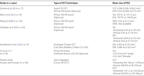
Most of the studies done on FAZ measurements of SCP and DCP used the RetiVue 100 XR Avanti as shown in Table 1 [1, 4, 5, 8, 12, 14] except Kuehlewein et al. [13] performed OCT Angiography with prototype swept source laser OCT (510 K clearance pending) from Carl Zeiss Meditec (Dublin, CA, USA). We used commer-cially available Zeiss Angioplex Cirrus HD OCT. Table 1
also highlights the consistent larger size of DCP FAZ in all the studies mentioned [4, 5, 8, 12–14]. Hence, the Fig. 2 Scatter plot distribution of values of horizontal and vertical
diameter of superficial capillary plexus
physio-anatomical characteristics of DCP FAZ in healthy eyes suggest the capillary perfusion of DCP. This has been clearly shown in most of the studies of OCT angi-ography in retinal vein occlusion and diabetic retinopa-thy where the DCP FAZ is affected in greater proportion than the SCP FAZ [4–8, 12, 15, 16]. This also suggest that DCP is susceptible to greater damage than SCP in retinal vascular disease or any ischemic insult on the macula.
The study limitation is the limited sample size. How-ever, a larger sample size would add strong normative data in these ethnic race. This study attempted to provide a normative data as it tried to describe a finding and not a phenomenon, establish a standard, created a platform for further research and cross sectional study with partici-pants from almost similar ethnicity with pigmented fundi. However, it has few advantages. Firstly, it compared the measurement with the contralateral eye and Secondly, included healthy young participants from middle east-ern, Indian and Filipino ethnic race with pigmented fundi. Thirdly, study participants had low refractive error (±0.5 D).
Conclusion
OCT-A is a useful imaging technology to evaluate the vascularity of superficial and deep capillary plexus. Nor-mative data will contribute to the average change in the FAZ diameter in the retinal vascular diseases. This study has shown that there is no difference in the FAZ
diameter of SCP and DCP between the contralateral eyes of healthy young adults. Based on this study, future development of diametric mapping of FAZ to the already available software of area mapping is another importance of this study which can be of clinical relevance in follow up of retinal vascular disease affecting the macula.
Authors’ contributions
NH collected data, analyzed and prepared the manuscript. AH contributed to concept of the study along with NH and prepared the manuscript. All authors read and approved the final manuscript.
Author details
1 Department of Ophthalmology, Al Zahra Hospital, PO Box 3499, Sharjah,
United Arab Emirates. 2 Al Buhairah Corniche, Sharjah, United Arab Emirates.
Acknowledgements
None.
Competing interests
The authors declare that they have no competing interests.
Consent for publication
The manuscript does not contain any data or image that reveal subject details.
Ethics approval and consent to participate
Informed consent was taken from all volunteers to undergo OCT angiography and to use the image for evaluation which is part of the routine in any case coming to the clinic. Institution review board (Ethics committee of Al Zahra Pvt Hospital, Sharjah/Dubai) permission was taken to use the data to perform the study and publish.
Received: 20 July 2016 Accepted: 28 October 2016 Table 1 OCT machine and size of FAZ measured by different studies in healthy eyes
SCP superficial capillary plexus, DCP deep capillary plexus, SVL superficial vascular layer, DVL deep vascular layer, SRVL superficial retinal vascular layer, DRVL deep retinal vascular layer
Study (n = eyes) Type of OCT/technique Mean size of FAZ
Samara et al. [4] (n = 17) Avanti SD OCT
(RTVue-XR Avanti; Optovue) SCP: 0.284 (0.206–0.362) mm
2
DCP: 0.410 (0.304–0.517) mm2
Wons et al. [5] (n = 19) RTVue 100 XR Avanti
(Optovue) SVL: 707.07 DVL: 797.97 ±± 141.75 µm 144.85 µm Wang et al.[8] (n = 105) RTVue 100 XR Avanti
(Optovue) SRVL: 0.35 ± 0.12 mm
2
DRVL: Not available Shahlaee et al. [9] (n = 34) RTVue 100 XR Avanti
(Optovue) SVL Horizontal: 0.59 ± 0.126 mm Vertical: 0.56 ± 0.118 mm DVL
Horizontal: 0.69 ± 0.123 mm Vertical: 0.63 ± 0.110 mm Kuehlewein et al. [10] (n = 19) Prototype SS laser OCT
(Carl Zeiss Meditec, Dublin CA, USA) SRL: 0.304 ± 0.132 mm
2
DRL: 0.486 ± 0.162 mm2
Yu et al. [11]
(n = 76) RTVue-XR Avanti(Software Version 2.0.5.39; Optovue) SVL CFZ: 0.42 mm2 (male)
0.52 mm2 (female)
Present study
Hussain and Hussain (n = 30) Zeiss Angioplex(Cirrus HD OCT) SCP Horizontal: 661.166 ± 119.99 µm Vertical: 660.033 ± 96.169 µm DCP
• We accept pre-submission inquiries
• Our selector tool helps you to find the most relevant journal • We provide round the clock customer support
• Convenient online submission • Thorough peer review
• Inclusion in PubMed and all major indexing services • Maximum visibility for your research
Submit your manuscript at www.biomedcentral.com/submit
Submit your next manuscript to BioMed Central
and we will help you at every step:
References
1. De Carlo TE, Romano A, Waheed NK, Duker JS. A review of optical coher-ence tomography angiography (OCTA). Int J Retina Vitreous. 2015;1:5. doi:10.1186/s40942-015-0005-8.
2. Jia Y, Bailey ST, Hwang TS, McClintic SM, Gao SS, Pennesi ME, et al. Quanti-tative optical coherence tomography angiography of vascular abnormali-ties in living human eye. PNAS. 2015. doi:10.1073/pnas.1500185112. 3. Mastropasqua R, Antonio LD, Staso AD, Agnifili L, Gregorio AD, Ciancaglini
M, Mastropasqua L. Optical coherence tomography angiography in retinal vascular diseases and choroidal neovascularisation. J Ophthalmol. 2015. doi:10.1155/2015/343515.
4. Samara WA, Shahlee A, Sridhar J, Khan MA, Ho AC, Hsu J. Quantita-tive optical coherence tomography angiography features and visual function in eyes with branch retinal vein occclusion. Am J Ophthalmol. 2016;166:76–83.
5. Wons J, Pfau M, Wirth MA, Freiberg FJ, Becker MD, Michels S. Optical coherence tomography angiography of the foveal avascular zone in retinal vein occlusion. Ophthalmologica. 2015. doi:10.1159/000445482. 6. Park JJ, Soetikno BT, Fawzi AA. Characterization of the middle capillary
plexus using optical coherence tomography angiography in healthy and diabetic eyes. Retina. 2016;0:1–12.
7. Scarinci F, Nesper PL, Fawzi AA. Deep retinal capillary non-perfusion is associated with photoreceptor disruption in diabetic macular edema. Am J Ophthalmology. 2016. doi:10.1016/j.ajo.2016.05.002.
8. Wang Q, Chan S, Yang JY, You B, Wang YX, Jonas JB, Wei WB. Vascular density in retina and choriocapillaries as measured by optical coher-ence tomography angiography. Am J Ophthlmol. 2016. doi:10.1016/j. ajo.2016.05.005.
9. Wu LZ, Huang ZS, Wu DZ, Chan E. Characteristics of the capillary free zone in the normal human macula. Jpn J Ophthalmol. 1985;29(4):406–11. 10. Wu LZ, Huang ZS, Wu DZ, Chan E. Characteristics of macular
microvascu-lature. Jpn J Ophthalmol. 1985;29(4):412–6.
11. Yap M, Gilchrist J, Weatherill J. Psychophysical measurement of foveal avascular zone. Ophthalmic Physiol Opt. 1987;7(4):405–10.
12. Shahlaee A, Petfianaki M, Hsu J, Ho AC. Measurement of foveal avascular zone dimensions and its reliability in healthy using optical coherence tomography angiography. Am J Ophthalmol. 2016;161(50–55):e1. 13. Kuehlewein L, Tepelus TC, An L, Durbin MK, Srinivas S, Sadda SR.
Noninva-sive visualization and analysis of the human parafoveal capillary network using swept source OCT optical angiography. Invest Ophthalmol Vis Sci. 2015;56:3984–8. doi:10.1167/iovs.15-16510.
14. Yu J, Jiang C, Wang X, Zhu L, Gu R, Xu H, Jia Y, Huang D, Sun X. Macu-lar perfusion in healthy chinese: an optical coherence tomography angiogram study. Invest Ophthalmol Vis Sci. 2015;56:3212–7. doi:10.1167/ iovs.14-16270.
15. Conrath J, Giogi R, Raccah D, Ridings B. Foveal avascular zone in diabetic retinopathy: quantitative and qualitative assess-ment. Ophthalmic Surg Lasers Imaging Retina. 2016;47(2):142–8. doi:10.3928/23258160-20160126-07.