B7x—from bench to bedside
Gurbakhash Kaur, 1 Murali Janakiram 1,2
To cite: Kaur G, Janakiram M. B7x—from bench to bedside. ESMO Open 2019;4:e000554. doi:10.1136/ esmoopen-2019-000554
Received 4 June 2019 Revised 12 August 2019 Accepted 14 August 2019
1Department of Medical
Oncology, Albert Einstein College of Medicine, New York city, New York, USA
2Department of Medicine,
University of Minnesota, Minneapolis, Minnesota, USA
Correspondence to Dr Murali Janakiram; mjanakir@ umn. edu © Author (s) (or their employer(s)) 2019. Re-use permitted under CC BY-NC. No commercial re-use. Published by BMJ on behalf of the European Society for Medical Oncology.
AbstrAct
B7x is an immune checkpoint molecule which belongs to the B7 family of ligands which includes PD-L1, PD-L2, B7-H3 and HHLA2. B7x belongs to the Immunoglobulin superfamily and its protein structure is similar to other members with a N terminus peptide, IgV and IgC like extracellular domain with four cysteine residues. Its receptor is yet to be identified. B7x inhibits T cell proliferation and expansion by IL-2 dependent and non-IL-2 dependent pathways. Even though high levels of B7x mRNA can be detected in most tissues its protein expression is highly limited suggesting significant post translational control. In vivo data, show that B7x plays an important role in limiting autoimmunity in the peripheral tissues and fine-tuning autoimmune responses. B7x is highly expressed in various cancers and in prostate cancer its expression is corelated with poorer outcomes. Local production of IL-6 and IL-10 in various cancers promotes B7x expression and tumor immune evasion. B7x is especially expressed in PD-L1 negative tumors suggesting that this may be an important method of immune evasion in these tumors. Currently drug development, targeting B7x through various mechanisms including monoclonal antibodies and antibody drug conjugates are in development in cancers and increasing B7x expression with fusion proteins in autoimmune diseases is underway.
IntroduCtIon
Immunotherapy has revolutionised the field of cancer therapeutics over the last decade. Immune checkpoint blockade (ICB) with cytotoxic T-lymphocyte antigen (CTLA-4), programmed death receptor (PD-1) and programmed death-ligand (PD-L1) antibodies has produced long and durable responses with a relatively low side effect profile. The clinical success has further fuelled a growing interest in developing newer checkpoint inhibitors. Since most patients are ineligible for ICB and even in those who receive CTLA-4, PD-1/PD-L1 therapy, only a small percentage respond. The third group of the B7 family, namely B7-H3, B7x and Human Endogenous Retrovirus-H Long Terminal Repeat-Associating Protein 2 (HHLA2), represents alternate immune checkpoints and has generated a considerable amount of interest. In this review, we will focus on B7x.
B7x (B7-H4/VTCN1//B7S1/B7 homolog 4) was discovered in 2003 using Expressed Sequence Tagged databases for proteins with homology to other members of the B7 family
(table 1). Human B7x (hB7x) is located on chro-mosome 1p12/13.1 and inhibits T-cell
prolifer-ation and cell cycle arrest.1 The genomic DNA
of hB7x contains six exons and five introns and
the coding region spans 849 base pairs.1 2 Exon
6 is used for alternative splicing to generate two different protein products, soluble B7x (sB7x) and cell surface B7x. A B7x pseudogene located on chromosome 20 p has also been described by
Choi et al.2 B7x is evolutionary conserved and
shares 87% of its amino acid (AA) sequence with mouse B7x, which is located on the F2 region of chromosome 3 and also codes for six exons
and five introns.1 2 The hB7x protein consists of
282 AA and shares varying level of similarity with
other members of the B7 family: B7‐1 (12%),
B7‐2 (13%), B7h (16%), PD‐L1 (18%), PD‐L2
(18%) and B7‐H3 (24%).1 3 4
StruCture
B7x belongs to the immunoglobulin super-family and its protein structure is similar to the other members of the B7 family. It consists of a N-terminus signal peptide, an IgV and IgC-like extracellular domain with four conserved cysteine residues and a
trans-membrane domain.1 Even though there
are reports that B7x is anchored to the cell
membrane via Gp1 linkage,4 treatment with
phosphatidylinositol-specific phospholipase
C did not change its expression levels.2 The
crystal structure of B7x IgV domain is similar to other B7 family members. The hB7x IgV
domain consisted of ‘β-sandwich’ folding
formed by a ‘back sheet’ (ABED strands) and
a ‘front sheet’ (C″C′CFGA′ strands) stabilised
by a disulfide bond between B and F strand.5
FunCtIon
B7x regulates T-cell proliferation and expan-sion via interleukin-2 (IL-2 dependent and non-IL-2)-dependent pathways. B7x expres-sion on EL4 cells inhibits CD8+ T-cell prolif-eration, cytokine secretion and inhibits
cytol-ytic activity.1 3 The inhibition of T-cell
prolifer-ation is by cell cycle arrest, rather than T-cell apoptosis and can only be partially reversed
by increased co-stimulation through CD28.3 4
Cell surface B7x suppresses Th1-derived and
copyright.
on September 12, 2020 by guest. Protected by
Table 1 Homology between hB7x and other members of the B7 family
HB7x1 (%)
B7-1 12
B7-2 13
B7h 16
PD-L1 18
PD-L2 18
B7-H3 24
hB7x, human B7x; PD-L, programmed death-ligand.
Th2-derived cytokines (interferon (IFN), IL-2, IL-4 and
IL-10) from naïve T cells.2 The underlying mechanism by
which B7x inhibits IL-2 production may be related to the downregulation of JunB, a component of the AP-1 tran-scription family that is usually activated after T-cell
acti-vation.4
B7x expreSSIon In normal and dISeaSe StateS
Normal tissue: B7x protein is not detected in most healthy tissues; however, high levels of B7x mRNA are observed in lung, liver, pancreas, spleen, thymus, kidney, prostate, testes and skeletal muscle suggesting post-transcriptional
regulation of its expression.2–4 6 B7x mRNA is expressed
on professional antigen presenting cells (APCs), bone marrow-derived dendritic cells (DCs), peritoneal macrophages and splenic B cells and broadly
distrib-uted in non-lymphoid tissue.4 Unlike other members
of the B7 family, B7x is not expressed on cells from the
haematopoietic origin.1 3 7 8 B7x expression is lost rapidly
in vitro culture.9 A soluble B7x-Ig protein binds to
acti-vated but not naive T cells and B-cell activation in mice with lipopolysaccharide, and IL-4 leads to B7x
downreg-ulation.4 B7x has been mostly shown to have cell surface
expression, but it can also translocate from the surface to
the nucleus.10
Autoimmunity: B7x has a vital role in suppression of autoimmune responses. Inhibition of B7x exacerbated
autoimmune encephalomyelitis (EAE) in mice.4
Treat-ment with B7xIg effectively ameliorated the progres-sion of relapsed and chronic EAE with decreased CD4 T cells within the central nervous system and spleen and a concurrent increase in the number of T regulatory cells
(Tregs).6 In a diabetes model, transfection of
diabeto-genic T cells into B7x-deficient mice resulted in a more aggressive disease state characterised by an earlier onset of disease with higher glucose levels due to increased
IFN-γ and IL-17 production. However, complete
abro-gation of diabetes was observed after introduction of diabetogenic T cells into mice known for
overexpres-sion of B7x, although evidence of insulitis remained.6
Similarly in an autoimmune kidney disease model, B7x knockout mice developed severe renal disease while
B7xIg decreased kidney damage and inflammation.11
These results show that B7x plays an important role in
suppressing autoimmunity in the periphery and for fine-tuning the response to inflammation.
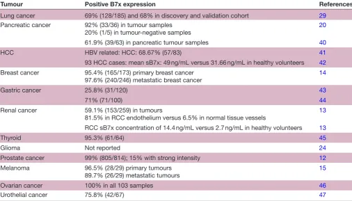
Cancer: Several observational studies show that the B7x protein is selectively expressed in higher levels in cancers including ovarian, renal cell cancer, pancreatic cancer, hepatocellular carcinoma (HCC), gastric cancer, lung cancer, glioma, breast, prostate cancer, urothelial cancer,
cervical cancer and melanoma12–21 (table 2). In vivo and
in vitro studies in lung and ovarian models have shown that both local cytokine production of Il-6, IL-10 and hypoxia promote B7x expression. In ovarian cancer cells, tumour-associated macrophage (TAM) stimulated Tregs to secrete IL-6 and IL-10, which, in turn, promote B7x
expression on APCs.19 22 In addition, production of IL-10
and TNF-α by the TAMs also increases B7x expression in
lung cancer models. IL-4 is known to suppress B7x
expres-sion,23 whereas IL-6, which is found in abundance in the
tumour microenvironment (TME) with IL-10, activates signal transducer and activator of transcription 3 which then binds to B7x promoter and enhances gene
expres-sion.24 Xenograft studies in a breast cancer model with
B7x deficiency protected from the development of lung metastasis, whereas a colon cancer model with high B7x expression had a sixfold increase in pulmonary
metas-tasis.25 26 B7x expression was upregulated in myeloid DCs,
plasmacytoid DCs, CD14+HLA-DRhi cells and CD14+
H-LA-DRlo/− cells in the peripheral blood of patients with
HCC compared with healthy donors. Furthermore, B7x expressed on early CD8+ tumour infiltrating lymphocytes (TILs) promoted T-cell exhaustion via Eomesodermin, a
transcription factor associated with T-cell exhaustion.27
These preclinical findings suggest that B7x expression in tumours helps in tumour immune evasion.
A large retrospective study conducted on B7x expres-sion on 948 prostate cancer samples showed that stronger intensity correlated with biochemical, clinical recurrence and death from prostate cancer. B7-H3 and B7x are highly expressed in human prostate cancer and was associated
with disease spread and poor outcomes.12 B7x expression
was evaluated in 259 renal cell carcinoma (RCC) cases and the protein was present in 59.1% of nephrectomy
specimens.13 In this study, higher B7x expression was
associated with adverse clinical and pathological features, including constitutional symptoms, tumour necrosis, and advanced tumour size, stage and grade. Interest-ingly, B7x was preferentially expressed on the endothe-lium of RCC tumour vasculature (81.5%) but not on
normal renal tissue vessels (6.5%).13 B7x was detected in
95.4% (165/173) of primary breast cancers and in 97.6% (240/246) of metastatic breast cancers, with the staining intensity being greater in invasive ductal carcinomas followed by invasive lobular carcinomas than in normal breast epithelium. Increased staining was associated with negative progesterone status and history of neoadjuvant chemotherapy, but unlike RCC there was no correlation
with higher expression and clinicopathological features.14
A meta-analysis of studies relating to B7x expression in ovarian cancer reported a pooled HR of 1.30 (95% CI:
copyright.
on September 12, 2020 by guest. Protected by
http://esmoopen.bmj.com/
Table 2 Human B7x expression in cancer
Tumour Positive B7x expression References
Lung cancer 69% (128/185) and 68% in discovery and validation cohort 29
Pancreatic cancer 92% (33/36) in tumour samples
20% (1/5) in tumour-negative samples 20
61.9% (39/63) in pancreatic tumour samples 40
HCC HBV related: HCC: 68.67% (57/83) 41
93 HCC cases: mean sB7x: 49 ng/mL versus 31.66 ng/mL in healthy volunteers 42
Breast cancer 95.4% (165/173) primary breast cancer
97.6% (240/246) metastatic breast cancer 14
Gastric cancer 25.8% (31/120) 43
71% (71/100) 44
Renal cancer 59.1% (153/259) in tumours
81.5% in RCC endothelium versus 6.5% in normal tissue vessels 13
RCC sB7x concentration of 14.4 ng/mL versus 2.7 ng/mL in healthy volunteers 13
Thyroid 95.3% (61/64) 45
Glioma Not reported 24
Prostate cancer 99% (805/814); 15% with strong intensity 12
Melanoma 96.5% (28/29) primary tumours
89.7% (26/29) metastatic tumours
15
Ovarian cancer 100% in all 103 samples 46
Urothelial cancer 75.8% (42/67) 47
HBV, Hepatitis B virus; HCC, hepatocellular carcinoma; RCC, renal cell carcinoma.
1.17 to 1.45, p < 0.05) between higher B7x expression and
worse progression-free survival.28 Hence, B7x protein is
widely expressed in various cancers, and in some cancers, it is associated with adverse clinical features.
Co-expression of B7x with other immune checkpoints
Since PD-L1 is expressed only in a minority of cancers, it stands to reason that the other members of the B7 family could be expressed or co-expressed to promote tumour immune evasion. In 392 primary non-small-cell lung cancers (NSCLC) samples, B7x was expressed in 69% of tumours. The co-expression of PD-L1 with B7x was infrequent at 6% and the majority (78%) of PD-L1-negative cases expressed B7x, HHLA2 or both. The triple-positive group (PD-L1, B7x and HHLA2) had
more TIL infiltration than the triple-negative group.29
In another study using multiplex quantitative immuno-fluorescence (QIF), PD-L1, B7-H3 and B7x expression was determined in 90 small-cell lung cancer (SCLC) samples. PD-L1, B7-H3 and B7x were expressed in 7.3%, 64.9% and 2.6%, respectively, of SCLC with limited co-expression and were not associated with the level of TILs. Elevated B7x expression was associated with shorter 5-year overall survival. The levels of CD3+, CD8+ and CD20+ TILs and the ratio of total/effector T cells were significantly lower in SCLC than in NSCLC. High levels of CD3+, but not CD8+ or CD20+ TILs, were
signif-icantly associated with longer survival.30 In
triple-nega-tive breast cancer, an immune cold microenvironment is defined by expression of B7x and a fibrotic stroma
signature while immunoreactive microenvironment was associated with CD8+ T cells, a type 1 IFN signature,
indoleamine-2,3-dioxygenase 1 and PD-L1.31 Hence,
B7x co-expression can define TME and B7x is expressed in PD-L1-negative tumours for immune evasion with important clinical implications.
B7x as a prognostic and predictive marker: Several studies have reported that B7x expression levels have predictive
and prognostic value.27 32–35 Detectable levels (>0.1 ng/
mL) of sB7x were observed in 53 patients with RCC compared with 18 controls. The median (range) observed concentration of sB7x for patients with RCC was 14.4 ng/ mL (0.1–56.9) in comparison to 2.7 ng/mL (0.2–37.1)
in the controls.32 In gastric cancer, the median
concen-trations of sB7x were significantly higher than those in healthy controls (16.85 vs 10.46 ng/mL; p= 0.008) and
high sB7x expression was associated with lower OS.35 A
meta-analysis of nine studies assessing B7x expression via enzyme-linked immunosorbent assay (two studies), immunohistochemistry (six studies) and QIF (one study) demonstrated that B7x was an unfavourable prognostic factor in NSCLC, as higher B7x expression was associ-ated with lymph node metastasis, advanced stage, poor differentiation and poor OS (HR=2.03, 95% CI=1.41 to
2.92, p<0.001).34 Another meta-analysis also reported that
B7x expression was associated with worse OS (HR=1.79,
95% CI 1.56 to 2.06, p<0.001) across many cancers.33
Based on these findings, B7x is expressed in many cancers and higher sB7x levels are associated with poor prognosis.
copyright.
on September 12, 2020 by guest. Protected by
drug development
The mounting evidence that B7x is expressed in a wide variety of human malignancies, and that its detection in either tumour samples or the blood serves as an adverse prognostic marker makes it an attractive drug target. B7x can be targeted through various mechanisms like mono-clonal-blocking antibodies (mAbs), single chain frag-ment variables (scFvs), antibody–drug conjugate (ADCs), CD3 bispecific antibodies (BiTE) and chimeric antigen
receptor T cells (CAR-Ts).9 36 37 Anti-B7x mAbs have been
demonstrated to inhibit tumour growth in vivo by blocking B7x-mediated immunosuppression and by killing tumour cells through antibody-dependent cell-mediated
cytotox-icity (ADCC).5 Anti-B7x scFvs have been shown to delay
the growth of ovarian cancer cell line, OVCAR5, in NSG
(NOD scid gamma mouse) mice.9 Since B7x is expressed
in breast cancer, a B7x scFv/CD3 BiTE has shown activity
in preclinical models.38 Leong et al generated a B7x ADC
with monomethyl auristatin that also led to tumour
regres-sion in triple-negative breast cancer xenograft models.37 In
addition, B7x-targeted CAR-T cells capable of recognising both human and murine B7x led to tumour regression in xenograft models; however, lethal toxicity was observed 6–8 weeks post-treatment. Post mortem analysis showed a case of on target/off toxicity as significant damage was observed in ductal and mucosal epithelial tissues with B7x
expres-sion.36
Currently, a phase Ia/Ib clinical trial (NCT03514121) with FPA150, a fully hB7x mAB with enhanced ADCC, in patients with advanced solid tumours is underway. The early results of this trial presented at the American Society of Clinical Oncology conference in 2019 reported a favourable side effect profile in 24 patients treated with FPA150 antibody in advanced solid cancers. Aside from Grade 1–2 diarrhoea and fatigue, the only grade 3 treat-ment related adverse event (TRAE) event reported was hypertension. Anti-tumour response has not yet been
reported.39 In rheumatoid arthritis, AMP-110, a fusion
protein containing extracellular domain of B7x plus Fc portion of IgG has been studied in two phase clinical trials (NCT01878123 and NCT02277574), although results have not been reported yet. The combination therapy of anti-B7x with PD-1 antibody blockade is a promising approach because B7x inhibition causes upregulation of PD-1 on CD8+ TILs. In murine models, combina-tion therapy has been proven to be more efficacious
than monotherapy with B7x and PD-1 alone.27 Hence,
targeting B7x in cancers and autoimmune diseases is an active area of drug development.
ConCluSIon
B7x is an immune checkpoint of the B7 family, inhibits T-cell proliferation and function, and has significant homology in protein structure to other members namely, PD-L1, B7-H3 and HHLA2. The receptor for B7x is yet to be discovered. Despite high mRNA expression in most tissues, its protein expression is very limited. Preclinical
models show that B7x is critical in regulating peripheral autoimmunity and autoimmune diseases can be reversed by upregulating B7x in animal models. Trials in human are testing this proof of concept. Due to its wide expression in cancer tissues, B7x is also an attractive target for cancer immunotherapy-blocking antibodies such as mAbs, scFvs, ADCs, CD3 BiTEs and CAR-Ts. Further research is needed to answer important questions regarding the receptor for B7x, conditions of upregulation of B7x in human tissues. Development of B7x-based therapeutics in autoimmune conditions and cancer is currently underway, and the results can be expected in the near future.
COI: The authors do not have any competing interests to report for this article.
Contributors GK and MJ: Writing, design, analysis and final submission. Funding The authors have not declared a specific grant for this research from any funding agency in the public, commercial or not-for-profit sectors.
Competing interests None declared. patient consent for publication Not required.
provenance and peer review Commissioned; externally peer reviewed. open access This is an open access article distributed in accordance with the Creative Commons Attribution Non Commercial (CC BY-NC 4.0) license, which permits others to distribute, remix, adapt, build upon this work non-commercially, and license their derivative works on different terms, provided the original work is properly cited, any changes made are indicated, and the use is non-commercial. See: http:// creativecommons. org/ licenses/ by- nc/ 4. 0/.
RefeRences
1. Zang X, Loke P'ng, Kim J, et al. B7x: a widely expressed B7 family member that inhibits T cell activation. Proc Natl Acad Sci U S A
2003;100:10388–92.
2. Choi I-H, Zhu G, Sica GL, et al. Genomic organization and expression analysis of B7-H4, an immune inhibitory molecule of the B7 family. J Immunol 2003;171:4650–4.
3. Sica GL, Choi IH, Zhu G, et al. B7-H4, a molecule of the B7 family, negatively regulates T cell immunity. Immunity 2003;18:849–61. 4. Prasad DVR, Richards S, Mai XM, et al. B7S1, a novel B7 family
member that negatively regulates T cell activation. Immunity
2003;18:863–73.
5. Jeon H, Vigdorovich V, Garrett-Thomson SC, et al. Structure and cancer immunotherapy of the B7 family member B7x. Cell Rep
2014;9:1089–98.
6. Wei J, Loke P'ng, Zang X, et al. Tissue-Specific expression of B7x protects from CD4 T cell-mediated autoimmunity. J Exp Med
2011;208:1683–94.
7. Lee JS, Scandiuzzi L, Ray A, et al. B7x in the periphery abrogates pancreas-specific damage mediated by self-reactive CD8 T cells. J.i.
2012;189:4165–74.
8. Hofmeyer KA, Scandiuzzi L, Ghosh K, et al. Tissue-Expressed B7x affects the immune response to and outcome of lethal pulmonary infection. J Immunol 2012;189:3054–63.
9. Dangaj D, Lanitis E, Zhao A, et al. Novel recombinant human B7-H4 antibodies overcome tumoral immune escape to potentiate T-cell antitumor responses. Cancer Res 2013;73:4820–9.
10. Zhang L, Wu H, Lu D, et al. The costimulatory molecule B7-H4 promote tumor progression and cell proliferation through translocating into nucleus. Oncogene 2013;32:5347–58.
11. Pawar RD, Goilav B, Xia Y, et al. B7x/B7-H4 modulates the adaptive immune response and ameliorates renal injury in antibody-mediated nephritis. Clin Exp Immunol 2015;179:329–43.
12. Zang X, Thompson RH, Al-Ahmadie HA, et al. B7-H3 and B7x are highly expressed in human prostate cancer and associated with disease spread and poor outcome. Proc Natl Acad Sci U S A
2007;104:19458–63.
13. Krambeck AE, Thompson RH, Dong H, et al. B7-H4 expression in renal cell carcinoma and tumor vasculature: associations with cancer progression and survival. Proc Natl Acad Sci U S A
2006;103:10391–6.
copyright.
on September 12, 2020 by guest. Protected by
http://esmoopen.bmj.com/
14. Tringler B, Zhuo S, Pilkington G, et al. B7-H4 is highly expressed in ductal and lobular breast cancer. Clin Cancer Res 2005;11:1842–8. 15. Quandt D, Fiedler E, Boettcher D, et al. B7-H4 expression in human
melanoma: its association with patients' survival and antitumor immune response. Clin Cancer Res 2011;17:3100–11.
16. Wang X, Wang T, Xu M, et al. B7-H4 overexpression impairs the immune response of T cells in human cervical carcinomas. Hum Immunol 2014;75:1203–9.
17. Shen L, Qian Y, Wu W, et al. B7-H4 is a prognostic biomarker for poor survival in patients with pancreatic cancer. Hum Pathol
2017;66:79–85.
18. Salceda S, Tang T, Kmet M, et al. The immunomodulatory protein B7-H4 is overexpressed in breast and ovarian cancers and promotes epithelial cell transformation. Exp Cell Res 2005;306:128–41. 19. Kryczek I, Zou L, Rodriguez P, et al. B7-H4 expression identifies
a novel suppressive macrophage population in human ovarian carcinoma. J Exp Med 2006;203:871–81.
20. Awadallah NS, Shroyer KR, Langer DA, et al. Detection of B7-H4 and p53 in pancreatic cancer. Pancreas 2008;36:200–6.
21. Simon I, Zhuo S, Corral L, et al. B7-H4 is a novel membrane-bound protein and a candidate serum and tissue biomarker for ovarian cancer. Cancer Res 2006;66:1570–5.
22. Podojil JR, Miller SD. Potential targeting of B7-H4 for the treatment of cancer. Immunol Rev 2017;276:40–51.
23. Chen C, Qu Q-X, Shen Y, et al. Induced expression of B7-H4 on the surface of lung cancer cell by the tumor-associated macrophages: a potential mechanism of immune escape. Cancer Lett 2012;317:99–105.
24. Yao Y, Ye H, Qi Z, et al. B7-H4(B7x)-mediated cross-talk between glioma-initiating cells and macrophages via the IL6/JAK/STAT3 pathway lead to poor prognosis in glioma patients. Clin Cancer Res
2016;22:2778–90.
25. Abadi YM, Jeon H, Ohaegbulam KC, et al. Host B7x promotes pulmonary metastasis of breast cancer. J.i. 2013;190:3806–14. 26. Ohaegbulam KC, Liu W, Jeon H, et al. Tumor-expressed immune
checkpoint B7x promotes cancer progression and antigen-specific CD8 T cell exhaustion and suppressive innate immune cells.
Oncotarget 2017;8:82740–53.
27. Li J, Lee Y, Li Y, et al. Co-inhibitory molecule B7 superfamily member 1 expressed by tumor-infiltrating myeloid cells induces dysfunction of anti-tumor CD8+ T cells. Immunity 2018;48:773–86.
28. Ye Y, Wang J-J, Li S-L, et al. Does B7-H4 expression correlate with clinicopathologic characteristics and survival in ovarian cancer?: a systematic review and PRISMA-compliant meta-analysis. Medicine
2018;97:e11821.
29. Cheng H, Borczuk A, Janakiram M, et al. Wide expression and significance of alternative immune checkpoint molecules, B7x and HHLA2, in PD-L1–negative human lung cancers. Clin Cancer Res
2018;24:1954–64.
30. Carvajal-Hausdorf D, Altan M, Velcheti V, et al. Expression and clinical significance of PD-L1, B7-H3, B7-H4 and TILs in human small cell lung cancer (SCLC). J Immunother Cancer 2019;7.
31. Gruosso T, Gigoux M, Manem VSK, et al. Spatially distinct tumor immune microenvironments stratify triple-negative breast cancers. J Clin Invest 2019;129:1785–800.
32. Thompson RH, Zang X, Lohse CM, et al. Serum-Soluble B7x is elevated in renal cell carcinoma patients and is associated with advanced stage. Cancer Res 2008;68:6054–8.
33. Song X, Shao Y, Gu W, et al. Prognostic role of high B7-H4 expression in patients with solid tumors: a meta-analysis. Oncotarget
2016;7:76523–33.
34. Tan Z, Shen W. Prognostic role of B7-H4 in patients with non-small cell lung cancer: a meta-analysis. Oncotarget 2017;8:27137–44. 35. Shi H, Ji M, Wu J, et al. Serum B7-H4 expression is a significant
prognostic indicator for patients with gastric cancer. World J Surg Oncol 2014;12:188.
36. Smith JB, Lanitis E, Dangaj D, et al. Tumor regression and delayed onset toxicity following B7-H4 CAR T cell therapy. Mol Ther
2016;24:1987–99.
37. Leong SR, Liang W-C, Wu Y, et al. An anti-B7-H4 antibody-drug conjugate for the treatment of breast cancer. Mol Pharm
2015;12:1717–29.
38. Iizuka A, Nonomura C, Ashizawa T, et al. A T-cell-engaging B7-H4/ CD3-bispecific Fab-scFv antibody targets human breast cancer. Clin Cancer Res 2019;25:2925–34.
39. Sachdev JC, Bauer TM, Chawla SP, et al. A phase I study of EpCAM/ CD3-bispecific antibody (MT110) in patients with advanced solid tumors. Available: http:// meetinglibrary. asco. org/ content/ 97493- 114 [Accessed 12 Jul 2019].
40. Chen Y, Sun J, Zhao H, Zhu D, et al. The coexpression and clinical significance of costimulatory molecules B7-H1, B7-H3, and B7-H4 in human pancreatic cancer. Onco Targets Ther 2014;7:eCollection 2014:1465–72.
41. Hong B, Qian Y, Zhang H, et al. Expression of B7-H4 and hepatitis B virus X in hepatitis B virus-related hepatocellular carcinoma. World J Gastroenterol 2016;22:4538–46.
42. Zhang C, Li Y, Wang Y. Diagnostic value of serum B7-H4 for hepatocellular carcinoma. J Surg Res 2015;197:301–6.
43. Arigami T, Uenosono Y, Ishigami S, et al. Clinical significance of the B7-H4 coregulatory molecule as a novel prognostic marker in gastric cancer. World J Surg 2011;35:2051–7.
44. Geng Y, Wang H, Lu C, et al. Expression of costimulatory molecules B7-H1, B7-H4 and Foxp3+ Tregs in gastric cancer and its clinical significance. Int J Clin Oncol 2015;20:273–81.
45. Zhu J, Chu B-F, Yang Y-P, et al. B7-H4 expression is associated with cancer progression and predicts patient survival in human thyroid cancer. Asian Pac J Cancer Prev 2013;14:3011–5.
46. Zang X, Sullivan PS, Soslow RA, et al. Tumor associated endothelial expression of B7-H3 predicts survival in ovarian carcinomas. Mod Pathol 2010;23:1104–12.
47. Fan M, Zhuang Q, Chen Y, et al. B7-H4 expression is correlated with tumor progression and clinical outcome in urothelial cell carcinoma.
Int J Clin Exp Pathol 2014;7:6768–75.
copyright.
on September 12, 2020 by guest. Protected by

![catena Poly[μ2 iodido diiodidobis(μ3 pyridine 2 thione κ3S:S:S)(μ2 pyridine 2 thione κ2S:S)tricopper(I)]](data:image/gif;base64,R0lGODlhAQABAIAAAP///wAAACH5BAEAAAAALAAAAAABAAEAAAICRAEAOw==)