Published online 2016 July 20. Research Article
Kienböck’s Disease; the Length of Capitate and Third Metacarpal
Bones
Davod Jafari,
1Hooman Shariatzadeh,
1and Ali Ajvadi
1,*1Bone and Joint Reconstruction Research Center, Shafa Orthopedic Hospital, Iran University of Medical Sciences, Tehran, IR Iran
*Corresponding author: Ali Ajvadi, Bone and Joint Research Center, Shafa Orthopedic Hospital, Iran University of Medical Sciences, Tehran, IR Iran. Tel: +98-2133542022, E-mail: aliajvadi@gmail.com
Received2016 February 28;Revised2016 April 26;Accepted2016 July 08.
Abstract
Background:The relationship between negative ulnar variance and Kienböck’s disease is unknown and does not justify all of the cases. The present study planed the hypothesis that maybe the pressure from distal structures to the lunate bone plays a role in the etiology.
Objectives: The current study aimed to investigate the possibility of a relationship between an increased length of the third metacarpal and the capitate with Kienböck’s disease .
Methods:The study compared the wrist posteroanterior (PA) X-ray images of 105 healthy individuals with those of 91 patients with Kienböck’s disease . Meticulous measurement criteria were defined in the present study to measure the third metacarpal and the capitate lengths. These lengths along with ulnar variance were measured on each X-ray. The Lichtman classification was used for staging. A new index, named capitate-index, was defined due to the linear relationship between the capitate and the third metacarpal lengths.
Results: Comparing the two groups, no meaningful difference was observed between the capitate and third metacarpal bone lengths in patients and the control group. Also, there was no significant difference in the capitate-index (capitate length/3rd metacarpal length) between the groups. Furthermore, no differences were observed comparing the patients with ulnar variance
=0 and patients with ulnar variance < 0 to the control group regarding the above parameters. The only significant difference was a higher negative ulnar variance among the patients with Kienböck’s disease .
Conclusions:Based on the above-mentioned findings, the existence of a relationship between the third metacarpal and the capitate lengths and the Kienböck’s disease is unlikely.
Keywords:Kienböck’s Disease, Lunate Bone, Capitate Bone, Metacarpal Bones
1. Background
Kienböck’s disease (KD) is the lunate bone osteonecro-sis that causes complete degeneration of the bone in the late stages leading to wrist osteoarthritis. Despite various surgical treatments performed on patients, studies could not find significant difference among these methods (1), showing that the etiology of KD is still unidentified.
Among the most important reasons discussed in the literature so far, negative ulnar variance, arterial ischemia, trauma and occupational factors are considered as the eti-ology of KD more than others (2). Nevertheless, none of these factors is proven or rejected. Despite the popularity of the lunate overload theory caused by the negative ul-nar variance, some cases cannot be explained by this the-ory. Two meta-analyses conducted in this field showed that there is not enough evidence to prove such a relationship (3,4).
2. Objectives
In an attempt to find another factor to be both in line with the lunate overload theory and justify zero or positive ulnar variance cases, the authors investigated the possibil-ity of a relationship between the third metacarpal and cap-itate lengths and KD.
3. Methods
The study was approved by the ethical committee of Iran University of Medical Sciences, and the X-rays of 111 patients with KD, underwent surgery in Shafa orthopedic hospital from 2009 to 2015 were reviewed. The diagnosis was made according to the X-ray and magnetic resonance imaging (MRI) findings. Among the patients, 91 cases with established KD and a good quality standard posteroante-rior (PA) X-ray images were included in the study.
The Lichtman classification was used for staging (5). Age matched, normal x-rays of 105 volunteers referring to
outpatient clinic of Shafa Hospital were used as the control group. Descriptive statistics of the patients and the control group are presented inTable 1.
According to the hospital protocol, the wrist PAX-ray is performed by Schreibman method (6) with a 90 degrees abduction of the shoulders, 90 degrees flexion of the el-bow, and a neutral position of the forearm.
Magnifier of the picture archiving and communica-tion system (PACS) is applied to find the bone markers more accurately.
For a few patients, the radiography had been taken as plane X-ray. For these cases a 2.5* loupe magnifying glass and a caliper with 0.1 mm accuracy was used to measure bone markers. To make sure that the measurements had identical calibration in both plane X-ray and PACS system measurements were compared in 20 patients X-rayed both systems. A constant difference was observed between the two methods that decreased to±0.15 mm with acceptable intra-observer reliability through defining a corrective co-efficient in the computer software.
3.1. Bony Landmarks
Three measurements were performed on each X-ray; the capitate and the third metacarpal lengths along their long axes and ulnar variance measurement through Palmer method (7).
While measuring the third metacarpal and the capi-tate lengths, authors noticed variations in the X-ray of dis-tal pole of the capitate and the proximal base of the third metacarpal which could disturb the measurements up to 1 mm in each region (Figure 1). Therefore, the intersection between the capitate and the metacarpal axes were used with the line that was farther from the carpometacarpal joint.
No exact landmark had been specified for these re-gions in the articles which measured the third metacarpal and capitate lengths (8-13). Therefore, a pilot study was conducted to define a bone marker with acceptable intra-observer reliability.
As shown inFigure 1, the third metacarpal and capi-tate anterior and posterior edges do not overlap most of the times and resemble two separate parallel lines. Of these lines, two central lines, closer to the carpometacarpal (CMC) joint, do not have clear-cut edges but farther lines are almost always clear. The intersection of the two bones axes with the line farther from the joint was determined as a reference point.
Measurements were performed twice by senior author on 20 X-rays (10 patients with Kienböck’s disease and 10 normal subjects) at intervals of one week to make sure about the intra-observer reliability of the reference points.
Figure 1.Reference Points to Measure Capitate and the 3rd Metacarpal Length
Near carpometacarpal joint in each bone there are two parallel lines resulted from anterior and posterior edges. The intersection between the capitate and the metacarpal axes with the line that was farther from the carpometacarpal joint was selected.
The variation between the two measurements was±0.16 mm which was acceptable (kappa value = 0.78).
Considering that the third metacarpal and the capitate length ratios in the present study (Figure 2andTable 2) and those of the other studies (8,9), follow a linear relationship and a meaningful regression formula, the current study defined a new index called capitate-index. Capitate-index was calculated from dividing the capitate length by the 3rd metacarpal length. This index was also used to compare the two groups.
CApitate Lenght, mm Fitted Values
50 60 70 80 Third Metacarpal Lenght, mm
Capi
tat
e
Lenght, mm
26
24
22
20
18
16
Figure 2.Linear Relationship Between the Third Metacarpal and the Capitate Length
in the Control Group
3.2. Statistical Analysis
Table 1.Demographic Data of Patients and Control Group
Gender Control Group (n = 105) Kienböck’s Disease (n = 91)
Grade 2 Grade 3a Grade 3b Grade 4
Male 63 17 21 22 4
Female 42 5 11 10 1
Total 105 22 32 32 5
Table 2.Linear Regression Equation Between Between the Capitate Length and the Third Metacarpal Length in the Control Group
Y X P Value R2 Linear Regression Equation
Capitate length Metacarpal length < 0.001 0.49 Y = 3.72 + 0.28X
A P-value less than 0.05 was considered statistically signif-icant.
4. Results
Measurements were performed on the wrist PAX-rays of 91 patients with KD and those of 105 individuals selected as a control group. The means of the capitate length, the third metacarpal length, the capitate-index, and the ulnar variance values were compared in both groups.
According to Table 3, no significant statistical dif-ference was found between the patients and the con-trol group in terms of the third metacarpal and capitate lengths. Also, there was no meaningful difference between the two groups regarding the capitate-index. The only sig-nificant difference was the negative ulnar variance in the Kienböck’s group.
Table 3.Comparing Measured Parameters
Variable Control Group (n
= 105) (Mean±
SD)
Kienböck (n = 91)
(Mean±SD)
P Value
Capitate length 22 (2.21) 21.94 (1.65) 0.83
Metacarpal length
64.50 (5.46) 64.40 (5.42) 0.89
Ulnar variance 0.26 (0.85) -1.26 (1.47) < 0.001a
Capitate-index 0.34 (0.03) 0.34 (0.02) 0.99
aThe only significant difference was the negative ulnar variance in the patients group.
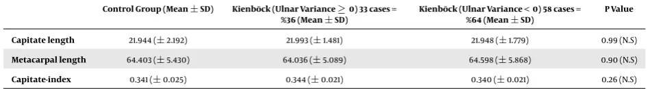
As illustrated inTable 4, there was no meaningful dif-ference between the control group and the two groups of patients with ulnar variance≥0 and ulnar variance < 0 in terms of the performed measurements.
5. Discussion
There was no consensus regarding the etiology of KD. Since 1928 that Hulten published his classic study on re-lationship between ulnar variance and KD (14), the lunate overload in ulnar minus cases were the most popular the-ory in this context (15,16). However this theory was ques-tioned by D’Hoore et al. reviewing 52 patients with KD. They found no significant statistical relationship between ulnar variance and KD (17).
Others studied the intraosseous pressure of the lunate with wrist motion and found greater pressure by 40 mm Hg in wrist extension that might be a risk factor for KD (18). Other investigators found that the radial slope and radial inclination had a significant effect on transmitted force to the lunate (19).
Following these lunate overloading theories as men-tioned above, the authors proposed the possibility of a re-lationship between an increased length of the capitate and the third metacarpal bones and the KD, which if proven, would justify the static and dynamic pressures on the lu-nate.
This idea arises from the studies that show the greater the capitate and the third metacarpal lengths, the stronger the grip strength (8). Thus, there is a possibility that in-creasing the third metacarpal and capitate lengths lead to an increase in static or dynamic pressures imposed by the 3rd ray on the lunate and vulnerability to KD. Although the capitate shortening procedures recommended in the joint level or ulnar plus patients (20-24), there was no study in-vestigating whether the capitate length of these patients was greater than those of normal individuals or not.
Table 4.Comparing the Control Group and Patients with Ulnar Variances
Control Group (Mean±SD) Kienböck (Ulnar Variance≥0) 33 cases =
%36 (Mean±SD)
Kienböck (Ulnar Variance < 0) 58 cases =
%64 (Mean±SD)
P Value
Capitate length 21.944 (±2.192) 21.993 (±1.481) 21.948 (±1.779) 0.99 (N.S)
Metacarpal length 64.403 (±5.430) 64.036 (±5.089) 64.598 (±5.868) 0.90 (N.S)
Capitate-index 0.341 (±0.025) 0.344 (±0.021) 0.340 (±0.021) 0.26 (N.S)
Abbreviation: N.S, Non-significant.
Given that lunate overload does not show all aspects of KD; perhaps it is time to focus more on the biological fac-tors such as vascular facfac-tors which are not directly related to trauma or over pressure on the lunate (25).
However, defining more meticulous reference points to measure the third metacarpal and capitate lengths is one of the strengths of the present study which was not no-ticed previously in normal wrist anatomic and radiologic studies (8-13).
Although these new reference points were checked in a pilot study and acceptable intraobserver reliability was observed, considering its importance in carpal height ra-tio and other wrist measurements; it is essential to inves-tigate larger samples and make sure about both interob-server and intraobinterob-server reliability of these parameters.
Acknowledgments
The authors thank Dr. Marjan Zohourian for language editing of the manuscript. Authors also wish to thank Dr. Doosti for help on statistical analysis and valuable advice.
Footnote
Authors’ Contribution: Study design: Davod Jafari;
manuscript writing: Hooman Shariatzadeh and Ali Ajvadi; data analysis: Ali Ajvadi; critical revision: Davod Jafari, Hooman Shariatzadeh and Ali Ajvadi; study supervision: Davod Jafari.
References
1. Innes L, Strauch RJ. Systematic review of the treatment of Kienbock’s disease in its early and late stages.J Hand Surg Am. 2010;35(5):713–7. doi:10.1016/j.jhsa.2010.02.002. [PubMed:20438990] 717 e1-4. 2. Stahl S, Stahl AS, Meisner C, Rahmanian-Schwarz A, Schaller HE, Lotter
O. A systematic review of the etiopathogenesis of Kienbock’s disease and a critical appraisal of its recognition as an occupational disease related to hand-arm vibration.BMC Musculoskelet Disord.2012;13:225. doi:10.1186/1471-2474-13-225. [PubMed:23171057].
3. Stahl S, Stahl AS, Meisner C, Hentschel PJ, Valina S, Luz O, et al. Critical analysis of causality between negative ulnar variance and Kienböck’s disease. Plast Reconstr Surg. 2013;132(4):899–909. doi:
10.1097/PRS.0b013e31829f4a2c. [PubMed:24076682].
4. Chung KC, Spilson MS, Kim MH. Is negative ulnar variance a risk factor for Kienbock’s disease? A meta-analysis.Ann Plast Surg.
2001;47(5):494–9. [PubMed:11716259].
5. Saunders BM, Lichtman D. A classification-based treatment algorithm for Kienböck’s disease : current and future considerations.Tech Hand Up Extrem Surg.2011;15(1):38–40. doi:10.1097/BTH.0b013e31820e82d2. [PubMed:21358524].
6. Schreibman KL, Freeland A, Gilula LA, Yin Y. Imaging of the hand and wrist.Orthop Clin North Am.1997;28(4):537–82. [PubMed:9257964]. 7. Palmer AK, Glisson RR, Werner FW. Ulnar variance determination.J
Hand Surg Am.1982;7(4):376–9. [PubMed:7119397].
8. Schuind FA, Linscheid RL, An KN, Chao EY. A normal data base of posteroanterior roentgenographic measurements of the wrist.J Bone Joint Surg Am.1992;74(9):1418–29. [PubMed:1429800].
9. Mohammed Ali MH. A normal data-base of posteroanterior radio-graphic measurements of the wrist in healthy Egyptians.Surg Radiol Anat. 2009;31(9):665–74. doi:10.1007/s00276-009-0500-4. [PubMed:
19352583].
10. Jafari D, Taheri H, Shariatzade H, Mazhar FN, Jalili A, Ghahramani M. Radiographic indices in one hundred fifty normal Iranian wrists.Med J Islam Repub Iran.2012;26(3):132–9. [PubMed:23482869].
11. Nattrass GR, King GJ, McMurtry RY, Brant RF. An alternative method for determination of the carpal height ratio.J Bone Joint Surg Am.
1994;76(1):88–94. [PubMed:8288669].
12. Wang YC, Tseng YC, Chang HY, Wang YJ, Chen CJ, Wu DY. Gender differ-ences in carpal height ratio in a taiwanese population.J Hand Surg Am.
2010;35(2):252–5. doi:10.1016/j.jhsa.2009.11.010. [PubMed:20141895]. 13. Stahelin A, Pfeiffer K, Sennwald G, Segmuller G. Determining carpal
collapse. An improved method.J Bone Joint Surg Am.1989;71(9):1400– 5. [PubMed:2793895].
14. Hulten O. Uber die entstehung und behandlung der lunatum-malaziemorbuskienbock. 76. Act Chir Scand; 1928. p. 121.
15. Lamas C, Carrera A, Proubasta I, Llusa M, Majo J, Mir X. The anatomy and vascularity of the lunate: considerations applied to Kienbock’s disease.Chir Main. 2007;26(1):13–20. doi:10.1016/j.main.2007.01.001. [PubMed:17418764].
16. Irisarri C. [Aetiology of Kienbock’s Disease]. Handchir Mikrochir Plast Chir. 2010;42(3):157–61. doi: 10.1055/s-0030-1253394. [PubMed:
20552544].
17. D’Hoore K, De Smet L, Verellen K, Vral J, Fabry G. Negative ulnar variance is not a risk factor for Kienbock’s disease.J Hand Surg Am. 1994;19(2):229–31. doi:10.1016/0363-5023(94)90010-8. [PubMed:
8201185].
18. Schiltenwolf M, Martini AK, Mau HC, Eversheim S, Brocai DR, Jensen CH. Further investigations of the intraosseous pressure char-acteristics in necrotic lunates (Kienbock’s disease).J Hand Surg Am. 1996;21(5):754–8. doi: 10.1016/S0363-5023(96)80187-0. [PubMed:
8891969].
20. Waitayawinyu T, Chin SH, Luria S, Trumble TE. Capitate short-ening osteotomy with vascularized bone grafting for the treat-ment of Kienbock’s disease in the ulnar positive wrist.J Hand Surg Am. 2008;33(8):1267–73. doi: 10.1016/j.jhsa.2008.04.006. [PubMed:
18929187].
21. Citlak A, Akgun U, Bulut T, Tahta M, Dirim Mete B, Sener M. Par-tial capitate shortening for Kienbock’s disease.J Hand Surg Eur Vol.
2015;40(9):957–60. doi:10.1177/1753193414562355. [PubMed:25432157]. 22. Moritomo H, Murase T, Yoshikawa H. Operative technique of a new decompression procedure for Kienböck’s disease : partial cap-itate shortening.Tech Hand Up Extrem Surg. 2004;8(2):110–5. doi:
10.1097/01.bth.0000126571.20944.47. [PubMed:16518122].
23. Gay AM, Parratte S, Glard Y, Mutaftschiev N, Legre R. Isolated capitate shortening osteotomy for the early stage of Kienböck’s disease with neutral ulnar variance.Plast Reconstr Surg. 2009;124(2):560–6. doi:
10.1097/PRS.0b013e3181addc50. [PubMed:19644275].
24. Afshar A, Mehdizadeh M, Khalkhali H. Short-Term Clinical Out-comes of Radial Shortening Osteotomy and Capitates Shortening Osteotomy in Kienböck’s disease.Arch Bone Jt Surg. 2015;3(3):173–8. [PubMed:26213706].