Research Article
Papsmear
Examination
for
Diagnosing
Pre
Cancer
Lesion
in
Invisible
Squamo
Columnar
Junction
Pemeriksaan
Papsmear
dan
IVA
untuk
Diagnosis
Lesi
Prakanker
pada
Tampilan
Sambungan
Skuamo
Kolumnar
Tidak
Tampak
Laila Nuranna, Sulaeman Daud, Gatot Purwoto, Hariyono Winarto, Kartiwa H Nuryanto
Division of Gynecology Oncology Department of Obstetrics and Gynecology Faculty of Medicine University of Indonesia/
Dr. Cipto Mangunkusumo General Hospital Jakarta
INTRODUCTION
Cervical cancer is the second most common cancer that affects women in the world.1-4 Apart from that,
it is the most common causes of death among can-cer, especially for women in developing countries.4
Based on the recent world’s estimation, there are
493,000 new cervical cancer cases occurred each year, whereas there are 409,400 (83%) cases happened in women in developing countries and only 84,400 (17%) cases in developed countries.2
Unfortunately, more than 80% of the cases are diagnosed at advanced stage when the 5-year survival rate is less than 40%.2
Abstract
Objective: To know the concealed pre-cancer lesion in women with invisible squamo-columnar junction (SCJ) by Papsmear examina-tion.
Method: This study was a descriptive cross-sectional design starting from August 2014 to March 2015 at several Public Health Cares in Jakarta. A total of 1,682 subjects were screened by Acetoacetate Visual Inspection (AVI) examination. After the data was collected, the process was continued by verification, editing, and coding. The descriptive analysis showed the percentage of SCJ in age distribu-tion, the percentage of AVI examination based on SCJ, and the per-centage of Papsmear examination in invisible SCJ according to nega-tive AVI result.
Result: There were 1,484 (88.2%) women with the visible SCJ and 198 (11.8%) women with invisible SCJ. The percentage of invisible SCJ in the menopausal women group was 122 (61,6%); meanwhile, in the non-menopausal women group, it was 76 (38.4%). Almost half of the percentage from visible SCJ was found in menopausal women group 45.8% (103/225 women). The positive AVI result was 4 (7.1%) in the menopausal women group and 52 (92.9%) in non-menopausal women group. The result of Papsmear examina-tion with invisible SCJ were 197 (100%) normal.
Conclusion: Almost half of visible SCJ was found in menopausal women group. Most of positive AVI result was found in the non-menopausal women group. All women with the invisible SCJ have a normal Papsmear result.
[Indones J Obstet Gynecol 2016; 4-3: 158-163]
Keywords: acetoacetate visual inspection, papsmear, pre-cancer le-sion, squamo-columnar junction
Abstrak
Tujuan: Untuk mengetahui kelainan yang tersembunyi pada keadaan sambungan skuamo-kolumnar (SSK) tidak tampak melalui pemerik-saan Papsmear.
Metode: Penelitian ini merupakan deskriptif potong lintang. Peneli-tian dilakukan pada periode Agustus 2014 sampai Maret 2015 di be-berapa puskesmas di Jakarta. Sebanyak 1.682 subjek yang dilakukan pemeriksaan IVA (Inspeksi Visual dengan Asam asetat). Setelah data dikumpulkan, akan dilakukan verifikasi data, editing, dan proses pengkodean. Analisis data deskriptif berupa variabel kategori yaitu persentase letak SSK berdasarkan distribusi usia, persentase hasil pe-meriksaan IVA berdasarkan SSK, dan persentase hasil pepe-meriksaan Papsmear pada SSK yang tidak tampak dari hasil pemeriksaan IVA negatif.
Hasil: Perempuan dengan SSK yang tampak 1.484 (88,2%), yang ti-dak tampak 198 (11,8%). Sambungan Skuamo-Kolumnar (SSK) yang tidak tampak pada perempuan yang sudah menopause sebanyak 122 (61,6%), sedangkan pada perempuan yang belum menopause se-banyak 76 (38,4%). Hampir setengahnya proporsi SSK yang tampak didapatkan pada kelompok perempuan yang sudah menopause 45,78% (103/225 perempuan). Hasil pemeriksaan IVA positif dida-patkan 4 (7,1%) pada kelompok perempuan menopause dan 52 (92,9%) pada kelompok perempuan yang belum menopause. Pada pemeriksaan Papsmear dengan SSK yang tidak tampak, persentase kelainan lesi prakanker yaitu sebesar 197 (100%) normal.
Kesimpulan: Hampir setengahnya SSK yang tampak ditemukan pada kelompok perempuan menopause. Sebagian besar IVA positif dite-mukan pada kelompok perempuan yang belum menopause. Seluruh perempuan dengan SSK yang tidak tampak memiliki hasil pemerik-saan Papsmear normal.
[Maj Obstet Ginekol Indones 2016; 4-3: 158-163]
Kata kunci: IVA, lesi prakanker, papsmear, sambungan skuamo-kolumnar
The main cause of high incidence of cervical can-cer in developing countries is due to the lack of effective screening programs to detect and manage the early stage of cervical cancer or pre-cancer le-sions of the stage.4 Of the various modality
screen-ing which have been studied, Papsmear has ability to decrease 70% of cervical cancer cases; however, this examination is difficult to implement in deve-loping countries like Indonesia. It seems that visual inspection of the cervix is the best techniques to apply, especially in areas with limitation of health facilities resources.2,4-7 This technique is known as
the VIA test (Visual Inspection with Acetic acid ap-plication). It is defined as a technique of cervical direct observation after being applied with acetic acid without the use of any magnifying tools.
Visual Inspection of Acetic acid examination de-pends on the Squamo-Columnar Junction (SCJ) condition in the cervical region. In childbearing age women and pregnant women, SCJ is located in the ecto-cervical that it can be seen on direct examina-tion. Whereas in postmenopausal women, SCJ is often located in the endo-cervical canal so it cannot be seen on direct observation. However, the per-centage of visible SCJ in menopausal women is 64.28% and invisible SCJ is 11.66%.8 This rate is
high for the menopausal women. Another study conducted by Dhaubhadel, et al. prospectively and descriptively in women aged 20-50 years, the re-sult showed negative VIA Papsmear test from all 46-50 year of group women.2 The study did not
describe the visibility of SCJ in the investigation. Therefore, it could make the false negative results. If the SCJ is not visible, it suggests doing the Pap smear test. Unfortunately, the problem is in the first-line health care facility, such as primary health centers with limited facilities, the invisible SCJ (in the second examination), particularly in meno-pausal women is justified without doing the VIA examination. Therefore, this study aims to deter-mine the percentage of hidden invisible SCJ in ab-normal cervical pre-cancer lesions through the Pap smear examination.
THEORITICAL OVERVIEW
Invasive cervical cancer is usually preceded by a long phase of pre-invasive lesions, which are mi-croscopically seen as precursor lesions developing from atypical cells to the various level of cervical intraepithelial neoplasia (CIN) before progression to invasive carcinoma. The epidemiological studies
have identified several risk factors that contribute to the development of the CIN and cervical cancer. The risk factors are Human Papilloma Viral (HPV) infection, sexual contact in early age, changing sex-ual partners, multi-parity, long-term of oral contra-ceptive use, smoking, low socioeconomic status, in-fection with Chlamydia trachomatis, micronutrient deficiency described by less intake of vegetables and fruits. The types of HPV 16, 18, 31, 33, 35, 39, 45, 51, 52, 56, 58, 59 and 68 are closely related to CIN and invasive cancer. Infection of one or more HPV types is suspected as the cause of cervical neo-plasia. Infection of one or more of the oncogenic HPV types will contribute to the entrance of com-bined viral genome into the host cell genome; this formation will lead to cell neoplasia and turn into various level of CIN and finally, it will further de-velop into cervical cancer.9-13
Application of 5% acetic acid is believed to cause a reversible coagulation or precipitation of the cell protein. This application also causes swelling of the epithelial tissue and cell dehydration. The color of normal squamous epithelium is pink; while, the co-lumnar epithelium will be in red color due to the reflection of light from the stroma in which its bot-tom is rich for blood vessels. If the epithelium con-tains a lot of protein cells, the acetic acid will coa-gulate this protein which will eliminate the color of the stroma. The result from this coagulation pro-duction is called as aceto-white area that can be seen with the naked eye and distinguished from the normal area which should be pink around it. Therefore, the effect of acetic acid depends on the amount of protein found in epithelial cells. Areas where the increased activity of the nucleus and DNA will change very clearly.9,13
for the CIN and early stage of cancer. This area can also be seen in other conditions due to the increase of core proteins, such as immature squamous me-taplasia, healing or regenerating epithelium (asso-ciated with inflammation), leucoplakia (hyperkera-tosis), and condyloma.9,12,13 The symptoms of
cer-vical cancer are vaginal bleeding, post-coital bleeding, vaginal discharge, lower abdominal pain, edema of lower extremity, obstructive uropathy, bowel obstruction, and also anemia.12,14
Aceto-white area in CIN and early stage of can-cer are more whitish, thicker, and opaque as well as clear boundary; while, the aceto-white area in immature squamous metaplasia, the inflammation and regenerating epithelium will make less pale, thin, translucent, and ill defined. Aceto-white area caused by inflammation and epithelial healing process is usually spread to the cervix, not only in the transformation zone. Besides, this color will be disappeared within a minute. Leucoplakia and con-dyloma turn the color into grayish white after the application of acetic acid. The effect of acetic acid (aceto-white area) is slower in CIN lesions and early invasive cancer than immature squamous metaplasia and inflammation. This effect occurs af-ter 3-5 minutes at CIN 2-3 and invasive cancer.9,13
METHODS
This cross-sectional design study is conducted in several primary health centers in Jakarta on the pe-riod of August 2014 to March 2015. The target population is all women who are married or have ever had sexual intercourse for VIA examination. We included women who did the VIA screening examination in primary health centers Jakarta in the period of August 2014 to March 2015. We ex-cluded pregnant women and women with history of total hysterectomy. From the calculation for-mula, the total number of subjects needed in this study is 1,658 subjects. In this study, we performed VIA examination on 1,682 women.
All women that met the criteria of the study were labeled and subsequently did the Papsmear examination and the results were interpreted by expert pathologist. The result of Papsmear exami-nation would be described with Bethesda system criteria in 2001. The data was analyzed where we conducted the descriptive statistics for categorical variables, namely the proportion of SCJ layout based on the age distribution, the proportion of the VIA examination results based on the location of
the SCJ of the age distribution, and the percentage of the Papsmear examination results of the SCJ which was not visible from the IVA. All data were shown in frequency and percentage.
RESULTS
Of 1,682 subjects, only 9 (0.5%), 311 (18.5%), 638 (37.9), 492 (29.3), 232 (13.8%) women were less than 20, 20-29, 30-39, 40-49, and more than 50 years old; respectively.
Table 1. The Characteristics of the Subjects
Characteristics n %
Age
< 20 yo 9 .5
20-29 yo 311 18.5
30-39 yo 638 37.9
40-49 yo 492 29.3
50 yo 232 13.8
Menopause state
Not yet 1457 86.6
Menopause 225 13.4
Contraception state
Not using contraception 354 21.0
Pill 127 7.6
Implant 43 2.6
Injection 603 35.9
IUD 470 27.9
MOW 27 1.6
Condom 58 3.4
SCJ
Invisible SCJ 198 11.8
Visible SCJ 1484 88.2
Total 1682 100.0
In this study, according to the age group, we di-vided into group who were still menstruation (pre-menopausal state) and had stopped menstruation (menopausal state). There were 1,457 (86.6%) wo-men who were still wo-menstruation (prewo-menopausal) and the others had stopped menstruation.
invi-sible SCJ based on age group for more than 50, 40-49, 30-39, 20-29, less than 20 years old was 127 (64.1%), 35 (17.7%), 30 (15.2%), 6 (3.0%), and 0 (0.0%); consecutively.
Invisible SCJ proportion in women who had ex-perienced menopause was 122 (61.6%) and 76 (38.4%) for premenopausal women. Almost half of visible SCJ obtained in the group of women who were menopausal (45.78% (103 women)).
Table 2 depicted the proportion of visible SCJ. For age group of 30-39, 40-49, 20-29, and less than 20 years old, there were 608 (41.0%), 457 (30.8%), 305 (20.6%), and 9 (0.6%); consecutively. The pro-portion of visible SCJ in menopausal women was 103 (6.9%) and 1,381 (93.1%) women in preme-nopausal state. However, in the premepreme-nopausal group, nearly half of 103 (45.77%) women had visible SCJ.
Table 2. The Proportion of Squamo-Columnar Junction
(SCJ)
Characteristic SCJ
Invisible SCJ % Visible SCJ % Age
< 20 yo 0 0.0 9 0.6
20-29 yo 6 3.0 305 20.6
30-39 yo 30 15.2 608 41.0
40-49 yo 35 17.7 457 30.8
50 yo 127 64.1 105 7.1
Menopause State
Not Yet 76 38.4 1381 93.1
Menopause 122 61.6 103 6.9
Table 3. Proportion of VIA Examination with Visible SCJ
Characteristic VIA
Negative % Positive %
Age
< 20 yo 8 0.6 1 1.8
20-29 yo 291 20.4 14 25.0
30-39 yo 589 41.2 19 33.9
40-49 yo 439 30.7 18 32.1
50 yo 101 7.1 4 7.1
Menopause State
Not Yet 1329 93.1 52 92.9
Menopause 99 6.9 4 7.1
In this study, the overall positive VIA test result was in 56 (3.98%) women. The proportion of posi-tive VIA test result according to the age group were 19 (33.9%) for 30-39 years old, 18 (32.1%) for 40-49 years old, 14 (25.0%) for 20- 29 years old, 4 (7.1%) for more than 50 years old, and 1 (1.8%) for less than 20 years old. While, the proportion of positive VIA test result was obtained for 4 (7.1%) in menopausal women and 52 (92.9%) in menstru-ating women (Table 3).
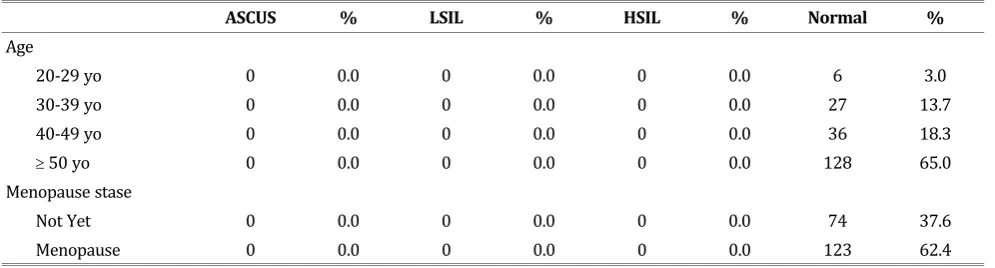
In the Pap test with SCJ which was not visible, every test presented the normal result (100%) (Ta-ble 4). In this study, invisi(Ta-ble SCJ was obtained in 198 subjects, but there was one subject that was not checked due to loss of data. However, this num-ber still met the minimum of required sample (196 subjects).
DISCUSSION
This study is based on the data from the examina-tion in several health centers in Jakarta. In this study, the total of 1,682 women were analyzed
Table 4. Proportion of Papsmear Test Result in Invisible SCJ
ASCUS % LSIL % HSIL % Normal %
Age
20-29 yo 0 0.0 0 0.0 0 0.0 6 3.0
30-39 yo 0 0.0 0 0.0 0 0.0 27 13.7
40-49 yo 0 0.0 0 0.0 0 0.0 36 18.3
50 yo 0 0.0 0 0.0 0 0.0 128 65.0
Menopause stase
Not Yet 0 0.0 0 0.0 0 0.0 74 37.6
based on their characteristics of age, history of con-traception, menopausal status, state of the SCJ, the results of the VIA examination and Papsmear. This study began between August 2014 and March 2015. Women with positive VIA test result were evaluated on subsequent visits. While the women had cervical abnormalities, such as cervicitis, it would be treated with topical antiseptics. If the doctor found other abnormalities, such as cervical polyps and suspicious cancer, the women would be sent to higher level of health care facility.
This study has strengths and limitations. The strengths of this study were the sample was taken by doctors and health workers who have had the training and experience from the Female Cancer Program (FCP) in identifying the SCJ, the abnor-malities in the cervical region, the procedures for VIA and conventional smear of Pap-smear exami-nation sampling. Additionally, in this study, one pa-thologist performed Papsmear examination. There were some flaws in this study among others, namely in the Pap-smear examination, sampling errors might occur that could affect the results.
In this study, there were 225 (13.37%) meno-pausal women. The proportion of invisible SCJ was among 198 (11.7%) women, whereas almost half of postmenopausal women had visible SCJ. In the study conducted by Nuranna, et al. in October 2007 until December 2010, it showed that of 3,791 post-menopausal women (16.49%), the proportion of invisible SCJ was on 2,680 (70.69%) women. More than half of the menopausal women had visible SCJ (2,437 (64.28%) women).8
In this study, in Pap-smear with invisible SCJ, all results showed normal (100%). Shwe, et al. con-ducted a study of cervical cytology in Myanmar from 2010 to 2011. Of 1,771 women screened, 762 women (43.0%) resulted in abnormal smear, 866 (48.9%) and 87 (4.9%) were diagnosed as inflam-mation and Atypical Squamous Cells of Undeter-mined Significance (ASCUS). There were 42 (2.3%) and 11 (0.6%) cases of Low Grade Squamous In-traepithelial Lesion (LSIL) and High Grade Squa-mous Intraepithelial Lesion (HSIL). The cases of squamous cell carcinoma (SCC) occurred on 3 (0.2%) women.15 While the study conducted by
Sengul, et al., there were 32,578 cases of Pap-smear examination performed and analyzed between Ja-nuary 2001 and April 2010. From the investigation which had carried out, the results showed that 1.18% of ASCUS; 0.39% of LSIL; 0.16% of HSIL;
0.07% of Atypical Glandular Cells of Undetermined Significance (AGUS); 0.02% of squamous cell carci-noma, and 0.006% of adenocarcinoma 0.006%.16
Abnormal cytology is more common in cases with older age, low parity, and period of perimeno-pause.16 However, this study did not have data on
SCJ picture.
In this study, all results were 100% normal cer-vical cytology at the invisible SCJ. However, this did not completely rule out pre-cancer lesions abnor-malities. Study by Pan, et al. explained that overall, the level of sensitivity, specificity, positive predic-tive value, negapredic-tive predicpredic-tive value, and accuracy of cervical cytology for detecting cervical intraepi-thelial neoplasia (NIS 2+) were respectively 81.0 %, 95.4%, 38.3%, 99.3% and 94.9%.17 A study held
by Pak, et al. which aimed to compare the history of previous Papsmear test result in patients with cervical adenocarcinoma and squamous cell carci-noma of the cervix. In patients with cervical ade-nocarcinoma, the false negative results of Pap-smear were very significant in the latest investiga-tion. As already mentioned above, the normal Papsmear results did not guarantee not to develop the cervical cancer. In general, 157 patients (41.8%) carried out repeated screening within 2 years. Fifty-five (14.6%) carried out repeated screening within 2-5 years and 80 (22.3%) over 5 years. A total of 16.8% did not have documentation for Pap-smear examination results. The false nega-tive results of Pap-smear for patients with cervical adenocarcinoma and squamous cell carcinoma were 9 (5.6%) and 2 (1.3%) patients.18 Kirschner,
et al. stated that the false negative of Pap-smear result was on 11 (9.8%) women.19
CONCLUSION
Almost half of invisible SCJ is found in the group of menopausal women and most visible SCJ is found in the group of women who have not menopausal yet. Positive VIA results are largely found in the group of women who have not menopausal yet. All the women with invisible SCJ have normal Papsmear examination results.
REFERENCES
1. Moore DH. Cervical Cancer. Clinical Expert Series, Am Col-lege Obstet Gynecol 2006; 107: 1152-3.
vi-sual inspection of cervix with acetic Acid. JNMA; n 2008; 47(170): 71-6.
3. Belinson JL, Pretorius RG, Zhang WH, et al. Cervical cancer screening by simple visual inspection after acetic acid. Obs-tet Gynecol 2001; 98(3): 441-4.
4. Ocviyanti D. Tes pap, tes HPV dan servikografi sebagai pe-meriksaan triase untuk tes IVA positif: upaya tindak lanjut deteksi dini kanker serviks pada fasilitas kesehatan dengan sumber daya terbatas beserta analisis sederhana efektivitas biayanya. Indones J Obstet Gynecol 2007; 31(4): 202-3. 5. Blumenthal PD, Gaffikin L, Deganus S,et al. Cervical cancer
prevention: safety, acceptability, and feasibility of a single-visit approach in Accra, Ghana. Am J Obstet Gynecol 2007; 196(4): 407.e1-8; discussion .e8-9.
6. Singh V, Sehgal A, Parashari A, et al. Early detection of cer-vical cancer through acetic acid application - an aided visual inspection. Sing Med J 2001; 42(8): 351.
7. Bharani B, Phatak S. Acetic acid visualization of the cervix an alternative to colposcopy in evaluation of cervix at risk. J Obstet Gynecol Ind 2005; 55: 530.
8. Nuranna L, Aziz MF, Irianti R, et al. The role of SCJ in VIA examination: can VIA still be done in menopause women? In: Nuranna L, editor. Asia-Oceania Research Organisation on Genital Infections and Neoplasia (AOGIN) Interim Meet-ing. Discovery Kartika Plaza Hotel, Bali: AOGIN; 2011. 9. RI DK. Buku Acuan Pencegahan Kanker Leher Rahim dan
Kanker Payudara. Jakarta: Direktorat Pengendalian Pe-nyakit Tidak Menular. Direktorat Jendral PP dan PL; 2007. 10. Benard VB, Eheman CR, Lawson HW, et al. Cervical screen-ing in the National Breast and Cervical Cancer Early Detec-tion Program, 1995-2001. Obstet Gynecol 2004; 103(3): 564-71.
11. Chong Q. The Abnormal-looking Cervix. The Singapore Fa-mily Physician 2003; 29(2): 56-9.
12. Andrijono. Kanker Serviks. 3rd ed. Fakultas Kedokteran
Uni-versitas Indonesia: Divisi Onkologi Ginekologi, Departemen Obstetri dan Ginekologi RSUPN-CM; 2010.
13. Sankaranarayanan R. A practical manual on visual screen-ing for cervical neoplasia. World Health Organization 2003. (accessed)‘.
14. Berek JS. Intraepithelial Disease of The Cervix, Vagina, and Vulva. In: Berek JS, ed. Berek & Novak’s Gynecology. Phila-delphia: Lippincott Williams & Wilkins; 2007: 570. 15. Shwe M, Harano T, Okada S, et al. Prevalence of high risk
human papillomavirus (HR-HPV) infection among women with normal and abnormal cervical cytology in Myanmar. Acta Medica Okayama 2014; 68(2): 79-87.
16. Sengul D, Altinay S, Oksuz H. Population-based cervical screening outcomes in Turkey over a period of approxi-mately nine and a half years with emphasis on results for women aged 30-34. Asian Pacific J Cancer Prev 2014; 15: 2069-74.
17. Pan Q, Hu S, Zhang X. Pooled analysis of performance of liquid based cytology in population-based cervical cancer screening studies in China. Cancer Cytopathol 2013; 121(9): 473-82.
18. Pak SC, Martens M, Bekkers R, et al. Pap smear screening history of women with squamous cell carcinoma and ade-nocarcinoma of the cervix. Au New Zealand J Obstet Gynae-col 2007; 47(6): 504-7.