Leukemia: A Systematic Review
Hossein Ayatollahi MD1, Maliheh Hasheminezhad MD PhD2,*, Arezoo Shajiee MSc3, Mohammad Hadi Sadeghian MD4, Ehsan Yazdandoust MSc5, Maryam Sheikhi MSc3*, Sepideh Shakeri MSc٣, Seyyede Fatemeh Shams MSc3
1. Cancer Molecular Pathology Research Center, Faculty of Medicine, Mashhad University of Medical Sciences, Mashhad, Iran
2. Hematology and Oncology Research Center, Shahid Sadoughi University of Medical sciences and Health Services, Yazd, Iran
3. Cancer Molecular Pathology Research Center, Faculty of Medicine, Mashhad University of Medical Sciences, Mashhad, Iran
4. Department of Hematology and Blood Bank, Cancer Molecular Pathology Research Center, Faculty of Medicine, Mashhad University of Medical Sciences, Mashhad, Iran
5. Department of Hematology and Blood Bank, Mashhad University of Medical Sciences, Mashhad, Iran
*Corresponding authors: Maliheh Hasheminezhad, Hematology and Oncology Research Center, Shahid Sadoughi University of Medical sciences and Health Services, Yazd, Iran. Email: mhasheminezhad@ssu.ac.ir & Maryam Sheikhi, Cancer Molecular Pathology Research Center, Mashhad University of Medical Sciences, Mashhad, Iran. E-mail:
sheikhim2@mums.ac.ir
Received: 6 September 2015 Accepted: 26 January 2016
Abstract
Background: Acute myeloid leukemia (AML) is a malignant disorder involving blood cells, characterized by obstructed or distorted differentiation of hematopoietic stem cells. In cytogenetically normal AML (CN AML), molecular abnormalities in NPM, FLT3, CEBPA, and BAALC genes are observed. Initially, high BAALC (Brain and Acute Leukemia Cytoplasmic gene) expression was introduced in a study on AML cases with trisomy 8. The present systematic review aimed to determine the prognostic value of BAALC expression in CN-AML patients within the age range of 1 month to 15 years.
Materials and Methods: In this systematic review, scientific databases including PubMed, Scopus, Embase, ISI, and Cochrane Library were searched. All the evaluated retrospective studies focused on the prognostic value of BAALC expression.
Results: Overall, BAALC expression was reported in 30-60% of AML patients (mean=45%). High expression of
BAALC gene was reported in M0, M1, and M2 subtypes of pediatric AML. In only one study, in addition to these subtypes, M4 was also reported (12.5% of cases).
Conclusion: This systematic review resulted in a significant association between high BAALC expression and poor response to chemotherapy in pediatric AML. However, OS (Overall Survival) and EFS (Event - Free Survival) findings were contradictory, relapse and death increase in the presence of high BAALC expression. Therefore, BAALC expression could be considered as a clinical prognostic marker for the treatment of AML patients. Further efficient prognostic indicators should be identified in order to gather information about therapeutic strategies for AML and risk analysis in these patients.
Keywords: BAALC, Expression, Pediatric AML and Prognosis.
Introduction
Acute myeloid leukemia (AML) as a malignancy of blood cells involves the differentiation of hematopoietic stem cells, characterized by obstructed or distorted differentiation of them. This condition results in abnormal relocation of immature cells and inhibition of growth and maturation of normal cells (1, 2). AML
accounts for one-fourth of all cases of acute leukemia in pediatric populations and is responsible for more than 50% of mortality in these patients (3, 4). With alterations in the prognostic factors, treatment or relapse conditions may vary, as well. Therefore, assessment of all determining prognostic markers such as
gender, age, race, and
molecular/cytogenetic abnormalities is essential. Cytogenetic information is generally accessible in 70-80% of pediatric AML patients. Moreover, the prognostic value of karyotypic abnormalities has been subcategorized into favorable or unfavorable groups associated with AML.
t(8;21), inv(16), t(15;17) are recognized as
favorable groups (3,5).
The most prevalent cytogenetic abnormalities in children are
t(8;21)(q22;q22), inv(16)(p13.1q22),
t(15;17)(q22;q21), 11q23/MLL,
t(1;22)(p13;q13), t(7;12)(q36;p13), and t(11;12)(p15;p13) which can be detected in
approximately half of pediatric AML patients (3,6,7). However, in cytogenetically normal AML (CN AML), molecular abnormalities in nucleophosmin (NPM1), fms related tyrosine kinase 3 (FLT3), CCAAT/Enhancer-Binding Protein Alpha (CEBPA), and Brain and Acute Leukemia Cytoplasmic (BAALC) genes have been reported (1, 8, 9, 10). Among these genes, BAALC, a gene located on the chromosomal region 8q22.3, can be regarded as a possible prognostic indicator in CN-AML (1, 10).
BAALC expression is limited to progenitor
cells, so no expression has been indicated in a mature bone marrow or white blood cells (WBC). Initially, high BAALC
expression was introduced in a study on AML patients with trisomy 8. As the results showed trisomy 8 had a negative prognostic correlation with AML (1). Moreover, according to the previous research, high BAALC expression is indicative of poor prognosis in patients and can be a significant risk factor in CN-AML (2). In addition, high BAALC
expression is associated with higher rates of primary resistance and relapse in these patients, resulting in poor overall survival (OS) (11, 12).
To the best of our knowledge, the prognostic value of BAALC expression in AML patients has not been systematically assessed. Therefore, this systematic review aimed to determine the prognostic value of
BAALC expression in CN-AML patients
within the age range of 1 month to 15 years.
Materials and Methods
Search strategy and article selection
In this systematic review, scientific databases including PubMed, Scopus, Embase, ISI, and Cochrane Library were searched, using the conformity keywords in various combinations: "BAALC" and "AML" or “Acute Myeloid Leukemia" and "pediatric" or "children" or "infants" and "prognosis". The keywords were planned to proper MeSH terms.
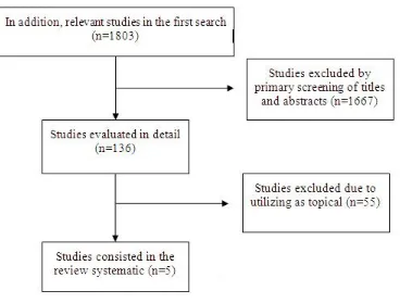
This study was confined to English language articles. The references of cohort studies in reference lists of all relevant studies were also searched. In the cross-reference search of cohort articles, reference lists of all relevant studies, as well as review articles were reviewed in order to identify studies which could not be found via computerized search. One reviewer thoroughly checked the article titles and abstracts to determine the accuracy of selected articles. The PRISMA Chart was adopted to summarize the results of the included studies (Figure 1). The final search was carried out in August 2015. The titles and abstracts of the articles were investigated and incoherent articles were omitted. The relevant articles were reviewed and assessed for this systematic review. The reference lists of included articles were investigated for rejecting the possibility of incorrect citations.
In the present study, the inclusion criteria were as follows: 1) revised English clinical
trials published before August 2015; 2)
survival information based on BAALC
expression status (high and low expression
rates); and 3) data related to patient
response to therapy including complete remission (CR), OS, event-free survival (EFS) and prognosis. All articles were evaluated based on the inclusion criteria and were compared according to their titles and abstracts. Then, full texts of the
articles, which met the inclusion criteria, were obtained.
The exclusion criteria were as follows: 1)
articles including less than ten patients; 2)
commentaries, review articles, case reports and non-English articles; 3) samples obtained from cell and tissue cultures; 4) inclusion of patients over 15 years of age;
and 5) flow cytometry data.
The collected data included information based on PRISMA guidelines. Data concerning the authors, year of publication, region of study, patient information (including: age range, median WBC count, blood blast %, and BM blast %) (Table 1), test and control groups, OS rate, relapse, and prognosis were extracted.
Results
Data provided from five selected articles are described in Table 1, which provides a detailed overview of these studies. The included studies were retrospective articles concerning the prognostic value of BAALC
gene expression in pediatric AML. The patient populations of the selected articles were from various countries and groups. All papers contained information about the question under study. Original retrospective articles including genetic tests, performed to evaluate patient prognosis, were also included.
Whole patients contributing in these studies were diagnosed as AML cases within the age range of 1 month to 15 years. The patient populations ranged between 40 and 294 cases. Based on the patient populations, the articles were divided into three groups: 20-100 patients (two papers) (14, 15) 101-200 patients (one paper) (16) and 201-300 patients (two papers) (17, 18) (Table 1).
BAALC expression was reported in
30-60% of patients (mean=45%). According to the results, just one article compared the changes of BAALC expression in two cytogenetic abnormal AML with
CN-AML (One group with t (8; 21) and the
other one with an inversion of chromosome 16 (16).
In all evaluated articles, BAALC
expression was assessed via reverse transcription quantitative polymerase chain reaction (RT-QPCR) (Table 1).
An overexpression of BAALC was
observed in the patients with t (8; 21) and
inv (16), which was more significant than that in the CN-AML group (16). In fact, in the CN-AML group, no significant Overexpression was observed.
The median age and WBC count were within the ranges of 3-9.8 and 12.9-97 ×109/L, respectively. Three of five papers compared age and median WBC count between cases with high and low BAALC
expression rates (14, 15, 16). As the results indicated, the median age in cases with high BAALC expression was higher than those with low expression. Moreover, WBC count in subjects with high BAALC
expression was lower than cases with low
BAALC expression (Table 1).
In this study, the correlation between
BAALC expression and the
French-American-British (FAB) classification of AML was investigated. Based on cellular differentiation M0, M1 and M2 subtypes of AML showed increased BAALC
expression. In three out of five studies (15, 16, 18), patients were categorized by FAB system according to high and low BAALC
expression. The results indicated that high
BAALC expression could be observed in
M0, M1 and M2 subtypes of AML. In only one study, in addition to these subtypes, M4 was also reported (12.5% of cases) (15). In two articles, percentages of bone marrow and blood blasts were investigated in the correlation with high BAALC
expression that showed increase in the presence of a gene overexpression (14, 15). Additionally, in the majority of articles, OS, EFS, CR, relapse, and follow-up were evaluated in terms of high or low
BAALC expression.
In five articles, the association between the expression status of BAALC gene and OS was reported (P=0.185 to P<0.001). The significance of this association was P≤0.001 in two papers (15, 16), P>0.001 in
two articles (14, 16), and negligible in one paper. Therefore, the relationship between
BAALC expression and OS was significant
in only two papers in cases with high
BAALC expression (14, 17) and
insignificant in other studies (15, 16, 18). The percent of OS, provided in only one paper, was decreased in AML patients with high BAALC expression (18). The mean follow-up duration was between 30 months and 12 years in the evaluated studies (table 1). The effect of BAALC
expression on the percent of EFS was reported in one article. According to the findings, the percentage of EFS was lower in cases with high gene expression, compared to those with low expression rates (18). Furthermore, EFS was calculated in four studies. The significance of the association between BAALC
expression and EFS ranged between P=0.82 and P=0.007 (P≤0.001 in two studies (12, 14) and P>0.001 in two other
articles (17, 18)). Therefore, it can be concluded that in only two papers, EFS rate was significant in cases with high gene expression rates (14,16), while in two other studies, EFS was insignificant (17,18). In one study, relapse was assessed according to high or low expression rates
of BAALC gene. High BAALC expression
was more common in patients with relapse. In addition, mortality rate increased in patients with high expression of this gene (15). Response to therapy was evaluated according to the rate of BAALC
expression. In two studies; low BAALC
expression was associated with good response to therapy while high expressed
BAALC gene patients showed a poor
response to treatment (14, 16). Additionally, CR was measured in these patients. It was shown an association between decreases of CR with high
BAALC expression (15).
Table I. Data extraction from included studies investigated the effect of BAALC expression in
pediatric AML patients. (NR: data not reported, Exp: Expression, WBC count: Median WBC Count
(109/L)
In all included studies Real-time quantitative polymerase chain reaction (RQ-PCR) were used.
Author Country Cases NO.
NO. of patients with high and low BAALC
expression Follow up (yr) Overall Median Age
(yr)
Median WBC count (109/L)
Mizushima et al.,2010 Japan 241 11-18 8 53.09 6.6
Staffas et al., 2011 Sweden 185 74-75 10-3 12.9-18.9 12
Hermkens et al., 2013 USA 294 NR NR NR 5
Aref et al., 2015 Egypt 49 24-23 7.0 -6.0 95-97 1
Hagag et al.,2015 Egypt 40 24-16 8.35-7.74 75.51-76.44 2.5
Figure 1: PRISMA Flow Diagram: the screening procedure of papers in this study.
Discussion
The prognostic value of high ERG, MN1,
BAALC, FLT3, and WT1 expression and
the correlation between these markers and AML have not been thoroughly evaluated in patients with pediatric AML. Nevertheless, the prognostic value of these genetic markers has been mainly evaluated in cases with CN-AML (8, 14).
Tanner et al., (2001) proposed that BAALC
gene expression is stage-particular and can be regarded as a new marker, which is mutual among progenitor cells in myeloid, lymphoid, and erythroid signaling pathways (19). Moreover, Baladus et al., (2003) showed that high BAALC
expression is obviously non-random among FAB subtypes and cytogenetic groups of AML, implying the role of BAALC protein in the leukemic phenotype (20). The association between BAALC
expression and poor prognosis in AML is another indicator of the role of this gene in
increased number of blasts (18). Eid et al., (2010) demonstrated a correlation between overexpression of BAALC and ERG genes with the percentage of blast cells in the blood and bone marrow, while other parameters were of no significant value in these patients (21).
Based on univariate analysis in the study by Eid et al., (2010) high rates of ERG and
BAALC expression are significantly
associated with poor EFS and shorter OS (18). Therefore, high BAALC expression can be a prognostic factor for poor EFS and OS (14, 21). However, high BAALC
expression is of no autonomous prognostic importance in pediatric CN-AML patients, as confirmed in a study by Hermkens and colleagues, (2013) (14).
According to the results of this systematic review, high BAALC expression was considerably related to poor response to chemotherapy in pediatric AML. Although
OS and EFS rates were insignificant and contradictory, but relapse and mortality rates showed a significant correlation. In fact, overexpression of BAALC gene led to increase of relapse and death rates. Therefore, in order to confirm the predictive value of this marker in OS and EFS, further research is required.
A major limitation of present review was the absence of prospective controlled studies and scarcity of research in this study. Considering the limited accessibility of data, it was not possible to perform univariate or multivariate analyses of solitary markers in all studies. Therefore, the findings can be applied in clinical prognostic measurements and therapy in AML patients. Recognition of further efficient prognostic indicators could be useful for obtaining further information about the therapeutic strategies and risk analysis in AML patients.
Conclusion
It was revealed that high BAALC
expression is a poor prognostic factor for remission success, therapy, relapse, and death in pediatric AML patients. However, only half of the evaluated articles indicated that high BAALC expression results in poor OS and EFS.
References
1. Ferrara F, Palmieri S, Leoni F. Clinically useful prognostic factors in acute myeloid leukemia. Crit Rev Oncol
Hematol. 2008; 66(3):181-93.
2. Weber S, Alpermann T, Dicker F, Jeromin S, Nadarajah N, Eder C, et al.
BAALC expression: a suitable marker for
prognostic risk stratification and detection of residual disease in cytogenetically normal acute myeloid leukemia. Blood
Cancer J. 2014 10; 4: 173.
3. Meshinchi S, Arceci RJ. Prognostic factors and risk-based therapy in pediatric
acute myeloid leukemia. Oncologist. 2007;
12(3):341-55.
4. Damiani D, Tiribelli M, Franzoni A, Michelutti A, Fabbro D, Cavallin M, et
al. BAALC overexpression retains its
negative prognostic role across all cytogenetic risk groups in acute myeloid
leukemia patients. Am J Hematol. 2013;
88(10):848-52.
5. Kumar CC. Genetic abnormalities and challenges in the treatment of acute
myeloid leukemia. Genes Cancer. 2011;
2(2):95-107.
6. Bou Samra E, Klein B, Commes T, Moreaux J. Development of gene expression-based risk score in cytogenetically normal acute myeloid leukemia patients. Oncotarget. 2012;
3(8):824-32.
7. De Rooij JD, Zwaan CM, van den Heuvel-Eibrink M. Pediatric AML: From Biology to Clinical Management. J Clin
Med. 2015 9; 4(1):127-49.
8. Heesch S, Neumann M, Schwartz S, Bartram I, Schlee C, Burmeister T, et al. Acute leukemias of ambiguous lineage in adults: molecular and clinical
characterization. Ann Hematol. 2013;
92(6):747-58.
9. Haferlach C, Kern W, Schindela S, Kohlmann A, Alpermann T, Schnittger S, et al. Gene expression of BAALC,
CDKN1B, ERG, and MN1 adds
independent prognostic information to cytogenetics and molecular mutations in adult acute myeloid leukemia. Genes
Chromosomes Cancer. 2012; 51(3):257
-65.
10. Langer C, Radmacher MD, Ruppert AS, Whitman SP, Paschka P, Mrózek K, et al. High BAALC expression associates with other molecular prognostic markers, poor outcome, and a distinct gene-expression signature in cytogenetically normal patients younger than 60 years with acute myeloid leukemia: a Cancer and Leukemia Group
B (CALGB) study. Blood. 2008. 1;
111(11):5371-9.
11. Rashed RA, Kadry DY, El Taweel M, Abd El, Wahab N, Abd El, et al .
Relation of BAALC and ERG Gene Expression with Overall Survival in Acute Myeloid Leukemia Cases. Asian Pac J
Cancer Prev. 2015; 16(17):7875-82.
12. Kaspers GJ, Zwaan CM. Pediatric acute myeloid leukemia: towards high-quality cure of all patients. Haematologica.
2007; 92(11):1519-32.
13. Smith ML, Hills RK, Grimwade D. Independent prognostic variables in acute
myeloid leukaemia. Blood Rev. 2011;
25(1):39-51.
14. Aref S, Al Khodary T, Zeed TA, El Sadiek A, El Menshawy N, Al Ashery R. The Prognostic Relevance of BAALC
and ERG Expression Levels in Cytogenetically Normal Pediatric Acute Myeloid Leukemia. Indian J Hematol
Blood Transfus. 2015; 31(1):21-8.
15. Hagag AA, El-Lateef AE. Prognostic value of brain and acute leukemia cytoplasmic gene expression in egyptian children with acute myeloid leukemia. Mediterr J Hematol Infect Dis.
2015; 20; 7(1).
16. Staffas A, Kanduri M, Hovland R, Rosenquist R, Ommen HB, Abrahamsson J, et al; Nordic Society of Pediatric Hematology and Oncology (NOPHO). Presence of FLT3-ITD and high BAALC
expression are independent prognostic markers in childhood acute myeloid
leukemia. Blood. 2011 24; 118(22).
17. Hermkens MC, van den Heuvel-Eibrink MM, Arentsen-Peters ST,
Baruchel A, Stary J, Reinhardt D, et al. The clinical relevance of BAALC and ERG
expression levels in pediatric AML.
Leukemia. 2013; 27(3):735-7.
18. Mizushima Y, Taki T, Shimada A, Yui Y, Hiraumi Y, Matsubara H, et al. Prognostic significance of the BAALC isoform pattern and CEBPA mutations in pediatric acute myeloid leukemia with normal karyotype: a study by the Japanese Childhood AML Cooperative Study
Group. Int J Hematol. 2010; 91(5):831-7.
19. Tanner SM, Austin JL, Leone G, Rush LJ, Plass C, Heinonen K, et al.
BAALC, the human member of a novel
mammalian neuroectoderm gene lineage, is implicated in hematopoiesis and acute leukemia. Proc Natl Acad Sci U S A. 2001
20; 98(24).
20. Baldus CD, Tanner SM, Ruppert AS, Whitman SP, Archer KJ, Marcucci G, et al. BAALC expression predicts clinical outcome of de novo acute myeloid leukemia patients with normal cytogenetics: a Cancer and Leukemia
Group B Study. Blood. 2003. 1;
102(5):1613-8.
21. Eid MA, Attia M, Abdou S, El-Shazly SF, Elahwal L, Farrag W, et al.
BAALC and ERG expression in acute
myeloid leukemia with normal karyotype: impact on prognosis. Int J Lab Hematol.
2010; 32(2):197-205.