Mari Auranen, MD, PhD*
Emil Ylikallio, MD, PhD*
Maria Shcherbii, MSc Anders Paetau, MD, PhD Sari Kiuru-Enari, MD,
PhD
Jussi P. Toppila, MD, PhD
Henna Tyynismaa, PhD
Correspondence to Dr. Auranen: mari.auranen@hus.fi or Dr. Ylikallio: emil.ylikallio@helsinki.fi
Supplemental data at Neurology.org/ng
CHCHD10
variant p.(Gly66Val) causes
axonal Charcot-Marie-Tooth disease
ABSTRACT
Objective:We describe the phenotype consistent with axonal Charcot-Marie-Tooth disease type 2 (CMT2) in 4 families with a c.197G.T (p.(Gly66Val)) variant inCHCHD10.
Methods:We sequenced theCHCHD10gene in a cohort of 107 families with CMT2 of unknown etiology. The patients were characterized by clinical examination and electroneuromyography. Muscle MRI and biopsy of the muscle or nerve were performed in selected cases. Neuropathologic autopsy was performed in 1 case.
Results:The c.197G.T variant inCHCHD10was found in 6 families, 4 of which included multiple individuals available for detailed clinical study. Variants in this gene have recently been associ-ated with amyotrophic lateral sclerosis-frontotemporal dementia, mitochondrial myopathy, or spinal muscular atrophy Jokela type (SMAJ), but not with CMT2. Our patients had a late-onset distal axonal neuropathy with motor predominance, progressing to involve sensory nerves. Neu-rophysiologic and neuropathologic studies confirmed the diagnosis of sensorimotor axonal neu-ropathy with no loss of anterior horn neurons. Muscle biopsies showed occasional cytochromec oxidase–negative fibers, combined with small amounts of mitochondrial DNA deletions.
Conclusions: CHCHD10c.197G.T (p.(Gly66Val)) is a cause of sensorimotor axonal neuropathy. This gene should be considered in patients presenting with a pure CMT2 phenotype, particularly when motor symptoms predominate.Neurol Genet2015;1:e1; doi: 10.1212/NXG.0000000000000003
GLOSSARY
ALS5amyotrophic lateral sclerosis;CMT25Charcot-Marie-Tooth disease type 2;COX5cytochromecoxidase;ENMG5
electroneuromyography;FTD5frontotemporal dementia;IHC5immunohistochemistry;MF5myelinated fibers;mtDNA5
mitochondrial DNA;SMAJ5spinal muscular atrophy Jokela type;WES5whole-exome sequencing.
Axonal Charcot-Marie-Tooth disease type 2 (CMT2) is a genetically heterogeneous group of
hereditary sensorimotor neuropathies. The
;
20 known disease genes for CMT2 take part in
diverse processes of axon maintenance, including cytoskeletal organization, axoplasmic
trans-port, and mitochondrial dynamics. The most common disease gene is
MFN2
, which is required
for mitochondrial fusion.
1However, the genetics of CMT2 is only partially understood, as
disease mutations are found in only 25% of cases.
2Abnormal mitochondrial function was recently also suggested as a pathogenic mechanism in
amyotrophic lateral sclerosis (ALS). Dominant variants in the 22q11.23-mapped gene
CHCHD10
,
encoding a protein localized to the mitochondrial intermembrane space and regulating
mitochon-drial crista architecture, were discovered in patients with familial ALS or frontotemporal dementia
(FTD).
3Muscle biopsies showed aberrant mitochondrial organization and abundant mitochondrial
DNA (mtDNA) deletions.
3This opened the possibility of mechanistic similarities between
ALS-FTD and CMT. The spectrum of
CHCHD10
disease has also expanded to include mitochondrial
*These authors contributed equally to the manuscript.
From the Research Programs Unit (M.A., E.Y., M.S., H.T.), Molecular Neurology, University of Helsinki; Clinical Neurosciences, Neurology (M.A., S.K.-E.), University of Helsinki and Helsinki University Hospital; Department of Pathology (A.P.), HUSLAB & University of Helsinki; Department of Clinical Neurophysiology (J.P.T.), Medical Imaging Center, Helsinki University Hospital; and Department of Medical Genetics (H.T.), Haartman Institute, University of Helsinki, Finland.
Funding information and disclosures are provided at the end of the article. Go to Neurology.org/ng for full disclosure forms. The Article Processing Charge was paid by the authors.
myopathy
4and late-onset spinal motor
neuro-nopathy,
5also known as spinal muscular
atro-phy Jokela type (SMAJ, OMIM #615048).
In this study, we establish a
CHCHD10
variant as the cause of CMT2 in 4 families.
Our results give further support to the
impor-tance of functional mitochondrial network in
the maintenance of peripheral nerves.
METHODS Patients.Our current study cohort of CMT2 pa-tients with unknown genetic etiology consists of 107 unrelated families. Here, we examined in detail 4 families in whom the CHCHD10variant c.197G.T (NM_001301339.1) was identi-fied. Diagnosis was based on clinical examination and electroneu-romyography (ENMG). Selected patients underwent muscle MRI or muscle and/or nerve biopsy. Detailed autopsy was per-formed in 1 patient. Blood samples were taken from patients and healthy family members, in accordance with the Declaration of Helsinki.
Standard protocol approvals, registrations, and patient consents.All participants gave informed consent, and the study was approved by the ethics review board of Helsinki University Hospital (dnro 399/E9/07).
Sequencing. Whole-exome sequencing (WES) and targeted gene panel sequencing to exclude variants in known CMT2 dis-ease genes had been previously performed for individuals from families 1 and 2 (WES for F1:IV-3, F1:III-9, F2:II-2, and F2:III-7, figure 16; targeted sequencing for F1:IV-3 and F2:II-2,7table
e-1 at Neurology.org/ng). In this study, we sequenced the coding exons and flanking intronic regions of theCHCHD10 gene in the index patient samples of our unsolved CMT2 cohort. The following oligonucleotide primers were used (forward followed by reverse): 59-AGCTGCTGGAAGGGAGATG-39, 59-CCGGAGAGATGGACGACC-39(exon 1); 59-TTAACCCTG CTTCCTCCCAC-39, 59-GGAAGCCTGCCTCTAAGTGA-39 (exon 2); 59-CAACTCCAAGCTGATCCTGC-39, 59-GAGTCT GCACCGACCTCTT-39 (exon 3); 59-ACCATGGTGAGTGA GTGGAC-39, 59-GCTACCCACAGTGCAGATTG-39(exon 4). Prediction of variant pathogenicity was obtained with PolyPhen-2 v.2.2.2r398 (http://genetics.bwh.harvard.edu/pph2/index.shtml),8
SIFT version 1.03 (http://sift.jcvi.org/),9 and MutationTaster
version 2 (http://mutationtaster.org/).10
Long range PCR.An 8-kb stretch of the human mtDNA was amplified with Phusion polymerase (Thermo Fisher Scientific, Waltham, MA) using 30 cycles of 98°C for 10 seconds and 68°C for 8 minutes, with primers 59-TAAAAATCTTTGAAA TAGGGCCCGTATTACC-39 (forward) and 59-CGGATA CAGTTCACTTTAGCTACCCCCAAGTG-39(reverse).
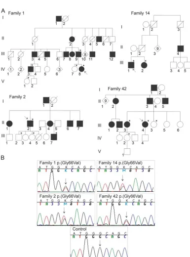
RESULTS Genetic findings.Our previous WES or tar-geted gene panel sequencing for families 1 and 2 did not identify potential disease-causing mutations (table e-1).6,7 The newly reported ALS gene, CHCHD10,3 was poorly covered by our WES and was not included in the targeted panel. By Sanger sequencing, we found a c.197G.T variant in CHCHD10
predicting a p.(Gly66Val) amino acid change to segregate with CMT2 in families 1 and 2. Within our CMT2 cohort, the entire CHCHD10 coding
region was sequenced, and we found the same variant in 4 additional index patients. Of these, 2 were from families with multiple members available for detailed study, families 14 and 42 (figure 1). The remaining 2 patients had been diagnosed with CMT2 elsewhere, with samples sent to us for DNA analysis, but detailed clinical information was not available. The p.(Gly66Val) variant was not found in 104 Finnish control individuals5 or in the 1000 Genomes database (http://www.1000genomes.org/, accessed January 2015). The variant was predicted to be
“probably damaging” by PolyPhen-2, “tolerated” by SIFT, and“disease causing”by MutationTaster.
Clinical findings.Detailed clinical data were available for 12 affected individuals from 4 families (table e-2). The typical presenting symptom was slowly progressive lower leg muscle weakness, and small hand muscles were affected later on in the disease course. The onset of symptoms varied from 30 to 56 years (mean 44). Clinical examination consis-tently showed loss of tendon reflexes, muscle weak-ness, and atrophy. Sensory abnormalities such as loss of sensation for vibration or cold were strong enough to be detected on clinical examination in 7 of 12 tients. Creatine kinase was normal in 4 of 12 pa-tients. Progression tended to be slow, and so far all patients are alive or died from a cause unrelated to neuropathy. No signs of upper motor neuron dis-ease, bulbar symptoms, or progressive cognitive problems were observed in the patients. Lower limb muscle MRI showed edema or fatty degeneration that was pronounced distally, particularly in calves, and milder in thighs.
Neurophysiologic findings. ENMG showed chronic motor neuropathy with regeneration in all studied patients. In families 1, 2, and 14, sensory findings (decreased sural nerve action potential amplitude) were evident in the first ENMG, consistent with typical CMT2 neuropathy (table e-2). In family 42, motor findings predominated and sensory abnormalities became evident only at a later age.
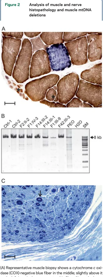
Neuropathologic findings.Muscle biopsies of selected patients demonstrated denervation atrophy: small and large group atrophy, some single atrophic fibers, variable fiber type grouping, some secondary myo-pathic changes, and end-stage atrophy in 1 case (F1:III-9) (figure 2A). Fibers deficient of cytochrome
coxidase (COX) staining (figure 2A) were detected in 2 patients (F1:III-8 and F2:II-2).
Nerve biopsy samples from the sural nerve of 4 patients showed unspecific axonal neuropathy, with thick (.7 mm) myelinated fibers (MF) most affected (figure 2C). In the available sural nerve biopsies from 3 patients, the perineurial sheath was slightly fibrotic at places, but no significant changes were observed in epineurium, perineurium, or ves-sels, and no amyloid could be demonstrated. In the
endoneurial compartment, the MF density varied between 2,500 and 3,600 MF/mm2and large MF represented between 5% and 25% of the total MF (normal values in our adult patients: approximately 7,000–9,000 MF/mm2 total count, large MF frac-tion 35%–45%). No marked hypertrophic changes, including marked sprouting, regenerative clusters, or demyelination features, were observed.
Figure 1 Pedigrees and sequencing
Neuropathologic autopsy was available for patient F1:III-9. He died of metastasized microcellular neu-roendocrine prostate cancer and also presented with epidural metastatic changes compressing the spinal cord at thoracic levels T5 to T6. Otherwise, the spinal cord was macroscopically normal; no anterior root atrophy was observed. Histologically, there was a marginal unspecific reduction of anterior horn motor neuron density, but changes typical for motor neuron disease/ALS were not observed. p62 and phosphory-lated TDP-43 immunohistochemistry (IHC) was also negative in the spinal cord and medullary hypoglossal nuclei. In the brain, no specific focal or otherwise diagnostic findings could be observed; IHC for
b-amyloid, phosphorylated tau, and p62 was negative in hippocampus and frontal cortex.
DISCUSSION Defects in CHCHD10 have emerged as an important cause of variable neurodegenerative phenotypes, underlying ALS-FTD,3SMAJ,5and mito-chondrial myopathy.4The c.197G.T (p.(Gly66Val)) variant has been previously described in patients with SMAJ5or ALS.11Some of our patients showed clinical overlap with SMAJ. For instance, cramps or fascicula-tions were common presenting symptoms in SMAJ5 and were among the main symptoms in 3 of our 12 patients. However, several lines of evidence suggested length-dependent degeneration of sensory and motor axons, i.e., CMT2, rather than SMAJ or ALS, in the patients described here. First, the typical initial symptom was distal lower limb muscle weakness, and predominantly distal muscle involvement was shown by MRI. In addition, clinical examination showed sensory impairment in 7 of 12 patients. Sensory impairment in patients with SMAJ was mild and had a later onset.5Second, ENMG revealed axonal neuropathy involving both sensory and motor fibers in 10 of 12 patients, including decreased sensory nerve action potential at first presentation in 9 of 12 patients. Third, axonal neuropathy was found in all 3 patients who underwent sural nerve biopsy. Fourth, autopsy excluded degeneration of anterior horn neurons. Therefore, our results demonstrate that the CHCHD10 p.(Gly66Val) variant can cause variable phenotypes ranging from ALS or lower motor
Figure 2 Analysis of muscle and nerve histopathology and muscle mtDNA deletions
(A) Representative muscle biopsy shows a cytochromec oxi-dase (COX)-negative blue fiber in the middle; slightly above it to the left is a single concave atrophic fiber. Above the lower margin in the middle is a small group atrophy with moderately atrophic fibers, and basally, in the left corner, a small group with very small fibers is seen (asterisks). Frozen section, COX-succinate dehydrogenase activity stain, original magni-fication 2003, scale bar 50mm. (B) Long range PCR on total muscle DNA was performed to detect mitochondrial DNA (mtDNA) deletions from patients and 2 control individuals without mtDNA deletions (Ctrl-1 and Ctrl-2). As a positive control, we used muscle DNA from a patient with progressive external ophthalmoplegia (PEO) caused by a variant in the
RRM2Bgene. The PCR primers were designed to overlap the 4977 bp common deletion between positions 13447-13459 and 8470-8482 and yielded a PCR product of;8 kb from undeleted mtDNA (arrow). The 8-kb band was readily detected in controls and patients. The patient with PEO had multiple smaller bands corresponding to deleted mtDNA
neuronopathy to sensorimotor axonopathy.CHCHD10
should thus be considered a candidate gene in patients with CMT2.
TheCHCHD10c.197G.T variant was found in
4.8% (6/126) of our cohort, when including solved and unsolved unrelated families.6,7Our patients are likely to have the same disease haplotype as the previously described patients with SMAJ or ALS, because of shared Finnish ethnicity. Such founder haplotypes are typical in Finland, which is a genetic isolate. Other factors than the haplotype are likely to modify the clinical pheno-type in the different patients who carry the same variant. The CHCHD10 protein localizes to the mitochondrial intermembrane space, where it enriches at cristae junc-tions and appears to be important for maintenance of cristae morphology and oxidative phosphorylation.3In our patients, the mitochondrial pathology in muscle was mild, with only occasional COX-negative fibers and small amounts of mtDNA deletions. In con-trast, p.(Gly58Arg) caused a pure myopathy,4while p.(Ser59Leu) led to a more aggressive ALS-FTD phenotype that included clear involvement of both the central and peripheral nervous system in addition to pathologic evidence of mitochondrial myopathy.3The disease spectrum of CHCHD10 resembles that of
MFN2, where CMT2 is the typical presentation but some patients have mitochondrial myopathy with mtDNA deletions12 or CNS involvement13 as addi-tional features. Peripheral neurons may be exquisitely sensitive to mitochondrial network defects owing to the length of their axons. Further functional studies are needed to fully understand the function of CHCHD10, the disease mechanisms of its variants, and the factors that modify the phenotypic outcome in the patients.
AUTHOR CONTRIBUTIONS
M.A. collected and analyzed patient data. E.Y. analyzed patient and sequencing data. M.S. performed sequencing and mtDNA analysis. A.P. performed neuropathology. S.K.-E. contributed to clinical analysis. J.P.T. performed and analyzed neurophysiology. M.A., E.Y., and H.T. designed the study and drafted the manuscript. All authors revised the manuscript.
STUDY FUNDING
Supported by Sigrid Jusélius Foundation, Academy of Finland, Univer-sity of Helsinki, Finska läkaresällskapet, Arvid and Greta Olin’s founda-tion, the Finnish Neuromuscular Disorders Associafounda-tion, and The Swedish Cultural Foundation in Finland.
DISCLOSURE
M. Auranen reports no disclosures. E. Ylikallio has received research sup-port from The Swedish Cultural Foundation in Finland. M. Shcherbii and A. Paetau report no disclosures. S. Kiuru-Enari has received travel
funding and/or speaker honoraria from Pfizer and research support from the Finnish Medical Society. J.P. Toppila reports no disclosures. H. Tyy-nismaa has patents for methods for diagnosis, treatment, and follow-up for dysfunction of the mitochondrial respiratory chain, and has received research support from the Academy of Finland, University of Helsinki, and the Sigrid Juselius Foundation. Go to Neurology.org/ng for full disclosure forms.
Received February 11, 2015. Accepted in final form March 4, 2015.
REFERENCES
1. Zuchner S, Mersiyanova IV, Muglia M, et al. Mutations in the mitochondrial GTPase mitofusin 2 cause Charcot-Marie-Tooth neuropathy type 2A. Nat Genet 2004;36: 449–451.
2. Rossor AM, Polke JM, Houlden H, Reilly MM. Clinical implications of genetic advances in Charcot-Marie-Tooth disease. Nat Rev Neurol 2013;9:562–571.
3. Bannwarth S, Ait-El-Mkadem S, Chaussenot A, et al. A mitochondrial origin for frontotemporal dementia and amyotrophic lateral sclerosis through CHCHD10 involve-ment. Brain 2014;137:2329–2345.
4. Ajroud-Driss S, Fecto F, Ajroud K, et al. Mutation in the novel nuclear-encoded mitochondrial protein CHCHD10 in a family with autosomal dominant mitochondrial myopathy. Neurogenetics 2015;16:1–9.
5. Penttila S, Jokela M, Bouquin H, Saukkonen AM, Toivanen J, Udd B. Late-onset spinal motor neuronopathy is caused by mutation in CHCHD10. Ann Neurol 2015; 77:163–172.
6. Auranen M, Ylikallio E, Toppila J, Somer M, Kiuru-Enari S, Tyynismaa H. Dominant GDAP1 founder muta-tion is a common cause of axonal Charcot-Marie-Tooth disease in Finland. Neurogenetics 2013;14:123–132. 7. Ylikallio E, Johari M, Konovalova S, et al. Targeted
next-generation sequencing reveals further genetic heterogeneity in axonal Charcot-Marie-Tooth neuropathy and a muta-tion in HSPB1. Eur J Hum Genet 2014;22:522–527. 8. Adzhubei IA, Schmidt S, Peshkin L, et al. A method and
server for predicting damaging missense mutations. Nat Methods 2010;7:248–249.
9. Kumar P, Henikoff S, Ng PC. Predicting the effects of coding non-synonymous variants on protein function using the SIFT algorithm. Nat Protoc 2009;4:1073–1081. 10. Schwarz JM, Cooper DN, Schuelke M, Seelow D. Muta-tionTaster2: mutation prediction for the deep-sequencing age. Nat Methods 2014;11:361–362.
11. Muller K, Andersen PM, Hubers A, et al. Two novel mutations in conserved codons indicate that CHCHD10 is a gene associated with motor neuron disease. Brain 2014;137:e309.
12. Rouzier C, Bannwarth S, Chaussenot A, et al. The MFN2 gene is responsible for mitochondrial DNA instability and optic atrophy“plus”phenotype. Brain 2012;135:23–34. 13. Chung KW, Kim SB, Park KD, et al. Early onset severe and
DOI 10.1212/NXG.0000000000000003
2015;1;
Neurol Genet
Mari Auranen, Emil Ylikallio, Maria Shcherbii, et al.
variant p.(Gly66Val) causes axonal Charcot-Marie-Tooth disease
CHCHD10
This information is current as of April 14, 2015
Services
Updated Information &
http://ng.neurology.org/content/1/1/e1.full.html
including high resolution figures, can be found at:
Supplementary Material
http://ng.neurology.org/content/suppl/2015/04/14/1.1.e1.DC1
Supplementary material can be found at:
References
http://ng.neurology.org/content/1/1/e1.full.html##ref-list-1
This article cites 13 articles, 0 of which you can access for free at:
Citations
http://ng.neurology.org/content/1/1/e1.full.html##otherarticles
This article has been cited by 12 HighWire-hosted articles:
Permissions & Licensing
http://ng.neurology.org/misc/about.xhtml#permissions
its entirety can be found online at:
Information about reproducing this article in parts (figures,tables) or in
Reprints
http://ng.neurology.org/misc/addir.xhtml#reprintsus
Information about ordering reprints can be found online:
Neurology. All rights reserved. Online ISSN: 2376-7839.
an open-access, online-only, continuous publication journal. Copyright © 2015 American Academy of is an official journal of the American Academy of Neurology. Published since April 2015, it is