Abstracts
12 2012 Volume 17 No 1 JEMDSA
Thursday, 19 April 2012: Oral presentations
TO1. Postmenopausal bone loss: an osteoimmunological
perspective
Roberto Pacifici
Garland Herndon Professor of Medicine; Director, Division of Endocrinology, Metabolism and Lipids, Emory University School of Medicine, Atlanta, Georgia, USA
TO2. Bone programming
Lisa Micklesfield,1,2 Shane Norris,1 John Pettifor1
1MRC/Wits Developmental Pathways for Health Research Unit and Department of Paediatrics, University of the Witwatersrand, Johannesburg 2UCT/MRC Research Unit for Exercise Science and Sports Medicine, University of Cape Town
Early growth has been associated with adult bone mass and fracture risk. Traditionally, birthweight has been used as a proxy for intrauterine growth, and is a significant predictor of postnatal, childhood, and adult bone density. More recently, results of a study from the Southampton Women’s Survey show that early intrauterine growth, measured using high-resolution ultrasound, determines bone mass at four years of age, while later intrauterine growth is more closely associated with bone mass at birth. Growth during the first years of life is important for skeletal development, and Birth to Twenty data have shown an association between weight and height at one year, and dual energy X-ray absorptiometry (DXA) scan bone mineral content in children aged 10 years. Findings from other cohort studies report similar findings between birthweight and growth in infancy, and bone mass later. To date, most evidence has been obtained using a DXA scan, which is known to be dependent on size. Peripheral quantitative computed tomography (pQCT) measures appendicular volumetric bone density, and is therefore not size-dependent, as well as bone geometry and trabecular bone mass. Results from most pQCT studies report a positive association between birthweight and bone size and strength, independent of current body size. However, bone mineral density appears to be associated with current weight and height more. This presentation will review the current literature investigating these relationships.
TO3. Bone mass and fractures in black and white South
African women
Magda Conradie, Stephen Hough
Division of Endocrinology, Department of Medicine, Stellenbosch University, Tygerberg
Ethnic differences in bone mass and fracture risk in South African adult black and white women will be reviewed. Generally, it is accepted that the shared genetic phenotype of black populations in America and on the African continent protect them against osteoporosis and consequent fragility fractures. It is also accepted that black populations in Africa will have similar patterns of bone gain and loss to African Americans. Studies in South Africa contest this belief. Exposure to adverse environmental factors may negatively impact on ultimate achievement of optimal bone mineral density (BMD), even in genetically protected black populations. Higher BMD values and lower fracture rates at all skeletal sites have been extensively documented in African-American females. Studies in South Africa, although limited, have consistently noted site-specific differences in bone mass and fracture risk in both pre- and postmenopausal black and white females. Vertebral BMD are either lower or similar in blacks, compared with whites, with recently documented similar
vertebral fracture rates in black and white females respectively (11.5 vs. 8.1%). A five-year longitudinal study found new morphometric vertebral deformities in a high percentage of black women over the age of 60 years (38%). BMD at the proximal femur is higher in South African black females. Recent data on South African hip fractures are not available. Adverse environmental factors in South African black females include low dietary calcium intake, limited physical activity (especially in the elderly), and frequent use of injectable contraception. Findings highlight the need to better define the role of clinical risk factors, to document hip fracture prevalence, and to develop local bone mass reference data that are appropriate for our black population.
TO4. Site-specific differences in bone mineral density in
black and white premenopausal South African women
K Dickie, S Chantler, JH Goedecke, EV Lambert, J Evans, Y Joffe, LK MicklesfieldUCT/MRC Research Unit for Exercise Science, University of Cape Town
Background: There is a paucity of data on the relative contribution of
body composition, lifestyle factors, and socio-economic status unique to different ethnic groups in South Africa, to bone mineral density (BMD). We examined differences in femoral neck (FN), total hip (TH) and lumbar spine (LS) BMD between black and white premenopausal South African women, and the associations between BMD and body composition, lifestyle factors, and SES, in these two ethnic groups.
Method: BMD and body composition were measured in 240 black
(27±7; 18-45 years) and 187 white (31±8; 18-45 years) women, using dual-energy X-ray absorptiometry. Questionnaires were administered to examine SES, physical activity, and dietary intake.
Results: After co-varying for age, FN and TH were higher in black
than white women (FN 0.882±0.128 vs. 0.827± 0.116 g/cm2, p-value < 0.001; TH 0.970±0.130 vs. 0.943± 0.124 g/cm2, p-value = 0.018). When adjusting for ethnic differences in body composition, LS was higher in white, than black, women. In black women, fat-free soft tissue mass, SES, and injectable contraceptive use, explained 33-42% of the variance in BMD at the hip sites, and 22% at the LS. In white women, fat-free soft tissue mass and leisure activity explained 24-30% of the variance in BMD at the hip sites, whereas fat mass, leisure activity, and oral contraceptive use, explained 11% of the variance at the LS.
Conclusion: FN and TH BMD were higher, but LS BMD was lower in
Abstracts
13 2012 Volume 17 No 1 JEMDSA
TO5. Longitudinal sex and ethnic differences in bone mass
acquisition between the ages of 9 and 18 years in
urban South Africa
Shane Norris,1 Lisa Micklesfield,1,2 John Pettifor1
1MRC/Wits Developmental Pathways for Health Research Unit and Department of Paediatrics, University of the Witwatersrand, Johannesburg
2UCT/MRC Research Unit for Exercise Science and Sports Medicine, University of Cape Town
Background: Bone mass acquisition during adolescence plays a
major role in determining peak bone mass in adulthood. Therefore, describing sex and ethnic bone mass longitudinally is critical in understanding factors that may influence this process. The aim of this study was to describe the pattern of bone mass acquisition in a longitudinal cohort of black and white boys and girls living in Johannesburg.
Method: Bone mass (whole body, lumbar spine and hip) was assessed
annually from dual energy X-ray absorptiometry (DXA) scans in a Bone Health sub-cohort (approximately 470 participants) of the Birth to Twenty cohort in Johannesburg, between the ages of 9 and 18 years. In addition to DXA-derived bone mass, several other parameters were measured, such as height, weight, and pubertal stage (Tanner’s pubertal development scale).
Results: Throughout the nine years of the study, black boys and girls
remained significantly shorter than their white peers (p-value < 0.01). After correcting for body size, hip bone mineral content throughout the adolescent period was significantly greater in black boys and girls, compared to white boys and girls (p-value > 0.05). However, there were no significant differences in lumbar spine bone mineral content between the sex and ethnic groups.
Conclusion: These longitudinal data provide local bone mass patterns
during adolescence, and highlight key similarities and differences in bone mineral content between black and white boys and girls. This valuable dataset provides a unique opportunity to further explore and identify key factors that influence growth, pubertal development, and skeletal maturity, and in turn, bone mass and fracture risk.
TO6. Association of body mass index with fracture risk and
bone mass in urban South African children: the Birth to
Twenty cohort
Kebashni Thandrayen, John Pettifor, Shane Norris
MRC/Wits Developmental Pathways for Health Research Unit, Department of Paediatrics, University of the Witwatersrand, Johannesburg
Worldwide prevalence of childhood obesity is increasing. This study aims to investigate the association of body mass index (BMI) with fracture risk and bone mass. Using the Bone Health cohort of the Birth to Twenty longitudinal study, we retrospectively obtained information of lifetime fractures until age 15 years, in 533 subjects. Whole body bone mineral content (BMC), bone area (BA), fat mass (FM) and lean mass (LM) [measured by dual energy X-ray absorptiometry (DXA) scans], physical activity, and anthropometric data, were obtained at ages 10 and 15 years. Non-fracturing black females were used as the control group. Individual anthropometric measurements [height for age Z score (HAZ) and BMI for age Z score (BAZ)] were calculated using the World Health Organization Anthroplus® software. White males who fractured were significantly taller (10 years, p-value < 0.01), more physically active (15 years, p-value < 0.05) and had
higher LM (10 years, p-value = 0.01; 15 years, p-value < 0.001), while white females who fractured were fatter (10 years, p-value = 0.05 and 15 years, p-value < 0.05), than their non-fracturing peers.Overweight or obese white females had a greater number of fractures by the age of 15 years. At the age of 10 years, obese subjects had a higher BA and BMC at most sites, and fracturing obese black males had a lower BMC Z score at the whole body, hip, and spine, compared to their non-fracturing obese peers. Increased adiposity in white females is associated with increased fracture risk. This association is not evident in black females, despite an increasing prevalence of overweight or obesity in both groups that needs further elucidation.
TO7. Demographic profile and risk factors for osteoporotic
hip fractures in the elderly in the Ethekwini municipality,
KwaZulu-Natal
Farhanah Paruk, Bilkish Cassim
Department of Geriatrics, College of Health Sciences, University of KwaZulu-Natal
Background: The aim was to determine the demographic profile
and risk factors for osteoporotic hip fractures in the public sector in eThekwini.
Method: Consecutive subjects aged ≥ 60 years, with minimal
trauma hip fractures, were prospectively enrolled from five public sector hospitals. Demographic details were documented, and a questionnaire for risk factors, administered.
Results: In the 277 enrolled subjects, the mean age was 75.8 ± 9.1
years, with a female:male ratio of 2.8:1. Men were significantly younger than women ( 71.7 ± 9.3 years vs. 77.4 ± 8.6 years, p-value < 0.0001). Indian subjects comprised 51.3%; African subjects, 32.1%; white subjects, 14.1%; and mixed ancestry subjects, 2.5%. Of those who completed the questionnaire (n = 200), hypertension was present in 112 (56%), diabetes mellitus in 56 (26.6%), arthritis in 55 (26.6%), and an underlying malignancy in 16 (8%). Forty-seven (23.5%) were smokers, 28 (14%) consumed alcohol weekly, 39 (19.5%) were below 57 kg, 72 (34%) had prior falls, and 14 (7%) gave a maternal history of falls or fractures. Despite a previous fragility fracture in 56 subjects (28%), only two subjects received specific treatment.
Conclusion: This study is the first to report hip fractures in all ethnic
groups in South Africa, and suggests that the Indian population may be at high risk. In contrast to developed countries, hip fractures appear to occur at a younger age, especially in men. This study underscores the need to include osteoporosis in the healthcare package. A limitation of the study is the possible disproportionate representation of the ethnic groups due to differences in utilisation of public-sector hospitals.
Abstracts
14 2012 Volume 17 No 1 JEMDSA
TO8. Functional outcomes and mortality rates in patients
with osteoporotic hip fractures in public sector hospitals
in Ethekwini, KwaZulu-Natal
Farhana Paruk, Bilkish Cassim
Department of Geriatrics, College of Health Sciences, University of KwaZulu-Natal
Background: The aim was to determine the functional outcome and
mortality rate in subjects presenting with osteoporotic hip fractures.
Method: In a longitudinal study, subjects aged 60 years and over
with minimal trauma hip fractures, were reviewed at three months to assess functional outcome and mortality.
Results: Two hundred patients were prospectively enrolled. The mean
age was 74.2 ± 8.8 years (range 60-113 years), with a female:male ratio of 2.6:1. Surgery was performed on 186 subjects (93%), with 14 (7%) being unfit for it. The average length of stay was 21.6 days (range 4-120 days), and average time to surgery was 10.4 days (range 1-37 days). At three months, 35 subjects (17.5%) had died, and of the 117 subjects reviewed, 27 (23.5%) were able to ambulant independently, 64 (54.7%) required help, 26 (22.3%) were bed-bound, and 106 (90.5 %) were unable to complete at least one instrumental activity of daily living (IADL). The quality of life (QOL) scale and Owestry disability index showed a twofold decline, and the patient global assessment (visual analogue scale) worsened from 0.9 to 5. Apart from a history of arthritis, there were no other predictors of mortality.
Conclusion: The early mortality and morbidity in this study is higher
than that reported internationally, and may be due to the significant delay before surgery, and lack of dedicated orthogeriatric facilities. Urgent healthcare reform is required to improve the management of osteoporosis and fragility fractures.
This study was supported by an unrestricted educational grant from Servier Laboratories (SA).
TO9. Fractures and bone mass in children (mini-review)
Kebashni Thandrayen, John Pettifor, Shane NorrisMRC/Wits Developmental Pathways for Health Research Unit, Department of Paediatrics, University of the Witwatersand, Johannesburg
Fracture rates in childhood are as high as those in the elderly. Studies have found a high incidence of fracture, with 27-40% of girls, and 42-51% of boys, sustaining at least one fracture during growth. The peak incidence of fractures in girls occurs between 9-14 years, and in boys between 13-14 years, corresponding to the ages of peak height velocity. In South Africa, black children have fracture rates that are approximately half those of their white peers.
Factors influencing childhood fracture risk include nutrition, breastfeeding duration, carbonated beverage consumption, sport participation, body composition, rate of pubertal development, computer and television use, bone mass, and heredity. Despite the inverse relationship observed between fracture risk and bone mass, higher bone mass associated with increased physical activity in children does not necessarily compensate for the increased exposure to injuries, and higher fracture risk. After adjusting for differences in body size, black children only have consistently higher bone mass at the hip, thus the mechanisms for the lower childhood fracture rates are currently not known. Recent findings have shown that South African black children have greater bone strength, as measured by peripheral quantitative computed tomography (pQCT). However,
the association between fracture risk and bone strength, or geometry, needs further investigation. Structural differences in bone geometry may provide protection against fractures in black South Africans. Whether or not the relative protection that black children have against fractures is going to alter with the change in lifestyles associated with the rapid acculturation that is occurring in South Africa, is unclear.
TO10. The anatomical basis of cortical porosity in children
CM Schnitzler,1 JM Mesquita21MRC/Wits Developmental Pathways for Health Research Unit, Department of Paediatrics
2Division of Orthopaedic Surgery, University of the Witwatersrand, Johannesburg
Debate I: Bone mineral density measurement is a very useful tool to monitor the response to therapy in osteoporosis
Graham Ellis (for), and Stephen Hough (against)
TO11. T cells: unexpected players in the mechanism of action
of parathyroid hormone
Roberto Pacifici
Garland Herndon Professor of Medicine; Director, Division of Endocrinology, Metabolism and Lipids, Emory University School of Medicine, Atlanta, Georgia, USA
TO12. Endocrine cross-talk between bone and fat: emerging
new roles for well-known players
Hanel Sadie van Gijsen,1 Nigel Crowther,2 Stephen Hough,1 William Ferris1 1Division of Endocrinology, Department of Medicine, Stellenbosch University, Tygerberg
2NHLS, University of the Witwatersrand
Abstracts
15 2012 Volume 17 No 1 JEMDSA
TO13. Expression of the tumour suppressor PDC4 during
osteoblastic differentiation of adipose-derived
stromal cells
Martine van den Heever, Hanel Sadie-van Gijsen, Stephen Hough, William Ferris
Department of Medicine, Stellenbosch University, Tygerberg
Background: The use of mesenchymal stromal cells (MSCs) has
been cited as a putative therapeutic option for tissue regeneration in the future. Adipose tissue is an abundant source of these cells, which have the propensity to be differentiated into an osteoblastic, chondrocytic, adipocytic, or myoblastic phenotype, for possible regenerative therapy of bone, cartilage, fat, and muscle. However, for successful therapeutic use, the cell cycle must be precisely regulated to avoid abhorrent cell function, such as uncontrolled proliferation or premature death. The expression of programmed cell death 4 (PDCD4) has been associated with the control of differentiation and proliferation in neoplastic and immortalised cells. However, the role for PDCD4 in the regulation of these cellular fates in primary MSCs had yet to be assessed. The aim was to examine the expression of PDCD4 in primary subcutaneous and visceral adipose-derived stromal cells of rats (ADSCs) during differentiation into osteoblasts.
Method: ADSCs were differentiated into mature osteoblasts, and
proliferation measured using tritiated thymidine incorporation after 0, 3, 7, 14, 21, and 28 days of differentiation. The state of differentiation was determined by Alizarin Red S staining of mineralised nodules, and real-time quantitative polymerase chain reaction (QRT-PRC) analyses of the bone markers, MSX2 and Runx2. PDCD4 mRNA expression, during differentiation, was measured by QRT-PRC. PDCD4 protein expression was measured by Western Blot analysis.
Results: Subcutaneous ADSCs, but not visceral ADSCs, differentiate
into osteoblasts after incubation in osteogenic media, as seen by calcified nodule formation and expression of bone markers. Preliminary data indicate that PDCD4 mRNA expression correlates with the state of differentiation, increasing during subcutaneous ADSC osteogenesis, but remaining low in non-differentiating visceral ADSCs. The proliferation profile of both cell types was similar.
Conclusion: Consequently, the tumour suppressor, PDCD4, is more
closely associated with an increase in osteoblastic differentiation, rather than a decrease in proliferation in ADSCs.
TO14. Sodium orthovanadate stimulates osteoblast
proliferation, but inhibits osteoblast function in vitro and
in vivo
Hanel Sadie-van Gijsen, Riana Eager, Micheline Sanderson, Stephen Hough, William Ferris
Division of Endocrinology, Department of Medicine, Stellenbosch University, Tygerberg
Background: We have previously shown that vanadate can stimulate
extracellular signal-regulated kinase activity and proliferation in culture-naive rat adipose-derived stromal cells (ADSCs), and also stimulates osteoblast proliferation in rats in vivo. We have also shown that ADSCs from rat subcutaneous adipose tissue can undergo osteoblast differentiation in vitro, but the effects of vanadate on the osteoblastic differentiation of ADSCs have not been investigated. Consequently, we wished to compare the effects of vanadate on osteoblast proliferation, differentiation, and function, between these in vitro and in vivo systems.
Method: For in vitro studies, ADSCs were isolated from the inguinal
adipose tissue of rats, expanded in number, and treated with osteoblast differentiation media (OM: standard culture media supplemented with dexamethasone, ascorbic acid, and glycerol-2-phosphate) in the presence and absence of vanadate for 28 days. The deposition of calcified matrix, as a marker of mature osteoblast function, was quantified by means of Alizarin Red S staining, and normalised to relative cell density, which was measured using crystal violet stain. For in vivo studies, rats were administered vanadate via the drinking water for nine weeks, after which time spaced tetracycline labelling was used to quantify bone deposition in the femur. Bone mineral density (BMD), bone formation rate (BFR), and surfaces covered by osteoblasts and osteoclasts, were also measured.
Results: In the in vitro studies, vanadate stimulated the proliferation
of differentiating osteoblasts, but inhibited the formation of calcified matrix. This lack of increased function with increased osteoblast number was paralleled in in vivo data showing that vanadate increased osteoblast surfaces in rat femora, while no concomitant increase in BFR, or BMD, was observed.
Conclusion: Vanadate induced an increase in osteoblast numbers
in vitro in rat ADSCs, as well as in vivo in rats, yet concomitantly decreased osteoblast activity. Further investigation examining osteoblastic marker expression is required to find whether this is due to inhibition of differentiation.
TO15. Osteopenia: how we created a “disease”, and how to
manage patients with osteopenia
Steven Cummings
Professor of Medicine and Epidemiology; Director, San Francisco Coordinating Center, CPMP Research Institute, University College of San Francisco, USA
Osteopenia and osteoporosis are not diseases. In 1992, a committee (the World Health Organization task force) rather arbitrarily defined femoral neck T-score < -2.5 as “osteoporosis”, and between -1 and -2.5 as “osteopenia”. By definition, about half of all postmenopausal women in any population have T-score “osteopenia.”
Drug treatment is probably not worthwhile for most women with osteopenia. The development of the WHO fracture risk assessment tool (FRAX), and calculations of absolute risk of fracture, have shown that, in general, women who are younger than 60 years with “osteopenia” have a low risk of fracture that does not warrant drug therapy. Additionally, several, but not all, trials, have found that treatments do not reduce the risk of non-spine fractures in women with femoral neck T-scores > -2.5. (In contrast, those with T-scores ≤ -2.5 have > 30% reduction in risk of non-spine fractures.) For women who have been taking alendronate or zoledronate, results from extensions of those trials indicate that women who have hip T-scores >-2.5 after three to five years on treatment, do not significantly benefit from continuation of treatment.
Abstracts
16 2012 Volume 17 No 1 JEMDSA
TO16. Human immunodeficiency virus and bone
Stephen HoughDivision of Endocrinology, Department of Medicine, Stellenbosch University, Tygerberg
The improved pharmacotherapy of patients with human immunodeficiency virus (HIV)/acquired immune deficiency syndrome (AIDS) has significantly reduced mortality, converting this disease into a chronic disorder that requires long term-treatment. As individuals live longer, and are subjected to the chronic sequelae of both the disease and its treatment, a number of complications have emerged, including metabolic bone disorders, such as osteoporosis, osteomalacia, osteonecrosis, and immune reconstitution inflam-matory syndrome-induced hypercalcaemia.
It has been suggested that more than 65% of patients with HIV have a low bone mineral density (BMD), and that the odds ratio for osteoporosis and fracture are 3.7 and 2.5 respectively. Yet the underlying pathogenesis, clinical relevance, and most appropriate management of this disorder, remains unclear. Some ascribe the low BMD largely to the low body mass which typically accompanies HIV/ AIDS.
Others implicate:
• The disease (pro-inflammatory cytokines which iosteo-clastogenesis, malnutrition which hosteoblastic bone formation, or even direct infection of osteoblasts (Ob) by the HIV virus and hOb apoptosis);
• Associated osteoporosis risk factors (alcohol, smoking, hypogonadism, and hypovitaminosis D); and or
• The deleterious effects of HIV treatment on bone.
The latter may involve RANKL-mediated increase in osteoclastic bone resorption by some protease inhibitors, but more likely to result from the effects of antiretroviral drugs on osteoprogenitor cells, with an increase in adipogenesis, at the expense of osteoblastogenesis. An inverse relationship between lipohypertrophy or metabolic syndrome and BMD has been reported by some, but not all, researchers. Recent meta-analyses show that bisphosphonates increase BMD, but fracture data will have to await further study.
TO17. Bone health and body composition in HIV-positive and
-negative urban black South African women
Matthew Hamill,1,2 Kate Ward,2 John Pettifor,1 Shane A Norris,1 Ann Prentice3
1MRC/Wits Developmental Pathways for Health Research Unit, University of the Witwatersrand, Johannesburg
2MRC Human Nutrition Research Cambridge, UK
3 MRC Human Nutrition Research Cambridge UK and MRC Keneba, The Gambia
Background: Human immunodeficiency virus (HIV) infection and
antiretroviral drugs (ARVs) are associated with low bone mineral density (BMD). The purpose of this study was to consider bone density and body composition (BC) in HIV-positive, urban women.
Method: Two hundred and forty-seven premenopausal [mean ?
standard deviation (SD) age 32.1 ?7.24 years], black women were recruited in Soweto, and underwent baseline dual energy X-ray absorptiometry (DXA) scans, and other assessments. Group 1: HIV-negative control (n = 98), Group 2: HIV-positive, preserved cluster of differentiation 4 (CD4) (mean CD4: 413?91; n = 74), Group 3: HIV-positive, low CD4 prior to ARV initiation, (mean CD4: 161?70; n = 75).
Results: Mean values ? SD in groups 1, 2 and 3 respectively, for height
(m) were similar (p-value > 0.05). Weight (kg) significantly (p-value
< 0.02) differed, with group 3 being the lightest (69.7 ?17.0, 72.0 ?17.4, and 62.3 ?15.2 respectively). No significant differences in BMD at the hip, lumbar spine, and whole body (p-value > 0.05) were found with and without adjustment (weight, height, and age), and no difference in fat/lean ratio (p-value > 0.05). Previous fractures were reported in 23.4%, 25.7% and 13.3%, respectively. After full adjustment for bone area, weight, height, and age, there were no significant BMD differences at any site.
Conclusion: HIV-positive women with low CD4 counts are significantly
lighter than positive women with preserved CD4 counts and HIV-negative women, but have no significant difference in BMD and fat or lean measures. Currently, the subjects are being followed-up longitudinally to assess the effects of ARV exposure.
TO18. Prevalence and predictors of low bone mineral density
in HIV-positive South Africans on antiretroviral therapy
JA Dave,1 K Cohen,2 G Maartens,2 L Micklesfield,3 NS Levitt11Division of Diabetic Medicine and Endocrinology, University of Cape Town 2Division of Pharmacology, University of Cape Town
3MRC/Wits Developmental Pathways for Health Research Unit, Faculty of Health Sciences, University of the Witwatersrand
Background: Human immunodeficiency virus (HIV) positive men
and women from developed countries have reduced bone mineral density (BMD). In these patients, antiretroviral therapy (ART) does not appear to play a significant role. Traditional risk factors are more prominent. The prevalence and predictors of low BMD in HIV-positive patients on ART in sub-Saharan Africa are unknown. The objective was to determine the prevalence and predictors of low BMD in HIV-positive patients.
Method: The method was a cross-sectional study in an ambulatory
HIV-infected cohort in Cape Town which examined the metabolic complications of ART. A randomly selected sub-sample of these patients underwent BMD assessment, using dual-energy-X-ray absorptiometry (DXA) scans. Only patients < 50 years old were included, and a low BMD was defined according to the NOFSA guidelines as a Z-score < -2.0.
Results: DXA scanning was performed in 422 patients who were
ART-naive (n = 190); ART-reg1 (on non-nucleoside reverse transcriptase inhibitor-based ART), (n = 145); ART-reg2 (on protease inhibitor-based ART), (n = 87); randomly selected from 1 019 patients (ART-naive 436, ART 583) in the parent study. A low BMD was detected in 52 (27%) naive patients, 45 (31%) reg1 patients, and 27 (31%) ART-reg2 patients. Of the 124 patients with a low BMD, 121 (29%) had a low BMD at the spine, and 21 (5%) patients had a low BMD at the hip. Patients with a low BMD had a lower body mass index (BMI) [26 (22,32) vs. 24 (21,27) kg/m2, p-value < 0.0001], but all other measures of body composition (waist circumference, waist:hip ratio, abdominal skinfold thickness, and calf skinfold thickness) were not significantly different. There was no difference between the groups with respect to time on ART, height, cluster of differentiation 4 count, age, smoking, alcohol use, and vitamin D status. However, on multivariate logistic regression analysis, low BMI, ART exposure, and male sex, were all independently associated with low BMD at the hip; and male sex, low BMI, and smoking, were all independently associated with low BMD at the spine.
Conclusion: ART exposure was only associated with a low BMD at the
Abstracts
17 2012 Volume 17 No 1 JEMDSA
Friday, 20 April 2012: Oral presentations
FO1. Androgens and bone
Roger BouillonProfessor of Medicine, Laboratory for Experimental Medicine and Endocrinology, Katholieke Universiteit Leuven, Belgium
FO2. Non-union and delayed union in osteoporotic
fractures: is there a place for anti-osteoporosis
medication?
Stanley Lipschitz
The Osteoporosis Clinic, Rosebank, Johannesburg
Osteoporosis is defined as a condition of increased fracture risk. It is estimated that about 10% of all fractures are complicated by delayed fracture healing. Delayed union healing time is longer than expected for the specific type of fracture. With non-union, the fracture fails to heal within a certain amount of time.
The US Food and Drug Administration’s definition of “non-union” is a fracture that is more than nine months old, and that has not shown radiographic signs of progression toward healing for three consecutive months.
A case study of a patient with delayed union of fracture will be presented. The presentation will cover an extensive overview of the epidemiology, pathophysiology, and clinical presentation, of delayed fracture union, in addition to a discussion on why this condition may be more prevalent, and more difficult to manage, in patients with osteoporosis.
Traditionally, the approach to such patients has been surgical, with variable success.
An extensive review of the literature regarding the different modes of action of currently available anti-osteoporosis medications, as well as the potential for beneficial or detrimental effect, will then follow.
Current literature is sparse regarding the use of medication to improve outcomes in patients with non-union or delayed union. There is some preclinical evidence to suggest that certain agents may be beneficial, but more research is required.
FO3. Fragility fractures and the bisphosphonates
Mac LukheleHead of Division of Orthopaedics, Department of Orthopaedic Surgery, University of the Witwatersrand, Johannesburg
In this presentation, we will examine available current evidence on atypical fractures of patients who are on long-term bisphosphonates, the suggested mechanism, the clinical and radiological features of these fractures, and the current recommended management strategy.
The main aim of management of osteoporosis is the prevention of fragility fractures, in particular hip and vertebral fractures which are associated with high morbidity and mortality. For over a decade, bisphosphonate treatment has become the cornerstone in the prevention and treatment of fragility fractures. Phase III clinical trials reported a significant decrease in the incidence of fractures of the hip and spine, with concomitant reduction in associated costs and healthcare utilisation. Both nitrogen- and non-nitrogen-based bisphosphonates increase bone mineral density by inhibiting the osteoclast resorption function.
Bisphosphonates have a relatively good safety record, and are generally tolerated by patients. Usually, gastrointestinal side-effects have been overcome by the development of long-acting bisphosphonates, together with intravenous administered ones. Patients can also develop flu-like symptoms, which usually resolve. Rare complications, such as renal dysfunction and jaw osteonecrosis, have been described.
In the past six years, there have been several cases of patients who developed atypical fragility fractures after taking bisphosphonates for a long time.
The characteristics of these atypical fragility fractures are that: • They are associated with minor trauma.
• They are located subtrochanteric, or on the shaft. • They are transverse.
• There is associated cortical thickening, with beaking of the cortex.
• The patient often has pre-injury thigh pain.
The pathophysiology of atypical subtrochanteric fractures, following long-term usage of bisphosphonates, is not known. Several possible mechanisms that work either alone, or together, have been suggested. Bisphosphonates may affect bone mineralisation density distribution by slowing bone turnover, which leads to more homogenous bone, which increases the risk of crack and fracture. Reduced bone turnover also increases the accumulation of microdamage, as cracks are not repaired. This reduces bone durability, thereby increasing susceptiblity to more cracks. Although it has not been replicated in any studies, bisphosphonates were reported to prevent and reduce collagen maturation in that way, decreasing bone durability.
Abstracts
18 2012 Volume 17 No 1 JEMDSA
FO4. Calcium homeostasis during pregnancy and lactation
John PettiforMRC/Wits Developmental Pathways for Health Research Unit, Department of Paediatrics, Faculty of Health Sciences, University of the Witwatersrand, Johannesburg
Pregnancy and lactation place considerable demands on maternal calcium homeostasis. It is estimated that the growing foetus requires an average of 100 mg calcium daily, spread over the full nine months, while lactation imposes further stress of some 300 mg daily for the duration of lactation. These increased calcium demands have to be met by increased maternal intestinal calcium absorption, reduced renal calcium excretion, or increased bone resorption. Based on these increased requirements, it has been customary to recommend increased maternal calcium intakes, during both pregnancy and lactation. However, are these recommendations necessary, or based on scientific evidence?
A number of studies have investigated the influence of calcium supplementation, both during pregnancy and lactation, on the degree of bone loss that occurs during these periods, and the rate of recovery following weaning. There is no convincing evidence that calcium supplementation ameliorates the degree of bone loss, or increases the rate of recovery during weaning. Furthermore, supplementation has no effect on breast milk calcium concentrations during lactation. However, a few studies suggest that during pregnancy, calcium supplementation may influence foetal, or neonatal, bone mass.
In conclusion, bone loss that occurs during pregnancy and lactation recovers completely following weaning, even in mothers whose habitual calcium intakes are well below the levels recommended in a number of developed countries. There is no evidence that calcium supplementation influences maternal bone loss or recovery. Thus, it is not recommended as a routine supplement during this period of calcium stress.
FO5. The association between vitamin D status and body
composition, serum lipids and HOMA-IR in urban
12-year-old South African children
Machuene Poopedi,1,2 Shane Norris,1 John Pettifor1
1MCR/Wits Developmental Pathways for Health Research Unit and Department of Paediatrics
2Division of Orthopaedic Surgery, University of the Witwatersrand, Johannesburg
Background: The aim was to determine the association between
plasma 25-hydroxyvitamin D [25(OH)D], and body composition, lipid profiles, and homeostatic model assessment in insulin resistance (HOMA-IR) in urban South African children.
Method: The method was a cross-sectional study of 261 children,
(black children, n = 197; and white children, n = 64), with a mean age of 12.7 years, who formed the Bone Health sub-cohort of the Birth to Twenty cohort. The evaluation included weight and height, body composition by dual-energy-X-ray absorptiometry (DXA) scan, serum 25(OH)D (Diasorin Liaison®), lipids and glucose (RX Randox Daytona®) and insulin (Immulite 1000®).
Results: Both ethnic groups were vitamin D-replete. White children
had greater 25(OH)D, height, weight, total cholesterol, triglycerides, low-density lipoprotein (LDL) cholesterol, lipoprotein, and total lean tissue/height, than their black peers. Black male children had greater 25(OH)D, and lower height, weight, body mass index (BMI), insulin,
and total fat tissue/height than black females. White males had greater 25(OH)D and total lean tissue/height, and lower insulin and total fat/height than their female peers. In black children, 25(OH)D correlated negatively with high-density lipoprotein (HDL) cholesterol (r = -0.17), and in black females, with triglycerides (r = -0.3). There were no significant correlation with anthropometry, body composition, or other lipid variables. In white children, 25(OH)D correlated negatively with total cholesterol (r = -0.3), triglycerides (r = -0.4), BMI (r = -0.3), and fat tissue/height (r = -0.4). In white female subjects only, 25(OH) D correlated negatively with HDL-cholesterol (r = -0.3), and HOMA-IR (r = -0.4).
Conclusion: The correlations of 25(OH)D with anthropometric
variables, body composition, lipids, and HOMA-IR, differ between ethnic and gender groups. This may be due to differences in fat mass distribution between the ethnic and gender groups. The authors had no conflict of interests.
FO6. The association between early life factors and bone
mass and size in 13-year-old urban South African
children
Lisa Micklesfield,1,2 Shane Norris,1 John Pettifor 1
1Developmental Pathways for Health Research Unit, Department of Paediatrics, Faculty of Health Sciences, University of Witwatersrand, Johannesburg
2UCT/MRC Research Unit for Exercise Science and Sports Medicine, UCT School of Health Sciences, University of Cape Town
Background: To determine if early growth is associated with bone
mass and size measured using peripheral quantitative computed tomography (pQCT) at 13 years of age.
Method: We examined the association between size [weight, height
and body mass index (BMI)] at two years of age, current height, fat and lean mass, and bone parameters in black boys (n = 170), and girls (n = 150), at 13 years of age.
Results: In boys, BMI at two years was associated with metaphyseal
(4%) tibia total and trabecular density, before and after adjusting for current size. Height at two years was associated with diaphyseal (38%) tibia total area, but not after adjusting for current size or puberty. In girls, metaphyseal (4%) radius total area was associated with weight at two years, but not after adjusting for current size, and then puberty. Weight at two years was inversely associated with 38% tibia total area in girls, before and after adjusting for current body size, and pubertal development.
Conclusion: pQCT-derived trabecular bone density was associated
Abstracts
19 2012 Volume 17 No 1 JEMDSA
FO7. Sex and ethnic differences in skeletal maturation
during adolescence
John Pettifor,1 Nicola Hawley,1,2 Noel Cameron,1,2 Shane Norris1 1MRC/Wits Developmental Pathways for Health Research Unit; and Department of Paediatrics, Faculty of Health Sciences, University of the Witwatersrand, Johannesburg
2School of Sports, Exercise and Health Sciences, Loughborough University, Loughborough, UK
Skeletal maturation during puberty plays a major role in determining bone mass and size accrual during adolescence, thus an understanding of sex and ethnic differences in the tempo of skeletal development is important when interpreting bone mass measurements during this period. The aim of this study was to determine the pattern of skeletal maturation in a longitudinal cohort of black and white boys and girls living in Johannesburg.
Skeletal maturity was assessed annually from hand-wrist radiographs of children (n ≈ 475) in the Bone Health sub-cohort of the Birth to Twenty cohort in Johannesburg from the age of 9 to 17 years, using the Tanner and Whitehouse III (TW3) RUS technique, and a computerised Greulich and Pyle method using BoneXpert ?. In girls, menarcheal age was determined by questionnaire.
Age of menarche was similar in black and white girls (mean 12.4 and 12.5 years respectively), and black and white girls? skeletal maturation advanced at similar rates through puberty. In black boys, skeletal maturity diverged from that of white boys from the age of 12 years, so that by 14 years, they were approximately 12 months behind, and remained so over the next three years.
Menarcheal age in black girls has shown marked secular trends over the last 50 years, with a reduction of 3.5 years, while less marked changes have occurred in white girls (0.6 years in 27 years). Black and white girls have shown a similar timing and rate of skeletal maturation during puberty. However, in black boys, skeletal maturation is delayed. These patterns should be taken into consideration when assessing bone mass measurements during adolescence.
Debate II: Kyphoplasty is an effective and safe way in which to manage the patient with symptomatic osteoporosis
Mac Lukhele (for), and Stan Lipschitz (against)
FO8. Meet the expert - bisphosphonates: when and how to
stop
Steven R Cummings
Professor of Medicine and Epidemiology; Director, San Francisco Coordinating Center, CPMC Research Institute, University College of San Francisco, USA
FO9. Novel treatments to manage osteoporosis
Steven CummingsProfessor of Medicine and Epidemiology; Director, San Francisco Coordinating Center, CPMC Research Institute, University College of San Francisoco, USA, and University of California, San Franscisco, USA
Several new treatments for osteoporosis are being developed that work using novel biological mechanisms, and which may have advantages over existing therapies. Denosumab has not yet been approved for use in South Africa, but it works via the receptor activator of the RANK pathway. Twice yearly, subcutaneous injections achieve maximum reduction in bone resorption, and improve compliance with treatment, and reduce the risk of all types of fractures, at least as effectively as bisphosphonates.
Odanacatib inhibits cathepsin K, reduces bone resorption more than formation, and appears to steadily increase bone mineral density (BMD), and perhaps add cortical bone. It is hoped that odanacatib might reduce non-spine fracture risk more than current therapies. Results of a pivotal anti-fracture trial should be available within the year.
Antisclerostin antibodies block sclerostin’s inhibition of osteoblastic bone formation. They dramatically increase bone formation, and decrease bone resorption. This therapy has the potential to “cure” osteoporosis. Anti-fracture trials will begin in 2012.
Daily intermittent nitroglycerin may work via osteocytes, including inhibition of sclerostin production. In a two-year trial, treatment increased bone formation, decreased resorption, and increased cortical bone width by 15-25% in the tibia and radius, more than has been observed using teriparatide, or any other therapy. Because it is generic and widely available, it will be difficult to prove its anti-fracture effects in a large expensive trial.
FO10. The placebo effect: is it real or placebo?
Graham EllisMediclinic Vergelegen, Somerset West
While there is widespread belief by medical practitioners that the “placebo effect” exists, the explanations for its existence and the magnitude of the effect are controversial, and difficult to measure. This uncertainty has spawned the burgeoning $60 billion-per-annum “alternative health” industry, in which 95% of alternative therapies have been ascribed to the placebo effect.
It is recognised that placebos have real physical effects, particularly on pain, mood, and insomnia. These effects are attributed to expectancy, whereby the anticipation of an effect will lead to a specific outcome, and conditioning, whereby the placebo effect is a conditioned response.
The placebo controlled, double-blind clinical trial has become the “gold standard” in the assessment of the efficacy of new therapeutic agents. The number of new therapeutic agents that make it through Phase II and III clinical trials is declining, and since 2007, approximately 50% of these agents have failed, due to their inability to prove any therapeutic advantage over placebo. There is evidence that the placebo effect in clinical trials is increasing.
The response that is seen in the placebo arm of a clinical trial is not always due to the placebo effect. Statistical regression to the mean predicts that patients selected for an abnormality will tend to improve. Many of the improvements attributed to the placebo effect may be no more than statistical regression, and many of the effects attributed to placebos may be no more than a statistical artifact. I ask: is the placebo effect a real, perceived, or statistical, effect?
FO11. The Bernard Pimstone Memorial Lecture.
Kisspeptin: puberty, reproduction and bone health
Bob MillarAbstracts
20 2012 Volume 17 No 1 JEMDSA
FO12. Menopausal hormone therapy in the management
of osteoporosis 10 years after the Women’s Health
Initiative study
Mike Davey
Westville Hospital, KwaZulu-Natal
The publication of the Women’s Health Initiative (WHI) study in 2002, called into doubt the use of hormone therapy for the management of menopausal problems. Specifically, the original publication concluded that the use of estrogen and progestin increased the risk of coronary heart disease death and non-fatal myocardial infarction, the primary end-points of this study. With reference to osteoporosis, the WHI reported a reduction in both vertebral and non-vertebral fractures, with the use of estrogen and progestin.
In the ensuing 10 years, the WHI has published more material. The original conclusion of increased risk of cardiac events has been modified to “no decrease in the risk of these events”. Re-analyses of this, and other studies, have suggested that there may be a decrease in overall cardiac events, and a decrease in overall mortality if hormone therapy is initiated within 10 years of the onset of menopause, giving rise to the “window of opportunity theory”.
Different types of hormones from those used in the WHI study, lower doses of hormones, and alternative modes of delivery, may further improve the risk:benefit ratio of hormone therapy.
Given the evidence that has emerged in the 10 years since the initial WHI publication, with appropriate individualisation of therapy, menopausal hormone therapy should still be considered as an available option when making treatment decisions for the prevention of bone loss, and decreasing fracture risk, in the recently menopausal patient.
FO13. Vitamin D is a multifunctional hormone
Roger BouillonProfessor of Medicine, Laboratory for Experimental Medicine and Endocrinology, Katholieke Universiteit Leuven, Belgium
The vitamin D endocrine system is essential for calcium and bone homeostasis. Absence of a functional vitamin D receptor (VDR) or cytochrome P27B1 (CYP27B1) creates a severe rachitic bone and growth plate phenotype in humans and mice, as in severe vitamin D deficiency. The intestine is the key target for VDR, as a high-calcium intake or selective VDR rescue in the intestine restores a normal bone and growth plate phenotype. Selective absence of VDR in osteoblasts does not create a bone phenotype when calcium intake is normal.
Tissue-specific deletions of VDR or CYP27B1 have now better defined the role of vitamin D in different tissues. The implications for humans are multiple. Rickets is still endemic in different parts of the world, and milder forms of vitamin D deficiency are present in more than a billion people worldwide, so that appropriate large-scale strategies are needed to correct this situation.
VDR is ubiquitously expressed, and about three per cent of the mouse or human genome is regulated by D-endo. The native immune defense system is activated by the vitamin D endocrine system, but VDR or vitamin D deficiency leads to increased sensitivity to autoimmune diseases, such as inflammatory bowel disease or
autoimmune diabetes, after exposure to predisposing factors. VDR-deficient mice do not have a spontaneous increase in cancer, but are more prone to oncogen, chemocarcinogen, or UVB-induced tumours. A wealth of observational studies in men also links a poor vitamin D nutritional status to increased risk of all major cancers. The vitamin D endocrine system also relates to the cardiovascular system, as VDR or CYP27B1 KO mice develop high renin hypertension, cardiac hypertrophy, and increased thrombogenicity. Observational studies in men also link poor vitamin D status to all aspects of the metabolic syndrome, and increased risk of cardiovascular diseases. The muscle development of VDR KO mice is delayed, and their fertility is impaired.
bcu ad for sapa 1 1/23/12 2:36 PM Page 1
Composite
Alli JEMDSAp.fh11 2/25/11 11:35 AM Page 1
Composite
Abstracts
23 2012 Volume 17 No 1 JEMDSA
Saturday 21 April 2012: Oral presentations
SaO1. Jackson revisited: the myth of pre-diabetes
Edwin GaleDiabetes and Metabolism Unit, University of Bristol, United Kingdom
SaO2. Glycated serum albumin, plasma fibronectin and
urinary type IV collagen as markers of incipient
nephropathy in African and Indian subjects with type 1
diabetes in KwaZulu-Natal
Anban Naidoo, Nicola Rodda, Sedeshan Govender, Sureka Maharaj, Imran Paruk, Fraser Pirie, Ayesha Motala
University of KwaZulu-Natal
SaO3. The prevalence of newly diagnosed diabetes, IGT,
and IFG in a population-based survey, and in an
HIV-positive cohort in Cape Town
JA Dave,1 N Peer,2 K Steyn, G Maartens,1 S West,3 EV Lambert,3 C Lombard,4 NS Levitt1
1Division of Diabetic Medicine and Endocrinology 2Division of Pharmacology
3Human Biology, University of Cape Town, Biostatistics and CDL Units MRC
Background: There is growing concern that exposure to antiretroviral
therapy (ART) will exacerbate the current rising prevalence of dysglycaemia. This is particularly pertinent to South Africa, which in global terms, has the greatest number of human immunodeficiency virus (HIV) positive people. The objective was to compare the prevalence of newly diagnosed diabetes, impaired glucose tolerance (IGT) and impaired fasting glucose (IFG) in the urban black population of Cape Town, and in a cohort of HIV-positive subjects from the same community.
Method: Two samples of similar age were studied, namely a cohort
of HIV-positive patients, who were ART-naive (n = 393) on non-nucleoside reverse transcriptase inhibitor-based ART (ART-Reg1) (n = 439), or protease inhibitor-based ART (ART-Reg2) (n = 108), attending a primary care HIV clinic, and a randomly selected cross-sectional sample (n = 880), from the same Cape Town townships, of unknown HIV status, who attend the primary care clinics. Demographic and anthropometric data were collected from all the patients, who also underwent oral glucose tolerance tests. The latest World Health Organization criteria were used to define categories of dysglycaemia. Logistic regression analysis assessed the independent effects of determinants on dysglycaemia (diabetes and IGT and IFG).
Results: The mean and standard deviation (SD) ages and body mass
index (BMI) were: naive: 33.6 (8.7), 25.9 (6.2); ART-Reg1: 36.1 (8.9), 26.8 (5.8); ART-Reg2: 36.7 (8.3), 27.5 (6); and cross-sectional 39.9 (10.0), 29.2 (8.4) years and kg/m2 respectively.
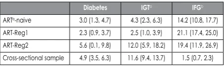
Prevalence and (95% CI) diabetes, IGT and IFG are listed in the Table I. Table I: Prevalence and (95% CI) diabetes, impaired glucose tolerance and impaired fasting glucose
Diabetes IGTa IFGb
ARTa-naive 3.0 (1.3, 4.7) 4.3 (2.3, 6.3) 14.2 (10.8, 17.7) ART-Reg1 2.3 (0.9, 3.7) 2.5 (1.0, 3.9) 21.1 (17.4, 25.0) ART-Reg2 5.6 (0.1, 9.8) 12.0 (5.9, 18.2) 19.4 (11.9, 26.9) Cross-sectional sample 4.9 (3.5, 6.3) 11.6 (9.4, 13.7) 1.5 (0.7, 2.3)
a = impaired glucose tolerance, b = impaired fasting glucose, c = antiretroviral therapy
On preliminary logistic regression analysis, using ART-naive as a reference group, age, male gender, BMI > 30 kg/m,2 and ART-Reg2 were positively associated with dysglycaemia, and being assumed HIV negative, i.e. the participants from the community survey, was protective.
Conclusion: Previously undiagnosed dysglycaemia is common in this
community, especially in subjects on regimen 2.
SaO4. Ethnic differences in ectopic fat and associations with
insulin sensitivity in black and white South African
women
JH Goedecke,1,2 C Weinreich,2 J Fan,2 J Hauksson,3 H Victor,2 K Utzschneider,4 D Keswell,2 NS Levitt,2 SE Kahn,4 T Olsson3 1Medical Research Council
2University of Cape Town 3Umeå University, Sweden
4VAPSHCS/University of Washington, USA
Abstracts
24 2012 Volume 17 No 1 JEMDSA
SaO5. Physical activity is positively associated with a lowered
metabolic risk in black South African women
K Dickie, S Chantler, LK Micklesfield, EV Lambert, J Evans, CL Jennings, Y Joffe, JH Goedecke
UCT/MRC Research Unit for Exercise Science and Sports Medicine, Department of Human Biology, University of Cape Town
Background: The objective was to compare body composition and
metabolic risk outcomes between sufficiently and insufficiently active black South African women.
Method: Physical activity (minutes per week) was estimated using
the validated Global Physical Activity Questionnaire (GPAQ). Body composition (dual-energy X-ray absorptiometry and computerised tomography), blood pressure, fasting glucose, insulin and lipid levels, were measured in 231 black South African women (26 ± 7 years).
Results: Based on the GPAQ analysis guide cut-offs [World Health
Organization (WHO), 2010], 61.1% (95% CI: 54.4-67.4%) of the women were considered active (n = 141), whereas 38.9% (95% CI: 32.6-45.6%) were considered insufficiently active (n = 90). Body mass index [24.9 (22.2-33.8) vs. 33.9 (25.4-38.7) kg/m2, p-value < 0.001]; body fat [35 (28.6-43.7) vs. 42.2 (35-46.3%), p-value < 0.001]; and fasting serum insulin levels [7.9 [5-14) vs. 9.8 (5.8-16.9) mU/l, p-value = 0.04) were significantly lower, whereas high-density lipoprotein (HDL) cholesterol levels were higher [1.3 (1.1-1.6) vs. 1.2 (1-1.6) mmol/l, p-value = 0.017) in the sufficiently active, compared to the insufficiently active group, respectively.
Conclusion: Women who meet the WHO criteria for physical activity
had lower body fat and fasting insulin levels, and higher HDL cholesterol values, than those who did not meet the criteria. The promotion of increased daily physical activity among black South African women should be encouraged to reduce metabolic risk.
SaO6. Somatostatin analogues as the first-line therapy in
acromegaly
Annamaria Colao
Department of Molecular and Clinical Endocrinology and Oncology, Federico II University of Naples, Italy
SaO7. Dipeptidyl peptidase-4 inhibition is cardioprotective
and improves glucose homeostasis in obese,
insulin-resistant rats
B Huisamen, A Genis, E Marais, A Lochner Faculty of Health Sciences, Stellenbosch University
Department Biomedical Sciences, Division Medical Physiology
Currently, therapy based on glucagon-like peptide-1 (GLP-1) is one of the most promising treatments for type 2 diabetes. Because GLP-1 is rapidly degraded by dipeptidyl-peptidase-4 (DPP-4), research has focused on DPP-4 inhibitors to raise levels of GLP-1 which are attenuated in type 2 diabetes. We tested whether treatment of obese, pre-diabetic rats with cardiovascular pathology, with a DPP-4 inhibitor (PFK275-055) was cardioprotective.
Obesity was effected in diet-induced obese (DIO) Wistar rats for 12 weeks, after which half of the control-fed and DIO rats were treated orally with 10mg/kg/day PFK275-055. After four weeks of treatment, in conjunction with the obesity-inducing diet, animals were sacrificed, the blood collected, the body weight and intraperitoneal (IP) fat weight recorded, pancreata harvested, and isolated hearts perfused (Langendorff perfusion: infarct development after regional
ischaemia, recovery after low-flow ischaemia). The kinase profile was determined in the reperfusion phase. Ventricular myocytes were prepared (standard collagenase perfusion) to determine insulin sensitivity via (3H)-2-deoxyglucose accumulation.
GLP-1 levels were attenuated in DIO, and restored by treatment. Insulin levels were 49% higher in DIO, and lowered by treatment. DIO suppressed pancreatic beta vs. alpha cell ratio, and treatment partially restored this. There were no effects on weight, IP fat or blood glucose levels. DIO animals: 47.7 ± 4.6% infarct of area at risk vs. control = 30.0 ±3.7 and DIO treated = 29.8 ± 3.1; p-value < 0.05, n = 6/group. The ratio of phosphorylation or total PKB/Akt and extracellular signal-regulated kinase 42 (ERK42) was attenuated in DIO, and improved after treatment. Cardiomyocytes did not show insulin sensitisation.
Treatment of pre-diabetic animals with a DPP-4 inhibitor was cardioprotective, improved glucose homeostasis and RISK pathway profile on reperfusion after ischaemia.
SaO8. Raising the bar: liraglutide (glucagon-like peptide-1
analogues) and composite end-points
MAK Omar
Centre for Diabetes and Endocrinology, Overport, Durban
Background: Cardiovascular disease (CVD) is the commonest cause
of mortality in subjects with Type 2 diabetes. Other well established CVD risk factors e.g. obesity, hypertension and dyslipidaemia often co-exist with diabetes. Hypoglycaemia is now also recognised as a CVD risk factor.
Method: Data from the Lead MT programme were analysed to
evaluate the effects of liraglutide, a GLP-1 analogue, and other anti hyperglycaemic agents, on glycaemic control and certain CVD endpoints. The Lead TM programme, comprising 6 phase 3 clinical trials involving over 4000 Type 2 diabetic subjects compared liraglutide as monotherapy and as combination therapy with other agents, viz: metformin, glimepiride, rosiglitazone, insulin glargine and exenatide. Composite end-point evaluation was based on meta-analysis using logistic regression across Lead studies 1-6.
Results: A decrease in HBAIC associated with loss of weight was
significantly more common in those on liraglutide (78%) and exenatide (72%) compared to the other treatment groups. Composite end-point (1) comprising HBAIC < 7% + no weight gain + no confirmed hypoglycaemia was more common in the liraglutide group (OR 2.0-10.3) compared to the other groups. Composite end-point (2) comprising HBAIC < 7%, systolic BP < 130mmHg and no weight gain was also more common in the liraglutide group.
Conclusion: Data from the Lead studies have shown that liraglutide
not only improves glycaemic control but has a beneficial effect on certain CVD composite end-points.
SaO9. The Bernard Pimstone Memorial Lecture:
manipulating GPCRs to treat endocrine diseases
Robert MillarEdinburgh, United Kingdom
SaO10. Treatment of type 1 diabetes: biologics to bionics
David NathanAbstracts
25 2012 Volume 17 No 1 JEMDSA
Saturday 21 April 2012: Poster presentations
SaP1. tissue-nonspecific alkaline phosphatase inhibition
impairs lipid accumulation in rat adipose-derived
stromal cells
CL Bartlett,1 NJ Crowther,2 FS Hough,1 WF Ferris1 1 Stellenbosch University
2University of Witwatersrand
Tissue-nonspecific alkaline phosphatase (TNAP) is found in many cell types throughout the body, including bone, liver, kidney, neural and adipose tissue. TNAP has an established function in bone mineralisation, where it serves to hydrolyse inorganic pyrophosphate (Ppi) into inorganic phosphate (Pi), which, in combination with Ca2+ ions, forms the hydroxyapatite crystals present in mineralised bone. TNAP also serves as a marker of osteoblastic differentiation in preosteoblastic cells. However, previous results obtained in immortalised 3T3-L1 pre-adipocytes have shown that TNAP is also involved in adipogenesis, surprisingly. Therefore, we questioned whether TNAP activity was required for lipid accumulation in primary adipose-derived mesenchymal stromal cells (ADSCs), and if any possible association between TNAP activity and adipogenesis was dependent on the depot from which the cells were obtained. Consequently, ADSCs from the inguinal subcutaneous and visceral perirenal depots were cultured in standard adipogenic differentiation medium, in the presence and absence of the TNAP-specific inhibitors, levamisole, histidine and L-homoarginine, in order to determine whether blocking TNAP would have an effect on adipogenesis. Cells from the subcutaneous and visceral depots were compared, as preadipocytes from these depots have previously been found to have different adipogenic potential. Lipid accumulation was used as an indicator of adipogenesis, and measured by staining with Oil Red O. Preliminary results indicate that lipid accumulation in rat ADSCs, from both visceral and subcutaneous depots, was similarly impaired in the presence of each of the three TNAP inhibitors, with a slight additive effect on the inhibition of lipid accumulation, observed when the inhibitors were used together. The obtained results indicate that TNAP may play a role in adipogenesis in ADSCs.
SaP2. Glucocorticoid receptor-
α
mNRA is downregulated in
gluteal adipose tissue of black South African women
and associates, with increased metabolic risk
JH Goedecke,1,2 RH Stimson,3 DEW Livingstone,3 P Hayes,3 K Adams,2 JA Dave,2 D Keswell,2 H Victor,2 EV Lambert,2 NS Levitt,2 SE Kahn,4 T Olsson5 1Medical Research Council, 2University of Cape Town, 3University of Edinburgh, Scotland, 4VAPSHCS/University of Washington, USA, 5Umeå University, SwedenIncreased capacity for glucocorticoid regeneration in subcutaneous adipose tissue (SAT) by 11β-hydroxysteroid dehydrogenase-1 (11HSD1) is associated with obesity and associated risk factors. We hypothesised that downregulation of SAT 11HSD1 and/or glucocorticoid receptor-α (GRα) may explain differences in body fat distribution and metabolic risk between black and white women. The study aimed to compare the expression of 11HSD1 and GRα, and glucocorticoid-responsive genes in gluteal SAT depots, and determine their relationships with body composition and metabolic risk factors in South African women. Body fat (DXA) and distribution (computerised tomography),insulin sensitivity (SI, intravenous glucose tolerance test), and the expression of 11HSD1, GRα, peroxisome proliferator-activated receptor γ (PPARγ), adiponectin, CD68 and tumour necrosis factor α (TNFα),
were measured in gluteal SAT of 56 normal-weight and obese black and white premenopausal South African women. 11HSD1 expression was increased with obesity in both black and white women (p-value < 0.001), but did not differ by ethnicity. In contrast, GRα mRNA levels were significantly lower in both normal weight and obese black, compared to white women (0.86 ± 0.25 vs. 1.31 ± 0.65 AU, and 0.52 ± 0.21 vs. 0.91 ± 0.26 AU, respectively, p-value < 0.01). Lower GRα expression in black women was associated with increased CD68 (r = -0.64, p-value < 0.001) and TNFα (r = -0.39, p-value < 0.01), reduced PPARγ (r = 0.84, p-value < 0.001) and adiponectin mRNA levels (r = 0.47, p-value < 0.001), as well as increased fat mass (r =- 0.61, p-value = 0.001) and serum triglycerides (r = -0.43, p-value = 0.022), and reduced high-density lipoprotein-cholesterol (r = 0.48, p-value = 0.010) and SI (r = 0.47, p-value = 0.016). In conclusion,expression of GRα is downregulated in gluteal SAT of black South African women, and associates with reduced adipogenic capacity and increased metabolic risk factors.
SaP3. The efficacy of Prosopis glandulosa as an
anti-diabetic treatment in rat models of diabetes and
insulin resistance
Cindy George, Barbara Huisamen Stellenbosch University
Background: In recognition of the increased incidence of diabetes
mellitus, untested agents are flooding the market. DiaviteTM (pods of Prosopis glandulosa) is marketed as a glucose-stabilising supplement. The aim of this study was to determine the efficacy of P. glandulosa as an anti-diabetic agent.
Method: Male Wistar rats were rendered type 1 diabetic with
streptozotocin (STZ) and insulin-resistant by diet. Half the animals were placed on P. glandulosa treatment (100 mg/kg/day) for eight weeks, and the remaining animals served as controls. At the time of sacrifice, blood was collected for glucose and insulin level determination, the pancreata of the STZ rats were harvested for histological analysis, and cardiomyocytes and skeletal muscle strips prepared for insulin sensitivity determination.
Results: In the type 1 diabetic model, P. glandulosa ingestion resulted
in significant increased insulin levels (p-value < 0.001), accompanied by decreased glucose levels (p-value < 0.05). Additionally, P. glandulosa ingestion resulted in increased small β-cells (p-value < 0.001) in the pancreata. Treatment also partially prevented the weight loss observed after STZ injection. In the insulin resistant model, P. glandulosa ingestion resulted in increased basal (p-value < 0.01) and insulin-stimulated (p-value < 0.05) glucose uptake in cardiomyocytes. It also increased insulin sensitivity (p-value < 0.05) in skeletal muscle from control animals.
Conclusion: This study showed that P. glandulosa treatment