R E S E A R C H
Open Access
Co-inhibition of HDAC and MLL-menin
interaction targets MLL-rearranged acute
myeloid leukemia cells via disruption of
DNA damage checkpoint and DNA repair
Jing Ye
1†, Jie Zha
1†, Yuanfei Shi
1†, Yin Li
2, Delin Yuan
1, Qinwei Chen
1, Fusheng Lin
1, Zhihong Fang
1, Yong Yu
3*,
Yun Dai
4*and Bing Xu
1*Abstract
While the aberrant translocation of the mixed-lineage leukemia (MLL) gene drives pathogenesis of acute myeloid leukemia (AML), it represents an independent predictor for poor prognosis of adult AML patients. Thus, small molecule inhibitors targeting menin-MLL fusion protein interaction have been emerging for the treatment of MLL-rearranged AML. As both inhibitors of histone deacetylase (HDAC) and menin-MLL interaction target the transcription-regulatory machinery involving epigenetic regulation of chromatin remodeling that governs the expression of genes involved in tumorigenesis, we hypothesized that these two classes of agents might interact to kill MLL-rearranged (MLL-r) AML cells. Here, we report that the combination treatment with subtoxic doses of the HDAC inhibitor chidamide and the menin-MLL interaction inhibitor MI-3 displayed a highly synergistic anti-tumor activity against human MLL-r AML cells in vitro and in vivo, but not those without this genetic aberration. Mechanistically, co-exposure to chidamide and MI-3 led to robust apoptosis in MLL-r AML cells, in association with loss of mitochondrial membrane potential and a sharp increase in ROS generation. Combined treatment also disrupted DNA damage checkpoint at the level of CHK1 and CHK2 kinases, rather than their upstream kinases (ATR and ATM), as well as DNA repair likely via homologous recombination (HR), but not non-homologous end joining (NHEJ). Genome-wide RNAseq revealed gene expression alterations involving several potential signaling pathways (e.g., cell cycle, DNA repair, MAPK, NF-κB) that might account for or contribute to the mechanisms of action underlying anti-leukemia activity of chidamide and MI-3 as a single agent and particularly in combination in MLL-r AML. Collectively, these findings provide a preclinical basis for further clinical investigation of this novel targeted strategy combining HDAC and Menin-MLL interaction inhibitors to improve therapeutic outcomes in a subset of patients with poor-prognostic MLL-r leukemia.
Keywords:MLL-rearrangement, Menin-MLL interaction inhibitor, HDAC inhibitor, DNA damage response, AML
© The Author(s). 2019Open AccessThis article is distributed under the terms of the Creative Commons Attribution 4.0 International License (http://creativecommons.org/licenses/by/4.0/), which permits unrestricted use, distribution, and reproduction in any medium, provided you give appropriate credit to the original author(s) and the source, provide a link to the Creative Commons license, and indicate if changes were made. The Creative Commons Public Domain Dedication waiver (http://creativecommons.org/publicdomain/zero/1.0/) applies to the data made available in this article, unless otherwise stated.
* Correspondence:yy3@tongji.du.cn;daiyun@jlu.edu.cn;
xubingzhangjian@126.com
†Jing Ye, Jie Zha and Yuanfei Shi contributed equally to this work. 3
Clinical and Translational Research Center of Shanghai First Maternity and Infant Hospital, School of Life Sciences and Technology, Tongji University, Shanghai, China
4Laboratory of Cancer Precision Medicine, The First Hospital of Jilin
University, Changchun, China
1Department of Hematology, The First Affiliated Hospital of Xiamen
University and Institute of Hematology, School of Medicine, Xiamen University, Xiamen, China
Introduction
Cytogenetic abnormalities are closely associated with clin-ical features and therapeutic responses in acute myeloid leukemia (AML) [1]. Chromosome 11q23 translocations occur in 10% of adult AML patients, while being even more frequent in pediatric cases (35%) [2–4]. In AML, most of 11q23 translocations led to fusion proteins involv-ing the mixed lineage leukemia (MLL) gene that encodes histone lysine methyltransferase 2A (KMT2A) [5], of which more than 70 translocation partners of MLL have been characterized so far [6, 7]. The clinical outcome of patients carrying MLL-rearrangement who often suffer from either failure of induction therapy or disease relapse remains extremely poor, while the response rate reported in adult MLL-rearranged (MLL-r) AML is approximately 40% [8]. Although dose intensification of chemotherapy might reduce the risk of relapse, it is however associated with the long-term adverse effects and a high rate of treatment-related mortality [9]. Therefore, a more effect-ive and less toxic therapy is urgently needed to treat this subset of AML with poor prognosis.
Chidamide, a novel histone deacetylase (HDAC) in-hibitor of the benzamide class that specifically inhibits HDAC1-3, has approved by the Chinese FDA for treat-ment of relapsed or refractory peripheral T cell lymph-oma (PTCL) [10,11]. Recently, several groups including ours have demonstrated that chidamide displays promis-ing activity against various cancer types, especially hematological malignancies including AML [12–14]. It has been well established that HDACs, of which at least 18 members have been characterized, play the key roles in the epigenetic regulation of gene expression through chromatin remodeling by inhibiting histone deacetyla-tion [15, 16]. Among them, HDAC1, 2 and 3 are fre-quently overexpressed in human leukemia [17], which could interact with MLL fusion partners and result in aberrant regulation of chromatin remodeling and thus the expression of tumor-driven genes [18]. Earlier stud-ies have demonstrated that chidamide synergizes with conventional chemotherapeutic agents in human leukemia cells by disrupting cell cycle progression and DNA damage responses, as well as promoting ROS-dependent apoptosis [19]. However, because the single-agent activity of chidamide towards relapsed or refrac-tory AML appears not to be satisfied, the combination strategy involving chidamide thus warrants investigation in this disease.
Menin, which is encoded by the multiple endocrine neoplasia 1 (MEN1) gene, is known to interact directly with the N-terminal domain of MLL. This interaction is required for the activation of target gene expression by the MLL-fusion proteins, thereby critical for their cap-ability to mediate transformation [20]. Menin is also in-volved in DNA damage response (DDR), particularly
DNA repair. For example, Menin is accumulated with CHK1 at the sites of double-strand break (DSB) [21]. Thus, the menin-MLL interaction has recently been con-sidered as a potential therapeutic target in MLL-r leukemia [22]. In this context, it raises a possibility that the specific inhibitors targeting the menin-MLL inter-action may block MLL fusion protein-mediated leukemic transformation by down-regulating the expression of MLL target genes, an event that is required for the onco-genic activity of MLL fusion proteins. Indeed, this type of small molecule inhibitors has displayed a single-agent anti-proliferative activity in vitro and in vivo in a model of MLL-r leukemia [23]. However, for most targeted agents, the single-agent therapy is unlikely to be curative in AML, primarily due to co-existing perturbations in-volving multiple leukemogenic signaling pathways in this highly heterogeneous disease. It is therefore anticipated that menin-MLL interaction inhibitors might be more effective in rational combination regimens.
To this end, we sought to examine whether and how the menMLL interaction inhibitor MI-3, which acts to in-hibit transcription of MLL target genes, would interact with the HDAC inhibitor chidamide, a class of epigenetic agents that regulate gene expression through chromatin remodel-ing, in MLL-r AML. Here, we reported that simultaneous inhibition of both HDAC and MLL-menin interaction ex-hibits a synergistic cytotoxic activity in vitro specifically against AML cells carrying MLL-rearrangement. This com-bination regimen was also active in vivo (e.g., delayed tumor progression and reduced tumor burden) in an MLL-r AML xenograft mouse model. Together, these findings argue that this novel regimen rationally combining the HDAC inhibi-tor chidamide and the MLL-menin inhibiinhibi-tor MI-3 might represent a promising option for treatment of MLL-r AML.
Results
Chidamide synergistically interacts with MI-3 to
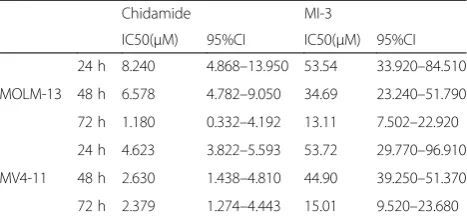
suppresses the growth of MLL-rearrangement AML cells We first examined the activity of chidamide and MI-3 alone and in combination to determine whether these two agents would synergistically interact with each other in inhibition of MLL-r AML cell viability. To this end, after exposing to a series of concentrations of chidamide or MI-3 for 24, 48, and 72 h, viability of MOLM-13 and MV4-11 cells, both lines carrying MLL-rearrangement, were determined by the CCK-8 analysis. As shown in Additional file1: Figure S1A–D, exposure to either agent resulted in a marked increase in cytotoxicity towards these MLL-r AML cells in a dose- and time-dependent manner. Based on the single-agent activity after treated for 24, 48, and 72 h, the IC50 values for chidamide and
each agent) resulted in a sharp increase in inhibition rate of cell viability in MOLM-13 (Fig. 1a) and MV4-11 cells (Fig. 1b) at 24, 48, and 72 h. Moreover, the combination index (CI) < 1.0 indicated a synergistic interaction be-tween chidamide (1.6–8.2 μM) and MI-3 (10.6–53.5 μM) in MOLM-13 (Fig. 1c), as well as between chida-mide (0.9–4.6μM) and MI-3 (10.7–53.7μM) in MV4-11 (Fig.1d). Values of fraction affected (Fa) and CI for each cell line after treatment for 24, 48, and 72 h were pro-vided in Additional file 2: Table S1, of which the CI values ranged from 0.4 to 0.8. In contrast, the human AML cell lines Kasumi-1 and KG1α without MLL-rearrangement were much less sensitive to this regimen combining chidamide and MI-3 in the same range of concentrations (Additional file 1: Figure S2A–C). To-gether, these results indicate that chidamide synergistic-ally interacts with MI-3 to reduce the viability of AML cells carrying MLL-rearrangement. They also raise a possibility that AML cells carrying MLL-rearrangement might be particularly susceptible to this combination regimen.
Co-exposure to chidamide and MI-3 induces apoptosis of MLL-rearrangement AML cells, in association with increased ROS generation and mitochondrial injury To validate the synergistic effect of the regimen combining chidamide and MI-3 on MLL-r AML cells, the colony for-mation assay was performed. As shown in Fig.2a, whereas chidamide (2.6μM) and MI-3 (13.9μM) displayed moder-ate single-agent activity, a significant reduction in colony formation was observed in MOLM-13 cells after combined treatment, compared with these two agents alone. Analo-gous results were obtained from MV4-11, another MLL-r AML cells (Additional file1: Figure S3A). Moreover, flow cytometry with Annexin V/PI staining was then performed to examine whether chidamide would interact with MI-3 to induce apoptosis in MLL-r cells. After exposing to chida-mide and MI-3 alone or in combination for 48 h, the per-centage of apoptotic (Annexin V-positive) cells was significantly increased in MOLM-13 (Fig.2b) and MV4-11 cells (Additional file 1: Figure S3B), compared to each
single agent. As loss of mitochondrial membrane potential (MMP) plays a crucial role in the initiation of intrinsic mitochondrion-dependent apoptotic cascade [25], we next examined the effect of chidamide and MI-3 individually or in combination on MMP. Consistent with the results for apoptosis, combined treatment with chidamide and MI-3 also induced loss of MMP, reflected by impaired mitochon-drial depolarization indicated by markedly decreased fluor-escence intensity ratio between JC-1 aggregate and monomer (Fig. 2c and Additional file 1: Figure S3C). To unveil the potential mechanism underlying the synergistic interaction between these two agents in the induction of apoptosis, flow cytometry was carried out to monitor intra-cellular ROS levels. After co-treated with chidamide and MI-3 for 48 h, a significant increase in ROS generation was observed in MOLM-13 (Fig. 2d) and MV4-11 cells (Add-itional file1: Figure S3D), compared to treatment with each single agent. Together, these results suggest that chidamide interacts synergistically with MI-3 to induce apoptosis of AML cells carrying MLL-rearrangement via promotion of ROS production and mitochondrial damage.
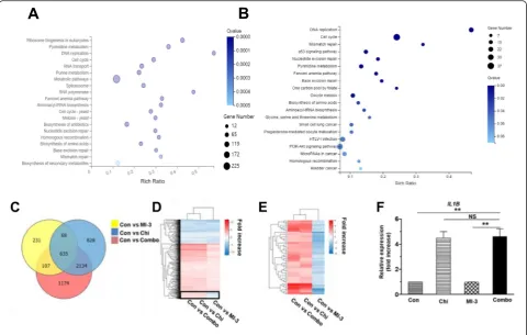
Co-treatment with chidamide and MI-3 alters genome-wide gene expression in MLL-rearrangement AML cells Anti-tumor activity of both HDAC inhibitor and menin-MLL interaction inhibitor involves transcriptional regu-lation of gene expression [26–29]. To further understand the mechanism of action underlying the synergistic interaction between chidamide and MI-3 in AML-carrying MLL-rearrangement, the RNAseq assay was then performed to profile genome-wide gene expression in MOLM-13 cells after treated with chidamide and MI-3 alone or in combination (see Additional file 2: Table S2 for a list of all genes). After combined treatment with chidamide and MI-3 for 24 h (Fig.3a) and 48 h (Fig.3b), the KEGG analysis indicated that the most significantly altered pathways involving cell cycle, DNA replication, and several DNA repair mechanisms [e.g., homologous recombination (HR), nucleotide excision repair, base ex-cision repair, mismatch repair, and Fanconi anemia pathway]. Further, the GESA analysis further revealed that most of these alterations stemmed from chidamide, rather than MI-3 (Additional file 1: Figure S4A–C). Moreover, the analysis of the dataset (48 h treatment) for the significantly downregulated or upregulated tran-scripts (log2FC ≥ 1,Q value≤ 0.001) revealed gene ex-pression profile (GEPs) for chidamide (blue) and MI-3 (yellow) alone or in combination (red) with an overlap of 635 genes (Fig. 3c). A majority of these overlapped genes displayed the same trends of expression (i.e., up-or downregulation) in MOLM-13 cells treated with MI-3 and chidamide alone or in combination (Fig. 3d). However, there was a small cluster of 59 genes (indicated by square in Fig. 3d) that were differentially expressed in
Table 1IC50 values of chidamide and MI-3 as single agent in AML cells
Chidamide MI-3
Fig. 1The HDAC inhibitor chidamide synergistically interacts with the menin-MLL interaction inhibitor MI-3 to inhibit cell viability in MLL-r AML cells.a,bHuman MLL-rearranged AML cell lines MOLM-13 (a) and MV4-11 cells (b) were exposed to the indicated concentrations (μM) of chidamide ± MI-3 (μM) for 24, 48, and 72 h, after which cell viability was examined using the CCK-8 assay. Values indicate mean ± SEM for at least three independent experiments performed in triplicate. *P< 0.05, **P< 0.01, and ***P< 0.001 for comparison with each single agent.c,dMOLM-13 (c) and MV4-11 (d) cells were treated as described in Additional file
2: Supplemental Table 1, followed by the analysis of cell viability as above, after which the CompuSyn analysis was performed to determine whether the interaction between these two agents is synergistic (CI value < 1.0)
MOLM-13 cells exposed to MI-3 (downregulation) versus chidamide alone or in combination (upregulation; Fig. 3e). The gene ontology (GO; Additional file1: Figure S4D) and KEGG (Additional file1: Figure S4E) analyses revealed that these genes were associated with several key survival signal-ing pathways (e.g., MAPK, NF-κB). They also suggested that cytokine signaling pathways (e.g., TNF), which is essential for the inflammatory reaction [30] and known to be involved in single-agent activity of chidamide [31], might also be in-volved in the interaction between chidamide and MI-3 in MLL-r AML cells. In this context, the real-time PCR analysis was performed to validate the expression of IL1Bthat en-codes the pro-inflammatory cytokine IL-1β, a representative gene selected from 59 overlapping genes described in Fig.3e. Consistent with the RNAseq results, exposure of MOLM-13 cells to chidamide in the absence or presence of MI-3 re-sulted in a marked increase in expression ofIL1B(Fig. 3f). However, qPCR failed to detect whether MI-3 alone down-regulated the expression ofIL1B, due to its low basal level in untreated cells. Together, these results suggest that the
mechanism of action underlying anti-leukemic activity of the combination treatment with chidamide and MI-3 might in-volve DDR in MLL-r AML cells. They also raise the possibil-ity that co-administration of chidamide might reactivate a set of genes that are silenced by MI-3.
Co-treatment with chidamide and MI-3 disrupts DNA damage response and results in DNA damage in AML-carrying MLL-rearrangement
We then performed a gene set enrichment analysis (GSEA) of differentially expressing genes in MOLM-13 cells after treated with chidamide and MI-3 alone or in combination. Four genes were identified, which met both of the following criteria, including (a) at least 2-fold downregu-lation by chidamide or MI-3 and (b) at least 4-fold down-regulation by combined treatment. These genes included SULT1A3 (sulfotransferase 1A3/1A4), AEBP1 (transcrip-tional repressor),CCNE2(cyclin E2), andATF5 (transcrip-tion factor; Fig.4a and Additional file1: Figure S5). Among them, SULT1A3 is known to catalyze sulfation of Fig. 2Co-treatment with chidamide and MI-3 induces robust apoptosis, in association with increased ROS generation and loss of mitochondrial membrane potential.aMOLM-13 cells were treated with 2.63μM chidamide ± 13.88μM MI-3 for 48 h, followed by the colony formation assay.
nitrotyrosine, which is associated with DNA damage [32, 33]. Since the RNAseq analysis after treatment for either 24 or 48 h (Additional file1: Figure S4A and S4B) suggested a potential role of DNA damage response (DDR) in the anti-tumor activity of the regimen combining chidamide and MI-3, qPCR analysis was performed to validate expression ofSULT1A3 as representative. Consistent with the results of the RNAseq analysis (Fig. 4a), exposure to either chida-mide or MI-3 was able to downregulateSULT1A3 expres-sion, while the combination treatment resulted in a greater reduction in its expression (Fig.4b). In this context, western blot analysis was further carried out to examine the DDR signaling pathway, which is orchestrated by ATM and ATR as well as their key downstream checkpoint kinases CHK1 and CHK2. As shown in Fig.4c and Additional file1: Figure S6, exposure to chidamide in the presence or absence of MI-3 led to increased acetylation of histone H3 in various AML cell lines, due to inhibition of its deacetylation
catalyzed by HDACs. Interestingly, treatment with MI-3 also modestly increased H3 acetylation in MOLM-13 cells (Fig.4c), but not in MV4-11 or Kasumi-1 cells (Additional file 1: Figure S6), suggesting a cell line-specific phenomenon. Notably, combined treatment with these two agents clearly induced phosphorylation (activation) of both ATM and ATR. In contrast, whereas exposure to either MI-3 or in a lesser extent chidamide attenuated phosphoryl-ation (activphosphoryl-ation) of both CHK1 and CHK2, these events were almost completely inhibited by co-administration of these two agents (Fig.4c). Combined treatment also mark-edly downregulated the expression of Rad51, an important DNA repair protein [34], while did not affect the levels of another DNA repair protein KU70. As a consequence, the combination treatment induced robust DNA damage, man-ifested by a sharp increase in expression of γH2A.X (Fig. 4c), a marker of DNA double-strand break [35]. Together, these results suggest that disruption of the DNA damage Fig. 3RNA sequencing reveals genome-wide gene expression profiles of MOLM-13 cells treated with chidamide and MI-3 alone or in combination.a,b MOLM-13 cells were treated with 2.6μM chidamide ± 13.9μM MI-3 for 24 h (a) or 48 h (b), after which total RNA was extracted and subjected to whole exome RNAseq that was performed in triplicate for each condition. The KEEG analysis reveals the annotations of the most enriched pathways of differentially expressing genes after combined treatment for 48 h, compared to untreated control.c–eAlternatively, a Venn diagram shows the number of genes and their relationship that were differentially expressed (log2FC≥1,Qvalue≤0.001) after treatment with chidamide and MI-3 alone or in combination, compared to untreated control (c). A heatmap shows hierarchical clustering of 635 genes significantly affected by all three treatments, including chidamide, MI-3, and combined treatment (d). A heatmap shows hierarchical clustering of 59 genes (indicated by square in panel B) that were upregulated by chidamide and combined treatment, but downregulated by MI-3 (e).fReal-time PCR analysis was performed to monitor expression ofIL1B, one of 59 genes shown in panelc, in MOLM-13 cells after treated with chidamide ± MI-3 for 48 h. The reaction was carried out in triplicate and relative expression levels were calculated as 2−△△CTafter normalization to
β -actin. Values indicate mean ± SEM for at least three independent experiments performed in triplicate (**P< 0.01, NS = not significant)
checkpoint through inactivation of CHK1 and CHK2, rather than their upstream kinases ATM and ATR, as well as interfere with the DNA repair machinery by downregulating DNA repair proteins (e.g., Rad51), might account for or at least contribute to the synergistic interaction between chi-damide and MI-3 in AML cells carrying MLL-rearrangement.
The regimen combining chidamide and MI-3 is active in vivo in a xenograft model of AML-carrying MLL-rearrangement
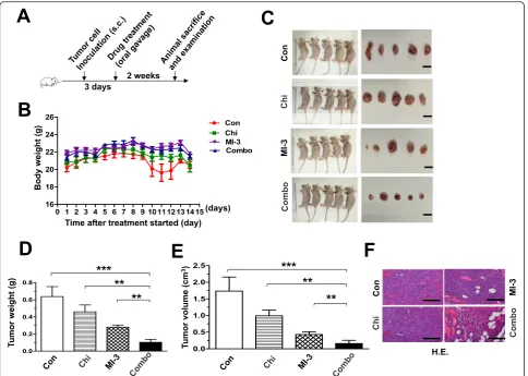
Last, anti-tumor activity of the regimen combining chida-mide and MI-3 was examined in a mouse xenograft model established by subcutaneous inoculation with MLL-r MOLM13 cells. After 3 days, mice were randomly divided into four groups, including vehicle control, chidamide, MI-3, and the combination treatment (Fig.5a). Although a moderate reduction in body weight was observed at days
10–12 after treatment with chidamide and MI-3 in com-bination, but rapidly recovered at day 14 (Fig.5b). Other-wise, no other signs of notable toxicity were observed. Significantly, combined treatment with chidamide and MI-3 resulted in a marked reduction in tumor burden, reflected by decreased volume and weight of tumor masses, compared to vehicle control as well as each indi-vidual agent (Fig.5c–e). These results were also confirmed by histological examination (Fig.5f). Together, these find-ings indicate that the combination regimen of chidamide and MI-3 is effective in vivo against MLL-r AML, while well tolerated.
Discussion
property [36]. This subset of AML is particularly aggres-sive and often associated with poor prognosis, primarily due to the lack of effective treatment [37, 38]. Menin acts as a key cofactor of oncogenic MLL fusion proteins [39]. For example, menin is required for the mainten-ance of HOX gene expression mediated by MLL fusion proteins [28]. In this context, small molecule inhibitors targeting the interaction between menin and MLL fusion proteins are recently emerging to treat AML carrying MLL-rearrangement. Indeed, these inhibitors are able to reverse MLL fusion protein-mediated leukemic trans-formation via downregulation of HOX genes. Therefore, this novel class of anti-cancer agents has recently attracted a lot of interests in treatment of poorly prog-nostic AML-bearing MLL-rearrangement. However, al-though earlier studies have demonstrated promising activity of several menin-MLL interaction inhibitors in vivo in MLL-r AML xenograft mouse models, thera-peutic responses remain limited. To this end, our
observations in the present study indicate that a rationale-based regimen combining these inhibitors (e.g., MI-3) and HDAC inhibitors (e.g., chidamide) might display signifi-cantly increased anti-tumor activity towards AML carry-ing MLL-rearrangement, compared to each scarry-ingle agent, at least in the preclinical setting.
The rationale for combining inhibitors of HDAC and menin-MLL interaction is laid on their properties target-ing transcription-regulatory machinery. On the one hand, HDACs regulate gene expression via chromatin remodeling [26]. They function to “tighten” or “close” chromatin structure by deacetylating nucleosomal his-tones (e.g., H3 and H4) to reduce the accessibility of transcription factors [27]. In contrast, HDAC inhibitors act to“loose”or“open”chromatin structure through in-hibition of histone deacetylation by HDACs, which al-lows transcription factors to access and binding to promoters of target genes, thereby initiating and pro-moting their expression [41–43]. Notably, the implication Fig. 5Co-administration of chidamide and MI-3 suppresses tumor growth in a xenograft model of MLL-r AML.aA scheme for the animal study that are described in detail in the“Materials and methods”section.bMice were weighed daily after tumor cell inoculation.c–eMice were sacrificed after drug treatment for 14 days, and then the images of mice and tumors were captured (c). Weight (d) and volume (e) of tumors were measured and calculated. Values indicate mean ± SEM for 6 mice/each group (**P< 0.01, ***P< 0.001).fTumors sections were prepared and stained by H&E for histological examination
of HDACs in AML has promoted the attempt to use the HDAC inhibitors to treat this disease [44, 45]. Moreover, leukemia-carrying MLL-rearrangement is highly susceptible to HDAC inhibition [46]. On the other hand, MLL methyl-ates histone H3 to upregulate expression of target genes, in-cluding several HOX genes [28, 29]. In MLL-rearranged AML, certain MLL fusion proteins display enhanced activ-ity to promote gene transcription by recruiting a transcrip-tional activation complex known as P-TEFb that consists of CDK9 and cyclin T1 [47,48]. The leukemia-driving activity of MLL fusion proteins relies on their interaction with menin, a protein encoded by the multiple endocrine neo-plasia (MEN1) gene [49]. Menin directly binds to the N-terminal domain of MLL in virtually all MLL fusion pro-teins, an event that is essential for leukemic transformation [50]. In this context, small molecule inhibitors (e.g., MI-3) specifically targeting the interaction between menin and MLL fusion proteins have recently been emerging to treat acute leukemias harboring MLL-rearrangement [51]. Therefore, as the mechanisms of action for both HDAC and menin-MLL interaction inhibitors involve transcription-regulatory machinery, a possibility then arose that these two classes of agents may interact synergistically in MLL-rearranged leukemia. Indeed, the results of the present study demonstrated a highly synergistic interaction between chidamide and MI-3 at their subtoxic dose ranges in human AML cells carrying MLL-rearrangement (e.g., MOLM-13 and MV4-11), but not in cells that did not har-bor this genetic abnormality. The anti-tumor activity of this combination regimen was further validated in vivo in a mouse xenograft model of human MLL-r AML. In addition, marked inhibition of the colony-forming activity of MLL-r AML cells by the combination treatment raises the possibility that this regimen might also suppress self-renewal of leukemic stem cells.
Because both HDAC and menin-MLL interaction inhibitors target general transcription-regulatory ma-chinery [40, 52], they may thus affect genome-wide ex-pression of numerous target genes. To this end, the RNAseq analysis revealed that exposure to chidamide and MI-3 alone influenced expression of 3665 and 1041 genes in MLL-r AML cells, respectively, while combined treatment resulted in altered expression of 4050 genes. Moreover, although treatment with chidamide and MI-3 alone or in combination led to global changes in gene expression, a set of 635 genes that were overlapped among these three treatment conditions was observed. Among them, a small cluster of 59 genes was oppositely regulated by MI-3 and chidamide (either alone or in combination). The functions of these differentially expressed genes were involved in several signaling pathways (e.g., MAPK, NF-κB) crucial for tumor cell survival and proliferation [30,53]. In addition, the roles of these genes also involved the inflammatory responses (e.g., the
TNF-signaling pathway). However, the effect of the combination treatment on inflammatory responses (e.g., expression of IL-1β, a well-established marker of inflammation) [54, 55] seems to solely stem from chidamide, rather than MI-3, thereby arguing against that this effect represents the pri-mary mechanism for the high synergy between these two agents. Therefore, an alternative strategy for further analysis of this RNAseq dataset might be required to identify other candidate pathways and targets responsible for or involved in the synergistic interaction between chidamide and MI-3 in MLL-r AML.
In the present study, bulk evidence indicates that the anti-tumor activity of the regimen combining chidamide and MI-3 towards MLL-r AML was associated with in-duction of apoptosis, primarily via the intrinsic mitochondrion-dependent pathway [36, 56]. First, while exposure to chidamide led to the loss of mitochondrial membrane potential, reflecting mitochondrial injury [57, 58], this event was significantly potentiated by co-administration of MI-3. Second, as mitochondria are considered as the main source of ROS in the cell [59, 60], a robust increase in ROS generation was observed in MLL-r AML cells exposed to both chidamide in the presence of MI-3. Last, flow cytometric analysis revealed that whereas treatment with either chidamide or MI-3 moderately increased apoptotic (Annexin V-positive) cells, this event was dramatically potentiated after co-administration of these two agents. Therefore, these ob-servations argue strongly that chidamide might interact with MI-3 to activate the mitochondrion-related apop-totic signaling cascade. However, due to the multifacial functions of HDAC inhibitors, a caveat that other mech-anisms might also contribute to the anti-leukemia effect of chidamide alone and even in combination with MI-3 in MLL-r AML cells could not be excluded.
together, these results suggest that this combination regi-men appears to target primarily on CHK1 and CHK2, ra-ther than their upstream kinases (ATR and ATM), while activation of the latter might reflect a feedback response to inactivation of CHK1 and CHK2 after co-exposing to chi-damide and MI-3. In addition, the combined treatment also downregulated Rad51, a DNA repair protein that plays a major role in homologous recombination (HR) repair of DSB [34]. However, it did not affect the levels of Ku70, an-other DNA repair protein critical for non-homologous end joining (NHEJ) repair of DSB [68]. These observations sug-gest that the regimen combining chidamide and MI-3 might selectively target DNA repair via HD, rather than NHEJ. Interestingly, the GSEA analysis of the RNAseq dataset identifiedSULT1A3as one of four genes that were downregulated by chidamide and MI-3 alone or in combin-ation, which was confirmed by real-time PCR analysis. SULT1A3 encodes an enzyme involved in the metabolism of nitrotyrosine, while increased levels of nitrotyrosine serve as a biomarker of oxidative stress that induces DNA dam-age [32, 33]. Thus, this finding raises the possibility that SULT1A3might represent a potential target that links oxi-dative stress (e.g., ROS) and DNA damage together in MLL-r AML cells co-treated with MI-3 and HDAC inhibi-tors. However, further studies are required to address this possibility.
In summary, the findings of the present study dem-onstrate a highly synergistic interaction between the HDAC inhibitor chidamide and the menin-MLL inter-action inhibitor MI-3 both in vitro and in vivo in AML cells with MLL-rearrangement. They also pro-vide epro-vidence for the potential mechanisms underlying the markedly increased anti-leukemia activity of this combination regimen, including ROS generation, apoptosis induction via the mitochondrion-dependent signaling pathway, and disruption of DDR (e.g., DNA damage checkpoint and DNA repair via HR). In addition, the genome-wide gene expression profile (GEP) by the RNAseq analysis might serve as a re-source for future studies to identify potential targets (e.g., SULT1A3) and pathways to further understand the mechanisms of action for these two classes of agents in AML carrying MLL-rearrangement. Due to the current lack of effective therapy for the treatment of MLL-rearranged AML, the strategy combining HDAC and menin-MLL interaction inhibitors war-rants further investigation in this poorly prognostic subset of AML.
Material and methods Reagents
The HDAC inhibitor chidamide (CS055, purity > 95%) was kindly provided by Chipscreen Bioscience Ltd.
(Shenzhen, China). The MLL-menin interaction inhibi-tor MI-3 (purity = 99.03%; Cat. No. S7619) was pur-chased from Selleck Chemicals (Houston, TX, USA). The reagents were dissolved in dimethyl sulfoxide (DMSO; Invitrogen, Carlsbad, CA, USA) as 10 mM stock solution and stored at−20oC, which was then di-luted to the required concentrations with cell culture medium prior to experiments.
Cell culture and sorting
Human AML cell lines MOLM-13 and MV4-11 carrying MLL-rearrangement were purchased from ATCC (Rockefeller, MD, USA) and cultured at 37 °C in a 5% CO2incubator in Iscove’s modified Dulbecco’s medium
(IMDM) and RPMI-1640 medium (HyClone, Thermo Scientific, Logan, UT, USA) supplemented with 10% fetal bovine serum (FBS, Gibco, Thermo Scientific, Grand Island, NY, USA), respectively. For sorting CD34+/CD38−cells, cells were stained with hCD34-APC (eBioscience, Thermo Scientific, San Diego, CA, USA) and hCD38-PE (eBioscience, Thermo Scientific, San Diego, CA, USA) for 30 min at 4 °C. After washed twice with PBS containing 1% FBS, CD34+/CD38− cells were sorted using FACS Aria IIU (Biosciences, Franklin Lakes, NJ, USA).
Analysis of cell proliferation and viability
Cytotoxicity was determined by using the Cell Count-ing Kit-8 (CCK-8, Dojindo, Kumamoto, Japan) [24]. Briefly, 2 × 10⁴/well cells were seeded in 100 μl medium on 96-well plates and treated with indicated concentrations of chidamide and MI-3 alone or in combination for 24, 48, and 72 h. The CCK-8 reagent (10 μl/well) was then added and incubated for add-itional 2 h, after which the absorbance at 450 nm was detected by a microplate reader (ELx800, BioTek, Wi-nooski, VT, USA). The data from three independent experiments in triplicate was presented as percentage of viable cells by comparing to untreated control. IC50 values were determined using the SPSS 20.0 software.
Analysis of apoptosis
Cells were cultured and treated chidamide with or with-out MI-3 for 24, 48, or 72 h as described above, followed by double staining with Annexin V-FITC and PI (eBioscience, Thermo Scientific, San Diego, CA, USA) for 15 min at room temperature in dark as per the man-ufacturer’s instruction. Cells were then analyzed by flow cytometry (FACS Fortessa, BD Biosciences, Franklin Lakes, NJ, USA). The percentage of Annexin V-positive (apoptotic) cells was determined.
Analysis of mitochondrial membrane potential
2 × 10⁵/ml cells were seeded in 24-well plates and treated with chidamide and MI-3 alone or in combin-ation at indicated concentrcombin-ations. After 24 h incubcombin-ation, cells were harvested and stained with 2 μM Rhodamine 123 (Byeotime, Shanghai, China) for 30 min at 37 oC. After washed, mitochondrial membrane potential (MMP) was analyzed by flow cytometry (FACS Fortessa).
Measurement of ROS
Intracellular ROS levels were determined using the Reactive Oxygen Species Assay Kit (no. S0033, Beyotime, Shanghai, China) as per the manufacturer’s instruction. Briefly, after treated with indicated concentrations of chidamide and MI-3 alone or in combination for 24 h, cells were harvested and stained with 10 μM 2′,7′-dichlorofluorescein diacetate (DCFH-DA, 1:1000 diluted in RPMI1640) for 20 min at 37 °C. After washed twice, intracellular ROS level was deter-mined by flow cytometry (FACS Fortessa).
Colony formation assay
2×10⁵/well cells at logarithmical growth phase were seeded in 24-well plates and treated with indicated con-centrations of chidamide and MI-3 alone or in combin-ation for 48 h. After washed, 500/well cells were then cultured in complete methylcellulose medium (R&D Sys-tems, Minneapolis, MN, USA) in 3.5 cm dishes for 14 days. Colonies consisting of at least 50 cells were counted and analyzed for colony-forming capability.
Western blot analysis
2×10⁵/well cells were treated with chidamide in the ab-sence or preab-sence of and/or MI-3 for 48 h, and the sub-jected to western blot analysis using indicated primary antibodies and secondary HRP-conjugated antibodies (1: 10,000, Abcam, Cambridge, UK). The primary antibodies included anti-caspase-3 (#9662S), anti-PARP (#9532S), anti-histone H3 (#4499S), anti-phospho-H3 (#53348S), anti-γH2A.X (#2577S), anti-RAD51 (#8875S), anti-KU70 (#4588S), anti-STAT3 (#9139 S), anti-Mcl-1 (#94296S), anti-phospho-p53 (#9286S), anti-p21(#2947S), phospho-ATM (#5803S), ATM (#2873S), anti-phospho-ATR (#2835S), anti-CHK1 (#2360), anti-CHK2 (#2662), anti-P-CHK1 (#2197S), and anti-P-CHK2 (#2348S) from Cell Signaling Technology (Boston, MA, USA). The primary antibodies were diluted with 5% fat-free milk-TBST. Anti-β-actin (1:1000, Cell Signaling Technology) was used as loading control. Blots were then detected using the ECL Western Blotting Detection Kit (GeneFlow, Staffordshire, UK).
RNA sequencing
Cells were incubated with chidamide with or without MI-3 for 24 or 48 h, after which total RNA was isolated as described previously [12]. RNA sequencing (RNAseq) was then carried out via a commercially available service (service ID# F18FTSSCWLJ1284, BGI, Huada Gene, Wuhan, China). Briefly, after total RNA was fragmented into short fragments and mRNA was enriched using oligo (dT) magnetic beads, followed by cDNA synthesis. Double-stranded cDNA was purified and enriched by PCR amplification, after which the library products were sequenced using BGIseq-500. The KEGG pathway and GO bioinformatics analyses were performed by the BGI, using the Dr. TOM approach, an in-house customized data mining system of the BGI. Altered (upregulated or downregulated) expression of genes was expressed as log2FC, which represents log-transformed fold change (log2FC = log2[B] − log2[A], while A and B represent values of gene expression for different treatment conditions).
Quantitative real-time PCR
After total RNA was extracted and mRNA purified, mRNA was converted to cDNA using the Trans-Scriptor First-Strand cDNA Synthesis SuperMix (TransScript, #AT301, Beijing, China). The assays-on-demand primers and probes and TaqMan Univer-sal Master Mix were used to examine gene expres-sion by the Roche LC480 Sequence Detection System (TransStart) according to the user’s manual. The house-keeping gene ACTB (encoding β-actin) was used as internal control. All reactions were car-ried out in triplicate and relative expression levels were calculated as 2−△△CT after normalization to the internal control. Each sample was analyzed inde-pendently three times.
Animal study
Statistical analysis
Values represent the mean ± SEM for at least three in-dependent experiments. All statistical analyses were car-ried out using the SPSS 20.0 and GraphPad Prism 5.0 softwares. Variables between two groups were compared using the two-tailed Student’s t test. Comparisons among multiple groups were performed using the one-way analysis of variance (ANOVA) followed by the Bonferroni post hoc test.P< 0.05 was considered as sta-tistically significant.
Additional files
Additional file 1:Figure S1.The HDAC inhibitor chidamide and the Menin-MLL interaction inhibitor MI-3 display dose- and time-dependent effects on viability of MLL-r AML cells.Figure S2.The regimen combining chidamide and MI-3 is not effective in non-MLL-r AML cells.Figure S3. Co-treatment with chidamide and MI-3 induces robust apoptosis, ROS gener-ation, and loss of mitochondrial membrane potential in MLL-r MV4-11 cells.
Figure S4.The KEGG analysis of the RNAseq data reveals involvement of multiple cell cycle and DNA repair pathways in the interaction between chi-damide and MI-3 in MLL-r AML cells.Figure S5.Genome-wide RNA sequen-cing identifies four genes that are differentially expressed in MOLM-13 cells treated with MI-3 vs. chidamide alone or in combination.Figure S6. Treat-ment with chidamide results in increased acetylation of histone H3 in both MLL-r and non-MLL-r AML cells. (DOCX 3896 kb)
Additional file 2:Table S1.List of all genes that were differentially expressed in cells exposed to MI-3, chidamide, or both, in which 635 overlapped genes shown in the Venn Diagram (Figure 3C) as well as 59 genes indicated by a square (Figure 3D) are highlighted in pink and yellow, respectively. (XLSX 1820 kb)
Abbreviations
AML:Acute myeloid leukemia; CI: Combination index; DDR: DNA damage response; HDAC: Histone deacetylase; MLL-r: MLL-rearrangement
Authors’contributions
JY, JZ, YS, YL, and DY performed in vitro experiments. QC and FL performed the animal study. JY, JZ, and ZF carried out the statistical analysis. YY and BX designed the study. JY, JZ, YL, YY, YD, and BX wrote the manuscript. All authors read and approved the final manuscript.
Funding
This work was supported by the National Natural Science Foundation of China (grant nos. 81800163, 81770126, 81570156, and 81770161), Foundation of Health and Family Planning Commission of in Fujian Province (grant no. 2017-2-99), and Fujian Natural Science Foundation of China (grant no. 2017J01354).
Availability of data and materials
The RNAseq datasets of the present study are available on request from the corresponding author.
Ethics approval and consent to participate
This study was approved by the Animal Care and Use Committee of Xiamen University.
Consent for publication
All authors have read and approved the manuscript.
Competing interests
The authors declare that they have no competing interest.
Author details
1Department of Hematology, The First Affiliated Hospital of Xiamen
University and Institute of Hematology, School of Medicine, Xiamen University, Xiamen, China.2Department of Oncology, The First Affiliated
Hospital of Jinan University, Jinan University, Guangzhou, China.3Clinical and
Translational Research Center of Shanghai First Maternity and Infant Hospital, School of Life Sciences and Technology, Tongji University, Shanghai, China.
4
Laboratory of Cancer Precision Medicine, The First Hospital of Jilin University, Changchun, China.
Received: 18 February 2019 Accepted: 5 August 2019
References
1. Kuykendall A, Duployez N, Boissel N, Lancet JE, Welch JS. Acute myeloid leukemia: the good, the bad, and the ugly. American Society of Clinical Oncology educational book. Am Soc Clin Oncol Annual Meeting. 2018;38: 555–73.
2. Kim HJ, Cho HI, Kim EC, Ko EK, See CJ, Park SY, et al. A study on 289 consecutive Korean patients with acute leukaemias revealed fluorescence in situ hybridization detects the MLL translocation without cytogenetic evidence both initially and during follow-up. Br J Haematol. 2002;119(4): 930–9.
3. Grimwade D, Hills RK, Moorman AV, Walker H, Chatters S, Goldstone AH, et al. Refinement of cytogenetic classification in acute myeloid leukemia: determination of prognostic significance of rare recurring chromosomal abnormalities among 5876 younger adult patients treated in the United Kingdom Medical Research Council trials. Blood. 2010;116(3):354–65. 4. BV B, Raimondi SC, Harbott J, Zimmermann M, Alonzo TA, Auvrignon A,
et al. Novel prognostic subgroups in childhood 11q23/MLL-rearranged acute myeloid leukemia: results of an international retrospective study. Blood. 2009;114(12):2489–96.
5. Marneth AE, Prange KHM, Al Hinai ASA, Bergevoet SM, Tesi N, Janssen-Megens EM, et al. C-terminal BRE overexpression in 11q23-rearranged and t(8;16) acute myeloid leukemia is caused by intragenic transcription initiation. Leukemia. 2018;32(3):828–36.
6. Muntean AG, Hess JL. The pathogenesis of mixed-lineage leukemia. Ann Rev Pathol. 2012;7:283–301.
7. Meyer C, Hofmann J, Burmeister T, Gröger D, Park TS, Emerenciano M, et al. The MLL recombinome of acute leukemias in 2013. Leukemia. 2013;27(11): 2165–76.
8. Zeisig BB, Kulasekararaj AG, Mufti GJ, So CW. SnapShot: acute myeloid leukemia. Cancer cell. 2012;22(5):698–698.e1.
9. Sheridan C. First new drug approval for AML in 15 years. Nature biotechnology. 2017;35(8):696–8.
10. Zhang H, Dong L, Chen Q, Kong L, Meng B, Wang H, Fu K, et al. Synergistic antitumor effect of histone deacetylase inhibitor and doxorubicin in peripheral T cell lymphoma. Leukemia Res. 2017;56:29–35.
11. Ellmeier W, Seiser C. Histone deacetylase function in CD4(+) T cells. Nature reviews. Immunology. 2018;18(10):617–34.
12. Li Y, Wang Y, Zhou Y, Li J, Chen K, Zhang L, et al. Cooperative effect of chidamide and chemotherapeutic drugs induce apoptosis by DNA damage accumulation and repair defects in acute myeloid leukemia stem and progenitor cells. Clinical epigenetics. 2017;9:83.
13. Gao S, Li X, Zang J, Xu W, Zhang Y. Preclinical and clinical studies of chidamide (CS055/HBI-8000), An orally available subtype-selective HDAC inhibitor for cancer therapy. Anti Cancer Agents Med Chem. 2017;17(6): 802–12.
14. Shi P, Zhang L, Chen K, Jiang Z, Deng M, Zha J, et al. Low-dose decitabine enhances chidamide-induced apoptosis in adult acute lymphoblast leukemia, especially for p16-deleted patients through DNA damage. Pharmacogenomics. 2017;18(13):1259–70.
15. Blagitko-Dorfs N, Schlosser P, Greve G, Pfeifer D, Meier R, Baude A, et al. Combination treatment of acute myeloid leukemia cells with DNMT and HDAC inhibitors: predominant synergistic gene downregulation associated with gene body demethylation. Leukemia. 2019;33(4):945–56.
16. Ratner M. Small biotech steers HDAC inhibitor to clinic. Nature biotechnology. 2014;32(9):853–4.
17. Yang H, Maddipoti S, Quesada A, Bohannan Z, Cabrero Calvo M, Colla S, et al. Analysis of class I and II histone deacetylase gene expression in human leukemia. Leuk Lymphoma. 2015;56(12):3426–33.
18. Chen J, Santillan DA, Koonce M, Wei W, Luo R, Thirman MJ, et al. Loss of MLL PHD finger 3 is necessary for MLL-ENL-induced hematopoietic stem cell immortalization. Cancer research. 2008;68(15):6199–207.
19. Li X, Yan X, Guo W, Huang X, Huang J, Yu M, et al. Chidamide in FLT3-ITD positive acute myeloid leukemia and the synergistic effect in combination with cytarabine. Biomed Pharmacother. 2017;90:699–704.
20. Karnik SK, Hughes CM, Gu X, Rozenblatt-Rosen O, McLean GW, Xiong Y, et al. Menin regulates pancreatic islet growth by promoting histone methylation and expression of genes encoding p27Kip1 and p18INK4c. Proc Natl Acad Sci U S A. 2005;102(41):14659–64.
21. Gallo A, Agnese S, Esposito I, Galgani M, Avvedimento VE. Menin stimulates homology-directed DNA repair. FEBS Letters. 2010;584(22):4531–6. 22. Borkin D, Pollock J, Kempinska K, Purohit T, Li X, Wen B, et al. Property
focused structure-based optimization of small molecule inhibitors of the protein-protein interaction between menin and mixed lineage leukemia (MLL). J Med Chem. 2016;59(3):892–913.
23. Borkin D, Klossowski S, Pollock J, Miao H, Linhares BM, Kempinska K, et al. Complexity of blocking bivalent protein-protein interactions: development of a highly potent inhibitor of the menin-mixed-lineage leukemia interaction. J Med Chem. 2018;61(11):4832–50.
24. Wang R, Zhang S, Chen X, Li N, Li J, Jia R, et al. CircNT5E acts as a sponge of miR-422a to promote glioblastoma tumorigenesis. Cancer Res. 2018; 78(17):4812–25.
25. Jarman PJ, Noakes F, Fairbanks S, Smitten K, Griffiths IK, Saeed HK, et al. Exploring the cytotoxicity, uptake, cellular response, and proteomics of mono- and dinuclear DNA light-switch complexes. J Am Chem Soc. 2019; 141(7):2925–37.
26. Garrido Castro P, van Roon EHJ, Pinhanços SS, Trentin L, Schneider P, Kerstjens M, et al. The HDAC inhibitor panobinostat (LBH589) exerts in vivo anti-leukaemic activity against MLL-rearranged acute lymphoblastic leukaemia and involves the RNF20/RNF40/WAC-H2B ubiquitination axis. Leukemia. 2018;32(2):323–31.
27. Shen Y, Wei W, Zhou DX. Histone acetylation enzymes coordinate metabolism and gene expression. Trends Plant Sci. 2015;20(10):614–21. 28. El Ashkar S, Schwaller J, Pieters T, Goossens S, Demeulemeester J, Christ F,
et al. LEDGF/p75 is dispensable for hematopoiesis but essential for MLL-rearranged leukemogenesis. Blood. 2018;131(1):95–107.
29. Yokoyama A. Molecular mechanisms of MLL-associated leukemia. Int J Hematol. 2015;101(4):352–61.
30. Xia Y, Shen S, Verma IM. NF-kappaB, an active player in human cancers. Cancer Immunol Res. 2014;2(9):823–30.
31. Liang K, Volk AG, Haug JS, Marshall SA, Woodfin AR, Bartom ET, et al. Therapeutic targeting of MLL degradation pathways in MLL-rearranged leukemia. Cell. 2017;168(1-2):59–72 e13.
32. Yasuda S, Yasuda T, Liu MY, Shetty S, Idell S, Boggaram V, et al. Sulfation of chlorotyrosine and nitrotyrosine by human lung endothelial and epithelial cells: Role of the human SULT1A3. Toxicol Appl Pharmacol. 2011;251(2):104–9.
33. Yasuda S, Idell S, Liu MC. Generation and release of nitrotyrosine O-sulfate by HepG2 human hepatoma cells upon SIN-1 stimulation: identification of SULT1A3 as the enzyme responsible. Biochem J. 2007;401(2):497–503. 34. King HO, Brend T, Payne HL, Wright A, Ward TA, Patel K, et al. RAD51 Is a
Selective DNA repair target to radiosensitize glioma stem cells. Stem Cell Rep. 2017;8(1):125–39.
35. Tsukuda T, Fleming AB, Nickoloff JA, Osley MA. Chromatin remodelling at a DNA double-strand break site in Saccharomyces cerevisiae. Nature. 2005; 438(7066):379–83.
36. Kühn MW, Armstrong SA. Designed to kill: novel menin-MLL inhibitors target MLL-rearranged leukemia. Cancer Cell. 2015;27(4):431–3.
37. Grembecka J, He S, Shi A, Purohit T, Muntean AG, Sorenson RJ, et al. Menin-MLL inhibitors reverse oncogenic activity of Menin-MLL fusion proteins in leukemia. Nat Chem Biol. 2012;8(3):277–84.
38. Borkin D, He S, Miao H, Kempinska K, Pollock J, Chase J, et al. Pharmacologic inhibition of the menin-MLL interaction blocks progression of MLL leukemia in vivo. Cancer Cell. 2015;27(4):589–602.
39. Chen Y, Jones KL, Anastassiadis K, Kranz A, Stewart AF, Grembecka J, et al. Distinct pathways affected by menin versus MLL1/MLL2 in MLL-rearranged acute myeloid leukemia. Exp Hematol. 2019;69:37–42.
40. Xu S, Aguilar A, Xu T, Zheng K, Huang L, Stuckey J, et al. Design of the first-in-class, highly potent irreversible inhibitor targeting the menin-MLL protein-protein interaction. Angewandte Chemie (International ed. in English. 2018;57(6):1601–5.
41. Eckschlager T1, Plch J2, Stiborova M3, Hrabeta J. Histone deacetylase inhibitors as anticancer drugs. Int J Mol Sci. 2017;18(7):pii: E1414.
42. De Souza C, Chatterji BP. HDAC inhibitors as novel anti-cancer therapeutics. Recent Pat Anti-Cancer Drug Discov. 2015;10(2):145–62.
43. Hull EE, Montgomery MR, Leyva KJ. HDAC Inhibitors as epigenetic regulators of the immune system: impacts on cancer therapy and inflammatory diseases. BioMed Res Int. 2016;2016:8797206.
44. Thomas X, et al. Randomized phase II study of clofarabine-based consolidation for younger adults with acute myeloid leukemia in first remission. J Clin Oncol. 2017;35(11):1223–30.
45. Zhou L, Chen S, Zhang Y, Kmieciak M, Leng Y, Li L, et al. The NAE inhibitor pevonedistat interacts with the HDAC inhibitor belinostat to target AML cells by disrupting the DDR. Blood. 2016.https://doi.org/10.1182/blood-2 015-06-653717.
46. Burbury KL, et al. MLL-aberrant leukemia: complete cytogenetic remission following treatment with a histone deacetylase inhibitor (HDACi). Ann Hematol. 2016;127(18):2219–30.
47. Smith E, Lin C, Shilatifard A. The super elongation complex (SEC) and MLL in development and disease. Genes Dev. 2011;25(7):661–72.
48. Minzel W, Venkatachalam A, Fink A, Hung E, Brachya G, Burstain I, et al. Small molecules co-targeting CKIalpha and the transcriptional kinases CDK7/9 control AML in preclinical models. Cell. 2018;175(1):171–85 e25. 49. Kim JH, Baddoo MC, Park EY, Stone JK, Park H, Butler TW, et al. SON and its
alternatively spliced isoforms control MLL complex-mediated H3K4me3 and transcription of leukemia-associated genes. Mol Cell. 2016;61(6):859–73. 50. He S, Malik B, Borkin D, Miao H, Shukla S, Kempinska K, et al. Menin-MLL
inhibitors block oncogenic transformation by MLL-fusion proteins in a fusion partner-independent manner. Leukemia. 2016;30(2):508–13. 51. Dafflon C, Craig VJ, Méreau H, Gräsel J, Schacher Engstler B, Hoffman G,
et al. Complementary activities of DOT1L and Menin inhibitors in MLL-rearranged leukemia. Leukemia. 2017;31(6):1269–77.
52. Guha M. HDAC inhibitors still need a home run, despite recent approval. Nat Rev Drug Discov. 2015;14(4):225–6.
53. Sun Y, Liu WZ, Liu T, Feng X, Yang N, Zhou HF. Signaling pathway of MAPK/ ERK in cell proliferation, differentiation, migration, senescence and apoptosis. J Recept Signal Transduct Res. 2015;35(6):600–4.
54. Palomo J, Dietrich D, Martin P, Palmer G, Gabay C. The interleukin (IL)-1 cytokine family—balance between agonists and antagonists in inflammatory diseases. Cytokine. 2015;76(1):25–37.
55. Striz I. Cytokines of the IL-1 family: recognized targets in chronic inflammation underrated in organ transplantations. Clin Sci (London, England : 1979). 2017;131(17):2241–56.
56. Xu Y, Zhang P, Liu Y. Chidamide tablets: HDAC inhibition to treat lymphoma. Drugs Today. 2017;53(3):167–76.
57. Qu X, Yu H, Jia B, Yu X, Cui Q, Liu Z, et al. Association of downregulated HDAC 2 with the impaired mitochondrial function and cytokine secretion in the monocytes/macrophages from gestational diabetes mellitus patients. Cell Biol Int. 2016;40(6):642–51.
58. Fu M, Shi W, Li Z, Liu H. Activation of mPTP-dependent mitochondrial apoptosis pathway by a novel pan HDAC inhibitor resminostat in hepatocellular carcinoma cells. Biochem Biophys Res Commun. 2016;477(4): 527–33.
59. Shadel GS, Horvath TL. Mitochondrial ROS signaling in organismal homeostasis. Cell. 2015;163(3):560–9.
60. Sabharwal SS, Schumacker PT. Mitochondrial ROS in cancer: initiators, amplifiers or an Achilles' heel? Nat Rev Cancer. 2014;14(11):709–21. 61. Kottemann MC, Bale AE. Characterization of DNA damage-dependent cell
cycle checkpoints in a menin-deficient model. DNA Repair. 2009;8(8):944–52. 62. Long J, Fang WY, Chang L, Gao WH, Shen Y, Jia MY, et al. Targeting HDAC3, a
new partner protein of AKT in the reversal of chemoresistance in acute myeloid leukemia via DNA damage response. Leukemia. 2017;31(12):2761–70. 63. Nikolova T, Kiweler N, Krämer OH. Interstrand Crosslink Repair as a Target for
HDAC Inhibition. Trends Pharmacol Sci. 2017;38(9):822–36.
64 Carrassa L, Damia G. DNA damage response inhibitors: Mechanisms and potential applications in cancer therapy. Cancer Treat Rev. 2017;60:139–51. 65. Berger ND, Stanley FKT, Moore S, Goodarzi AA. ATM-dependent pathways of
chromatin remodelling and oxidative DNA damage responses. Philos Trans R Soc Lond B Biol Sci. 2017;372(1731):pii: 20160283.
66. Qiu Z, Oleinick NL, Zhang J. ATR/CHK1 inhibitors and cancer therapy. Radiother Oncol. 2018;126(3):450–64.
68. Zan H, Tat C, Qiu Z, Taylor JR, Guerrero JA, Shen T, et al. Rad52 competes with Ku70/Ku86 for binding to S-region DSB ends to modulate antibody class-switch DNA recombination. Nat Commun. 2017;8:14244.
Publisher’s Note
Springer Nature remains neutral with regard to jurisdictional claims in published maps and institutional affiliations.