Open Access
Research article
Generation of a non-small cell lung cancer transcriptome
microarray
Austin Tanney*
1, Gavin R Oliver
1, Vadim Farztdinov
1, Richard D Kennedy
1,
Jude M Mulligan
1, Ciaran E Fulton
1, Susan M Farragher
1, John K Field
2,
Patrick G Johnston
3, D Paul Harkin
1, Vitali Proutski
1and Karl A Mulligan
1Address: 1Almac Diagnostics Ltd, 19 Seagoe Industrial Estate, Craigavon, BT63 5QD, UK, 2Roy Castle Lung Cancer Research Programme, The University of Liverpool Cancer Research Centre, 200 London Road, Liverpool, L3 9TA, UK and 3Centre for Cancer Research and Cell Biology, Queen's University of Belfast, 97 Lisburn Road, Belfast, BT9 7BL, UK
Email: Austin Tanney* - [email protected]; Gavin R Oliver - [email protected]; Vadim Farztdinov - [email protected]; Richard D Kennedy - [email protected]; Jude M Mulligan - [email protected]; Ciaran E Fulton - [email protected];
Susan M Farragher - [email protected]; John K Field - [email protected]; Patrick G Johnston - [email protected]; D Paul Harkin - [email protected]; Vitali Proutski - [email protected];
Karl A Mulligan - [email protected] * Corresponding author
Abstract
Background: Non-small cell lung cancer (NSCLC) is the leading cause of cancer mortality worldwide. At present no reliable biomarkers are available to guide the management of this condition. Microarray technology may allow appropriate biomarkers to be identified but present platforms are lacking disease focus and are thus likely to miss potentially vital information contained in patient tissue samples.
Methods: A combination of large-scale in-house sequencing, gene expression profiling and public sequence and gene expression data mining were used to characterise the transcriptome of NSCLC and the data used to generate a disease-focused microarray – the Lung Cancer DSA research tool.
Results: Built on the Affymetrix GeneChip platform, the Lung Cancer DSA research tool allows for interrogation of ~60,000 transcripts relevant to Lung Cancer, tens of thousands of which are unavailable on leading commercial microarrays.
Conclusion: We have developed the first high-density disease specific transcriptome microarray. We present the array design process and the results of experiments carried out to demonstrate the array's utility. This approach serves as a template for the development of other disease transcriptome microarrays, including non-neoplastic diseases.
Background
Lung cancer is the single largest cause of cancer mortality worldwide [1]. The majority of lung cancer cases (70–
85%) are non-small cell lung carcinoma (NSCLC), charac-terised by low treatment response rates and poor overall prognosis with 5-year survival of 15% and survival rate
Published: 30 May 2008
BMC Medical Genomics 2008, 1:20 doi:10.1186/1755-8794-1-20
Received: 19 December 2007 Accepted: 30 May 2008
This article is available from: http://www.biomedcentral.com/1755-8794/1/20
© 2008 Tanney et al; licensee BioMed Central Ltd.
rarely exceeding one year [2]. Accurate staging of the dis-ease is critical for the identification of the best treatment modalities and means of managing the disease [3]. How-ever, the standard Tumour, Nodes, Metastasis (TNM) sys-tem used by pathologists in this respect is imprecise and limited information on prognosis [4] and the potential benefit from adjuvant or neo-adjuvant chemotherapy.
In recent years there has been an increasing focus on understanding the molecular basis of NSCLC [5]. A number of genes including p53, RRM1 and ERCC1 have been identified as being important in the development and progression of NSCLC [6,7]. A cluster of tumour sup-pressor genes at region 3p21.3 have been indicated as inhibiting growth of lung cancer cells, and deletions in this region are commonly seen in lung and other cancers [8,9]. Other studies have examined the utility of molecu-lar markers like EGFR and HER2 status in predicting patient survival and response to chemotherapy [1,10-12] and suggested that NSCLC is a prime candidate for tar-geted/individualised therapy [13-16].
Recent developments in the field of molecular genetics have highlighted the complexity of the transcriptome, questioning many of the fundamental assumptions about genes and their regulation [17-20]. This complexity is pri-marily due to processes such as the alternative splicing and polyadenylation of coding transcripts and the pres-ence of coding and antisense transcripts, with non-coding transcripts thought to make up a substantial pro-portion of the human transcriptome [21,22]. The mam-malian transcriptome has been estimated to consist of as many as 107 RNAs [23] and it has been suggested that the transcript rather than the gene should be regarded as the operational unit of the genome [24]. This is of particular relevance to lung tissue, which has been shown to have a high degree of transcriptome complexity relative to other tissues [25,26].
Commonly used gene expression profiling tools are sub-optimal for the study of lung tissue as they focus on well-characterized genes, resulting in the omission of a signifi-cant amount of information potentially of biological importance within the lung transcriptome. An expression array capable of detecting the entire human transcriptome would yield substantial benefits; however, this is unach-ievable with the current capacity limitations of microarray technology.
Therefore, focusing on the generation of gene expression arrays comprehensively representing the transcriptomes of individual tissues and diseases is a more feasible approach with current technology platforms. Using this approach we have characterized the transcriptome of Non-Small Cell Lung Carcinoma (NSCLC) and normal
lung tissue and used this information to develop a disease focused microarray – the Lung Cancer DSA research tool. We believe that this platform enables comprehensive tran-scriptome level expression profiling of lung tissue sam-ples.
Methods
Array generationcDNA library generation
The NSCLC tumor libraries were generated from a set of frozen tissue samples including ~60% adenocarcinomas and ~40% squamous cell carcinomas and representing all TNM stages, derived from 65 male and female patients. The normal library was generated from a set of frozen nor-mal lung tissues obtained from 19 nor-male and fenor-male donors representing a mix of ethnicities and an age range of 32–65 years. All samples were collected following con-sultation with the University of Liverpool Committee on Research Ethics and with the written consent of all partic-ipating patients.
Samples of frozen lung tissue were homogenized in RNA STAT-60 (Tel-Test), and RNA was extracted according to the manufacturer's instructions. Equal amounts of good quality total RNAs were pooled and mRNA was isolated using the μMACS mRNA isolation kit (Miltenyi Biotec) as described by the manufacturer. Non-radiolabeled lung cDNA libraries were constructed from 3 μg of mRNA using the CloneMiner™ cDNA library construction kit (Invitrogen) according to manufacturer's instructions. The titer and average insert size in each cDNA library was determined according to the manufacturer's instructions and plasmid preparations of individual clones were car-ried out using a modified Montáge® alkaline lysis method (Millipore) that incorporates MultiScreen® Plasmid384 Miniprep clearing plates for centrifugal lysate clearing.
Sequencing of lung cDNA libraries
Cycle sequencing reactions were performed in 10 μl vol-umes using a 1/16 dilution of Big Dye Terminator v3.1 ready reaction mix in Big Dye sequencing buffer (Applied Biosystems Inc.), 5 μM M13 primer and 100 ng template DNA. Cycle sequencing was performed for 40 cycles at 95°C for 10 sec; 50°C for 5 sec and 60°C for 2 min 30 sec. Excess dye terminators were removed using CleanSEQ (Agencourt Biosciences). Sequence plates were analyzed on Applied Biosystems 3730/3730 × l DNA Analyzers using Applied Biosystems Sequence Analysis software.
Retrieval of public lung sequences
Libraries annotated as originating from pooled tissue were excluded.
Inclusion of gene expression data
A total of 870 annotated microarray profiles generated on HG-U133 Plus 2 arrays were retrieved from the Interna-tional Genomics Consortium (expO) website [29] and processed using custom Perl scripts. 60 profiles originat-ing from lung cancer samples were identified and probesets called present by the Affymetrix MAS5 algo-rithm in ≥ 10% of those were selected. In-house expres-sion profiling of 5 normal and 5 tumor lung frozen samples was performed on HG-U133 Plus 2 arrays using standard Affymetrix protocols. Profiles were screened for MAS5 present calls and probesets called present at least once were combined with the probesets selected from expO data. Full-length sequences corresponding to the selected Affymetrix probeset identifiers were downloaded from the Affymetrix website [30] and separated into poly-adenylated and non-polypoly-adenylated sequence groupings using Paracel Filtering Package.
Literature mining
A non-redundant list of genes previously associated with lung cancer was composed by means of Pubmed searches, subsequent literature review and by use of GeneGO's Met-acore curated knowledgebase [31]. IDs of literature-derived genes were used to retrieve corresponding full-length mRNA sequences from the RefSeq [32,33], EMBL nucleotide sequence [34,35] and Ensembl [36] databases. The complete antisense complements to retrieved sequences were generated computationally.
Processing of public and in-house sequences
The 5' ESTs were filtered using Paracel Filtering Package (Paracel Inc.). Mitochondrial, bacterial and ribosomal contaminants as well as vector sequences, polyA tails, ambiguous end-regions and ESTs shorter than 100-bases were removed. Masking was performed for low-complex-ity regions (LCRs) and repeat sequences. The 3' ESTs were converted in to sense orientation using SeqUtil (Paracel Inc.) and filtered similarly to 5' ESTs, except 3' ESTs not containing polyA tails (8 or more consecutive adenine bases) were removed and masking of repeat sequences and LCRs was not performed in order to facilitate subse-quent identification of alternative polyadenylation.
Paracel Transcript Assembler (PTA) (Paracel Inc.), a mod-ified version of the CAP3 program [37] was used with default settings to assemble filtered 5' ESTs. The filtered 3' ESTs were also assembled using PTA but with the sequence-clipping option disabled and annotation of LCRs and repeats enabled in order to prevent spurious clustering and modification of the input sequences and to
facilitate subsequent identification of alternative polyade-nylation.
Detection of internal priming in 3' contigs and singlets
3' derived contigs and singlets were BLASTed against the RefSeq and EMBL databases. All BLAST analyses reported here were performed using Paracel Blast with e-value < 0.1. BLAST results and sequence files were processed using custom Perl scripts. Only same orientation alignments with sequence identity ≥ 95% over at least 100 bp (80 bp for singlets) and with <5 mismatches at their 3' extremity were considered. Alignment end-points (i.e. the last target base position matching the query sequence) were deter-mined. Multiple alignment end-points occurring within a 300 bp window were clustered to produce a single end-point. The regions of target database sequences immedi-ately downstream of the alignment end-points were ana-lyzed for the presence of potential internal priming sites i.e. 8 or more adenine bases in a 10-base window.
Detection of alternative polyadenylation in contigs derived from 3' sequencing
All 3' ESTs used in the assembly were BLASTed against the contigs resulting from the assembly. Only same orienta-tion alignments with sequence identity ≥ 95% and <26 mismatches at the 5' extremity and <5 mismatches at the 3' extremity of the query sequence were considered. Align-ment points were determined and multiple end-points within a 300 bp window were clustered to produce a single end-point. Alignments ending <300 bp from a contig's 3' end were disregarded. The contig regions immediately downstream of the end-points were ana-lyzed for the presence of potential internal priming sites and disregarded if internal priming appeared likely. Oth-erwise contigs were cleaved to produce alternatively poly-adenylated forms.
3'extension of sequences
polya-denylated or splice forms. Sequences not producing sig-nificant alignments to any public database sequences were included in their original form.
Contigs derived from the 5' EST assembly and non-polya-denylated sequences derived from expression data were BLASTed against sequences from RefSeq, EMBL and the 3' portion of UTRdb [38,39] databases. Alignments with ≥ 90% identity over ≥ 50% of the query sequence were selected and processed using custom Perl scripts in order to identify 3' extensions.
Sequence pruning and array design
All sequences were grouped and ranked according to their origin, quality and certainty of 3' end completeness. The 300-base long 3'-terminal regions of sequences from lower priority groupings were iteratively BLASTed against corresponding regions of sequences from all higher prior-ity groupings and those producing alignments with ≥ 90% identity over ≥ 115 bp were removed. [See Additional file 1].
The design of standard 11 × 25-mer probe probesets was carried out by Affymetrix within the 300-base region at the 3' end of selected sequences. The subsection of this region to which probes were actually designed is referred to henceforth as a target sequence. Sequences were removed from the design if creation of at least 8 probes was not possible. All standard HG-U133 Plus 2 normali-zation and housekeeping control probesets were included in the design and custom versions designed within the last 300 bases were also requested.
Sequence content analysis of the Lung Cancer DSA
Sequence annotation
Lung Cancer DSA research tool sequence content was annotated by blasting all target sequences against a series of public databases. The databases utilized (in order of priority) were RefSeq, human EMBL, human DDBJ and Unigene [40]. Target sequences were blasted and the high-est prioritized database sequence they aligned to with ≥ 90% identity over ≥ 50% of their length was used for annotation. Annotation was performed by retrieval of a target sequence's corresponding public database accession number and publicly available annotation information associated with it. Where sequences did not produce a sat-isfactory alignment to any of these databases, alignments to the human genome were performed.
Derivation of unique content list
HG-U133 Plus 2 full sequences and probes were down-loaded from the Affymetrix website [30]. Agilent and Illu-mina probes were downloaded from their respective manufacturers' websites [41,42]. Agilent and Illumina full sequences were unavailable from the manufacturers who
recommended downloading representative sequences from public databases based on array annotation. Array annotation was retrieved from the manufacturers' web-sites and public sequence accession numbers representing the probesets on the Illumina and Agilent arrays were used to retrieve sequences where possible. All Illumina sequences were obtained using the Batch Entrez [43] nucleotide retrieval function at the NCBI website. A majority of Agilent sequences were retrieved in an identi-cal fashion. A further subset of Agilent sequences was extracted using predicted transcript files (release 46) retrieved from Ensembl [44] and the DFCI's human gene index release 17.0 [45].
The Lung Cancer DSA probesets were blasted against the full sequences used in design of the generic arrays. Where 6 or more probes from a probeset (usually 11 probes) aligned to the same sequence with 100% identity over their entire length, the probeset was considered 'common' to a generic array. Full sequences representing probesets not considered common at this stage were extracted and generic array probes blasted against them. For the HG-U133 Plus 2 array, 6 or more probes from a probeset (usu-ally 11 probes) aligned to the same sequence with 100% identity over their entire length was considered a positive result. Since the Agilent and Illumina platforms utilize single probes rather than probesets, a single probe align-ment of 100% identity across its entire length was consid-ered a positive result. Where one or more of the 3 generic arrays produced a positive result, the Lung Cancer DSA probeset representing the full sequence was again consid-ered 'common'. Thus, the 'unique' grouping represents probesets that do not bear significant similarity to a generic array's full sequence, nor do they show significant similarity between their own full sequences and the generic arrays' probes.
Gene Ontology analysis
or annotating in reverse orientation to RefSeq sequences were created and used by the script to further subdivide the 9 categories.
Technical assessment experiment
Tissue collection and RNA isolation
Frozen pairs of lung squamous cell carcinoma and adja-cent normal lung tissue originating from a single donor were obtained from Asterand (Detroit, MI). All sample pairs were processed immediately and under identical conditions. All Asterand samples are collected following written patient consent and ethical review board approval [47].
Total RNA was isolated from frozen samples using Stat-60 (Tel Test, Friendswood, TX). Following RNA isolation, the frozen samples were subjected to DNase treatment using the RNase-free DNase set (Qiagen) and then purified and concentrated using the RNeasy MinElute Cleanup Kit (Qiagen).
Target preparation, hybridization and Affymetrix GeneChip analysis Target preparation was performed using the WT-Ovation™ RNA Amplification System (NuGEN Technologies, San Carlos, CA). To ensure sufficient cDNA yield, ten replicate amplifications were performed from each starting RNA sample using 10 ng and 50 ng of total RNA from the fro-zen samples. The amplified cDNA was fragmented and labeled using the FL-Ovation™ cDNA Biotin Module V2. Randomly selected pairs of fragmented cDNA samples were pooled and 5 μg of cDNA hybridized to Lung Cancer DSA arrays. Arrays were then washed and stained using Affymetrix fluidics script EukGE-WS2-v4 and scanned using the Affymetrix GeneChip Scanner 3000 for data acquisition. All kits, reagents and equipment were used according to manufacturer's instructions.
The yield of total RNA and amplified cDNA was assessed using the Eppendorf Biophotometer. The quality of the total RNA, cDNA and fragmented targets was determined using the Agilent 2100 Bioanalyser according to manufac-turer's instructions.
Gene expression analysis
Quality of samples and data was assessed on the basis of parameters extracted from GCOS report (RPT) files and detection of array outliers performed with the dChip soft-ware [48] Version 2007.
Gene expression analysis was carried out with dChip soft-ware and Matlab® (Version 2007a) with Bioinformatics and Statistics Toolboxes. Data pre-processing was carried out with dChip Invariant Set Normalization and PM-only Model Based Expression summarization. The MAS5.0
algorithm with the default significance threshold (α = 0.05) was used to define present calls for probesets.
Coefficient of variance and correlation coefficient were calculated only for probesets consistently called present in all tumor and all normal replicates. Coefficient of varia-tion for replicates in normal and tumor groups was calcu-lated as a median value of the ratios of standard deviation of intensity to the median intensity for all qualifying probesets across replicates, and multiplied by 100 to pro-duce the Coefficient of Variance. The correlation coeffi-cient was calculated as the average value of the Spearman correlation coefficients for all pairs of replicates within normal and tumor groups. Autocorrelations were excluded.
Selection of differentially expressed probesets (DEPS) between the replicates of tumor and normal samples was performed using the following criteria:
(1) Fold change (FC) filter: |log2(FC)| > log2(1 + 3*μCV) = log2(1.2), where μCV is the median of the coefficients of variation of expression intensity (CVi) for all probesets on the array. CVi for each probeset was calculated as a ratio of the pooled standard deviation of probeset intensity to the median probeset intensity:
, where μi is the median
probeset intensity for all replicates in both normal and tumor groups; σi1 and σi2 are the standard deviations of probeset intensity and n1 and n2 are the numbers of repli-cates in normal and tumor groups, respectively.
(2) Low expression difference filter: |E - B| > [AvBg + 3* σ(Bg)] = 38, where E and B are average expression inten-sities in tumor and normal groups, respectively; AvBg is the average background value for all profiles in tumor and normal groups; and σ(Bg) is standard deviation of back-ground values for all profiles in tumor and normal groups.
(3) Present call filter: present in all replicates of an over-expressed group (tumor or normal).
(4) Student's t-test filter: p < 0.001.
Perl and Matlab scripts used for data processing and anal-ysis are available from the authors upon request.
CVi
i
n i n i
n n
= 1 ( 1 1− ) 1++(−2 1− ) 2 1 2 2
μ
Results and Discussion
Generation of the Lung Cancer DSA
The transcriptome of NSCLC and normal lung tissue was characterized using three sources of information
(Figure 1)
(i) In-house and publicly available sequence data
Non-normalized cDNA libraries were generated and sequenced separately for NSCLC tumor and normal lung tissue. 403,494 expressed sequence tags (ESTs) were gen-erated in-house from the tumor libraries and supple-mented by 336,280 human lung ESTs from 291 publicly available libraries in order to optimally represent the diversity of the NSCLC transcriptome. These 3' and 5' derived sequences were included in the array design proc-ess along with 166,411 3' ESTs generated in-house from the normal lung library. The 3' and 5' derived sequences were filtered and assembled separately producing a total of 41,497 contigs and 89,695 singlet sequences for the array design process.
(ii)Gene expression data derived from in-house experiments and public databases
Data from 70 publicly available and in-house generated lung normal and cancer microarray profiles were analyzed in order to identify transcripts reliably detected as expressed in lung tissue. As a result, 17,128 polyade-nylated and 21,687 non-polyadepolyade-nylated publicly
availa-ble sequences corresponding to the detected probesets were selected for the array design process.
(iii) Literature mining
In addition to the above empirical approaches we per-formed literature mining, which resulted in the inclusion of 1,445 full-length mRNA sequences previously impli-cated in lung cancer. This grouping contained both known and putative transcripts. Analysis revealed that 96% of these had already been identified by the empirical means described, demonstrating the comprehensive nature of the approach. Due to their potential impor-tance, the literature-derived sequences were retained as a distinct grouping in order that they be excluded from pruning. We also included their computationally derived antisense complements.
The sequence data obtained from these approaches was assembled and analyzed for internal priming, alternative polyadenylation and potential 3' termini extension of 5' sequences
Identification of alternative polyadenylation
The advantage of 3' sequencing is that it allows the capture of genuine 3' sequence termini and the detection of alter-native polyadenylation. This important information is usually lost as a result of applying conventional sequence assembly methods. In order to prevent this loss, contigs
Schematic representation of the generation of the Lung Cancer DSA research tool showing the parallel approaches (in-house sequencing, public sequence database mining and gene expression profiling) used to characterize the NSCLC transcriptome, from which the Lung Cancer DSA was designed
Figure 1
derived from 3' in-house sequences were examined, using a custom computational pipeline, for potential instances of alternative polyadenylation. 732 such instances were identified and alternatively polyadenylated forms of such contigs were also included in the array design process. The combined approach of 3' sequencing and computational identification of alternative polyadenylation ensured that the final array represented all detectable 3' splice/polyade-nylation variants identified by our methods. This group of sequences potentially contains tissue or disease specific variants vital to focused NSCLC studies and not repre-sented on generic microarrays.
Sequence pruning and array design
All selected sequences were grouped and ranked according to their origin, quality and certainty of 3' end complete-ness and pruned against one another to remove redun-dancy [See Additional file 2]. The literature-derived lung cancer related sequences were given the highest priority and were not pruned. The resulting non-redundant set of sequences was considered to represent the NSCLC tran-scriptome and 3' target regions of these sequences were submitted to Affymetrix for probe design and array man-ufacture.
Sequence content assessment of the Lung Cancer DSA The resulting Lung Cancer DSA research tool contains 59,927 probesets representing transcripts expressed in NSCLC and normal lung tissue, and a further 489 normal-ization, hybridization and housekeeping control probesets, including all 162 standard Affymetrix HG-U133 Plus 2 GeneChip (HG-HG-U133 Plus 2) controls [See Additional file 2]. The majority of the array content (53%) was derived from cDNA sequencing (Figure 2A). This was followed by expression data-derived sequences (32%) and public database sequences identified while extending 3' termini of 5' EST assembly and expression data derived sequences (10%). The remaining 5% of the array content consists of literature-derived sequences previously impli-cated in lung cancer and their antisense complements.
Analysis of the array content demonstrated that 42% of transcripts on the Lung Cancer DSA research tool have no significant homology with sequences from NCBI's Refer-ence SequRefer-ence Database, RefSeq in either orientation and 13% represent sequences transcribed in antisense
orienta-tion to annotated RefSeq transcripts (Figure 2B). Further analysis identified a total of 6,206 transcripts, represented on the Lung Cancer DSA research tool in both the sense and corresponding antisense orientation.
Comparison of the content of the Lung Cancer DSA research tool with the 3 leading commercial human arrays demonstrated that there were 18,635 transcripts, 27,777 transcripts and 31,211 transcripts not detectable by the Affymetrix HG U-133 Plus2 array, Agilent Whole Human Genome Array and the Illumina Human 6 array respec-tively (Table 1). In addition, a core set of 15,541 Lung Cancer DSA research tool unique transcripts were not
A) Origin of the content of the completed Lung Cancer DSA, showing that the majority of the array content (53%) was derived from sequencing data, followed by expression profiling derived data (32%)
Figure 2
A) Origin of the content of the completed Lung Cancer DSA, showing that the majority of the array content (53%) was derived from sequencing data, followed by expression profiling derived data (32%). B) Comparison of the Lung Cancer DSA sequence content with the RefSeq mRNA data-base.
Table 1: Comparison of the Lung Cancer DSA content with the Affymetrix Plus2, Agilent Whole Human Genome and Illumina Human 6 arrays.
Commercial gene expression microarray Lung Cancer DSA research tool unique transcripts
Affymetrix HG-U133 Plus2 array 18635
Agilent Whole Human Genome array 27777
Illumina Human 6 array 31211
detectable by any of the 3 commercial arrays, thus demon-strating the increased coverage of the lung transcriptome by the Lung Cancer DSA research tools.
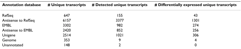
Examination of these 15,541 transcripts revealed that 4% (647) had significant homology with RefSeq transcripts, 40% (6,157) had significant homology with RefSeq in antisense orientation and 21% (3,302) and 16% (2,420) had significant homology with EMBL in sense and anti-sense orientations respectively (Table 2). For the remain-ing transcripts 16% (2,514) had significant homology with Unigene and 2% (353) with the human genome, while 148 demonstrated no homology with any of the databases. The large number of antisense sequences included within this grouping is notable. Antisense tran-scription is a subject which is now receiving increased attention in scientific literature and these transcripts are underrepresented on current generic microarrays. Anti-sense transcription has been associated with many impor-tant regulatory functions and its consideration when designing the Lung Cancer DSA research tool has ensured inclusion of probesets potentially vital in applications such as classifier generation.
Technical assessment experiment
To assess the biological relevance of the content of the Lung Cancer DSA research tool, five technical replicates of two RNAs extracted from a single patient matched normal and NSCLC frozen tissue were profiled on the arrays. The results demonstrated that in total 35,625 transcripts were consistently detected by the Lung Cancer DSA research tool as being expressed in either the normal or tumor tis-sue. These included 6398 (41%) of the Lung Cancer DSA research tool unique transcripts with 155 RefSeq, 982 human EMBL and 1,021 Unigene transcripts being detected in the sense orientation (Table 2). In addition we observed significant detection of antisense transcripts, with 3,437 (55%) of the RefSeq sense-antisense (SA) tran-script pairs detected as being expressed in either the nor-mal or tumor tissue.
Comparing the transcript expression levels between the normal and tumor lung tissue identified 2,148 of the unique transcripts as being differentially expressed and
thus potentially important to the underlying biology of this disease (Table 2). These included RefSeq sense tran-scripts to predicted proteins with associated functions in processes such as apoptosis, cell cycle control, cell prolif-eration and DNA damage repair [See Additional file 3]. In addition there was extensive differential expression of sense transcripts with homology to the coding DNA sequence (cds) of known genes and antisense transcripts (Table 2).
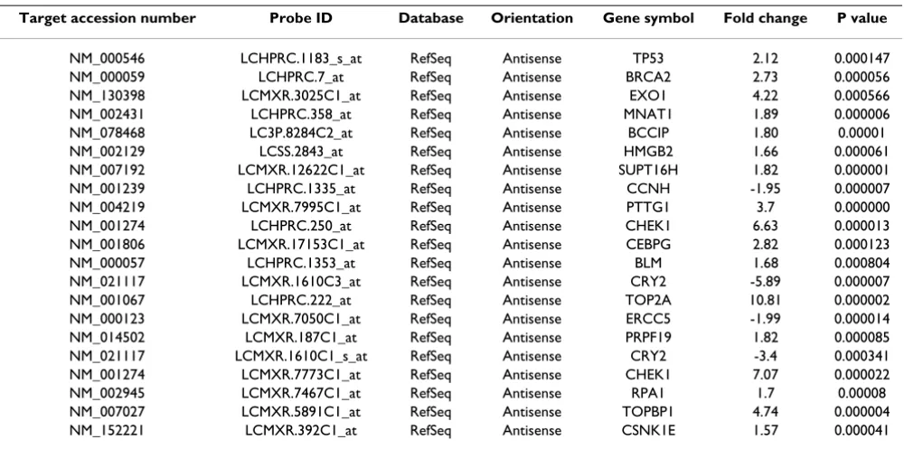
To further investigate the relevance of the detected and differentially expressed unique content to the biology of NSCLC, the annotation associated with these transcripts was assessed by Gene Ontology mining for implicated roles in processes linked to cancer. This clearly demon-strated association of these transcripts with the main cel-lular processes linked to cancer, including proliferation, apoptosis and DNA damage repair (Table 3). An example of one such group, DNA repair, is given in Table 4 where 21 antisense transcripts differentially expressed between the normal and tumor samples were identified.
Conclusion
The recently published results of the ENCODE consor-tium's pilot project indicate that the majority of the human genome is transcribed and that a minority of tran-scriptional activity leads directly to protein production [19]. This and other evidence has highlighted the com-plexity of the transcriptome and the need for better tools to study it [23,49]. Since studying the entire human tran-scriptome is not possible using current technologies, we propose a practical alternative of developing a range of microarray research tools capable of interrogating tran-scriptomes of individual disease settings
Given the importance of NSCLC as a disease and the extent of genomics research in this area, we have endeav-oured to characterise the transcriptome of NSCLC by means of in-house sequencing, mining of public sequence databases, gene expression profiling and literature min-ing. This information was used to design the Lung Cancer DSA representing ~60,000 transcripts empirically shown to be expressed in NSCLC and normal lung tissue
Table 2: Experimental detection of the Lung Cancer DSA unique content in matched normal tumor lung tissue.
Annotation database # Unique transcripts # Detected unique transcripts # Differentially expressed unique transcripts
RefSeq 647 155 43
Antisense to RefSeq 6157 3377 1301
EMBL 3302 982 274
Antisense to EMBL 2420 852 256
Unigene 2514 1021 306
Genome 353 9 4
When the Lung Cancer DSA is compared with the three most commonly used generic gene expression microar-rays, a significant proportion of the array is unique in rela-tion to each array as shown in Table 1. 15,541 Lung Cancer DSA research tool transcripts are not detectable by the Affymetrix HG U-133 Plus2 array, the Agilent Whole Human Genome Array or the Illumina Human 6 array, clearly demonstrating that the transcriptome of NSCLC is better represented by the Lung Cancer DSA research tool. This enables researchers using the Lung Cancer DSA to interrogate over 15,000 additional transcripts, all relevant to NSCLC.
The technical assessment experiment shows that data gen-erated by the Lung Cancer DSA is highly reproducible and
exhibits extremely good correlation and low coefficient of variance, similar to those obtained from generic Affyme-trix arrays in our own [See Additional file 4] and in previ-ously published studies [50].
A large part of the Lung Cancer DSA content is not repre-sented in the RefSeq database of well annotated mRNAs. Importantly, 46.3% of the non-RefSeq transcripts were reliably detected in the technical assessment experiment using samples taken from a single patient.
An even greater proportion (58%) of transcripts antisense to annotated RefSeq sequences were also consistently detected, further highlighting the magnitude of antisense transcription that is not fully annotated or understood.
Table 3: Gene Ontology mining of the experimentally detected unique Lung DSA transcripts associated annotation.
GO process Unique detected
transcripts
Unique detected RefSeq antisense transcripts
Unique differentially expressed transcripts
Unique differentially expressed RefSeq antisense transcripts
Angiogenesis 26 18 13 9
Apoptosis 311 186 112 77
DNA repair 119 65 32 21
Cell migration 71 34 27 17
Proliferation 287 186 117 85
Immunology/Inflammation 196 118 83 55
Developmental genes 12 8 10 7
Cell cycle control 384 239 139 111
Cell Signaling Pathway 55 40 33 25
Other 3851 2270 1382 910
Unknown 2461 820 770 293
Table 4: RefSeq antisense transcripts differentially expressed between the normal and tumor lung tissue, from the unique Lung Cancer DSA research tool content.
Target accession number Probe ID Database Orientation Gene symbol Fold change P value
NM_000546 LCHPRC.1183_s_at RefSeq Antisense TP53 2.12 0.000147
NM_000059 LCHPRC.7_at RefSeq Antisense BRCA2 2.73 0.000056
NM_130398 LCMXR.3025C1_at RefSeq Antisense EXO1 4.22 0.000566
NM_002431 LCHPRC.358_at RefSeq Antisense MNAT1 1.89 0.000006
NM_078468 LC3P.8284C2_at RefSeq Antisense BCCIP 1.80 0.00001
NM_002129 LCSS.2843_at RefSeq Antisense HMGB2 1.66 0.000061
NM_007192 LCMXR.12622C1_at RefSeq Antisense SUPT16H 1.82 0.000001
NM_001239 LCHPRC.1335_at RefSeq Antisense CCNH -1.95 0.000007
NM_004219 LCMXR.7995C1_at RefSeq Antisense PTTG1 3.7 0.000000
NM_001274 LCHPRC.250_at RefSeq Antisense CHEK1 6.63 0.000013
NM_001806 LCMXR.17153C1_at RefSeq Antisense CEBPG 2.82 0.000123
NM_000057 LCHPRC.1353_at RefSeq Antisense BLM 1.68 0.000804
NM_021117 LCMXR.1610C3_at RefSeq Antisense CRY2 -5.89 0.000007
NM_001067 LCHPRC.222_at RefSeq Antisense TOP2A 10.81 0.000002
NM_000123 LCMXR.7050C1_at RefSeq Antisense ERCC5 -1.99 0.000014
NM_014502 LCMXR.187C1_at RefSeq Antisense PRPF19 1.82 0.000085
NM_021117 LCMXR.1610C1_s_at RefSeq Antisense CRY2 -3.4 0.000341
NM_001274 LCMXR.7773C1_at RefSeq Antisense CHEK1 7.07 0.000022
NM_002945 LCMXR.7467C1_at RefSeq Antisense RPA1 1.7 0.00008
NM_007027 LCMXR.5891C1_at RefSeq Antisense TOPBP1 4.74 0.000004
Yelin et al [51] and Chen et al [52] predicted that there were 2667 and 5880 sense-antisense (SA) pairs tran-scribed from the human genome respectively. Our results from the analysis of a single tissue type and disease setting would suggest that the total number of SA pairs in the human transcriptome may be considerably higher.
Interestingly, based on the hypothesis that antisense tran-scription is the norm rather than the exception, we included in the array design 1143 sequences artificially created as antisense to those derived by literature and pathway mining. Of these 1143 sequences, 412 were con-sistently detected in the technical assessment experiment with 198 of these being differentially expressed, lending weight to the growing body of evidence that antisense transcription is extremely widespread [51,53-55].
Gene ontology mining served to demonstrate the poten-tial importance of the unique Lung Cancer DSA content. Major cancer related GO process categories were highly represented by uniquely detected or differentially expressed transcripts and significant proportions of these transcripts were shown to bear antisense homology to well characterised RefSeq sequences (Table 4). 21 anti-sense transcripts linked to DNA damage repair were differ-entially expressed between the normal and tumor samples. These included antisense transcripts homolo-gous to BRCA2, CHEK1 and TP53. Interestingly, TP53 antisense mRNA has previously been identified in human cells [51]. Antisense sequences are believed to function in gene regulation by modulating sense mRNA transcription, maturation, transport, stability and translation. The exten-sive detection of differentially expressed antisense tran-scripts between the normal and tumor tissue supports a role for these sequences in NSCLC pathogenesis. Moreo-ver, a conventional generic microarray would not have detected these potentially important transcripts.
The Lung Cancer DSA research tool presented in this com-munication represents a powerful and practical tool for both basic and translational research that better reflects the complexity of the NSCLC transcriptome than com-monly used microarrays. We believe it to have applica-tions in areas such as pathway analysis, biomarker and drug target discovery and multivariate prognostic and pre-dictive classifier generation. Its disease-focused design and novel, relevant content could ultimately lead to a bet-ter understanding of the underlying biology of NSCLC.
In addition, this methodology serves as a template for the development of other disease transcriptome focused microarrays, including non-neoplastic diseases.
Competing interests
The majority of the authors are employees of Almac Diag-nostics – a commercial entity – and accordingly we declare potential financial competing interests.
Authors' contributions
AT participated in the DSA research tool and study design, drafted the manuscript and was involved in coordinating the study. GRO participated in and performed design of the DSA research tool, performed the sequence content analysis and helped to draft the manuscript. VF performed gene expression analysis and participated in sample qual-ity control and helped to draft the manuscript. RDK was involved in the study design and coordination. JMM designed and performed the microarray expression exper-imental work for the technical validation of the DSA research tool. CEF coordinated the sequencing of the lung EST libraries. SMF carried out RNA extractions and cDNA library generation
JKF was involved in the study design. PGJ was involved in the initial conceptualisation and design of the DSA research tool. DPH was involved in study design and the initial conceptualisation and design of the DSA research tool. VP was involved in the study design and coordina-tion and helped to draft the manuscript. KAM was involved in the initial conceptualisation and design of the DSA research tool, participated in study design and coor-dination and helped to draft the manuscript.
Additional material
Additional file 1
Sequence content source breakdown for Lung Cancer DSA (table). Click here for file
[http://www.biomedcentral.com/content/supplementary/1755-8794-1-20-S1.doc]
Additional file 2
Lung Cancer DSA technical specifications (table) Click here for file
[http://www.biomedcentral.com/content/supplementary/1755-8794-1-20-S2.doc]
Additional file 3
RefSeq transcripts differentially expressed (table). Click here for file
[http://www.biomedcentral.com/content/supplementary/1755-8794-1-20-S3.doc]
Additional file 4
Reproducibility and reliability for technical study (table). Click here for file
Acknowledgements
This study was entirely funded by Almac Diagnostics. We thank D. McIl-waine, M. Totten, L. Campbell, K. Lavery and P. Hey for their work on the sequencing of the cDNA libraries. All microarray data has been submitted to ArrayExpress
The accession number for the data is E-TABM-387
References
1. Gray J, Simon G, Bepler G: Molecular predictors of chemother-apy response in non-small-cell lung cancer. Expert Rev Antican-cer Ther 2007, 7(4):545-9.
2. Rosell R, Cobo M, Isla D, Sanchez JM, Taron M, Altavilla G, Santarpia M, Moran T, Catot S, Etxaniz O: Applications of genomics in NSCLC. Lung Cancer 2005, 50(Suppl 2):S33-40.
3. Barker JM, Silvestri GA: Lung cancer staging. Curr Opin Pulm Med
2002, 8(4):287-93.
4. Kameyama K, Huang CL, Liu D, Okamoto T, Hayashi E, Yamamoto Y, Yokomise H: Problems related to TNM staging: patients with stage III non-small cell lung cancer. J Thorac Cardiovasc Surg
2002, 124(3):503-10.
5. Nguyen DM, Schrump DS: Lung cancer staging in the genomics era. Thorac Surg Clin 2006, 16(4):329-37.
6. Sun Y: p53 and its downstream proteins as molecular targets of cancer. Mol Carcinog 2006, 45(6):409-15.
7. Zheng Z, Chen T, Li X, Haura E, Sharma A, Bepler G: DNA synthe-sis and repair genes RRM1 and ERCC1 in lung cancer. N Engl J Med 2007, 356(8):800-8.
8. Oh JJ, Razfar A, Delgado I, Reed RA, Malkina A, Boctor B, Slamon DJ: 3p21.3 tumor suppressor gene H37/Luca15/RBM5 inhibits growth of human lung cancer cells through cell cycle arrest and apoptosis. Cancer Res 2006, 66(7):3419-27.
9. Hesson LB, Cooper WN, Latif F: Evaluation of the 3p21.3 tumour-suppressor gene cluster. Oncogene in press. 2007, May 28;
10. Uramoto H, Mitsudomi T: Which biomarker predicts benefit from EGFR-TKI treatment for patients with lung cancer? Br J Cancer 2007, 96(6):857-63.
11. Rosell R, Taron M, Camps C, López-Vivanco G: Influence of genetic markers on survival in non-small cell lung cancer.
Drugs Today (Barc) 2003, 39(10):775-86.
12. Cappuzzo F, Ligorio C, Toschi L, Rossi E, Trisolini R, Paioli D, Magrini E, Finocchiaro G, Bartolini S, Cancellieri A, Hirsch FR, Crino L, Varella-Garcia M: EGFR and HER2 gene copy number and response to first-line chemotherapy in patients with advanced non-small cell lung cancer (NSCLC). J Thorac Oncol
2007, 2(5):423-9.
13. Rosell R, Felip E, Garcia-Campelo R, Balaña C: The biology of non-small-cell lung cancer: identifying new targets for rational therapy. Lung Cancer 2004, 46(2):135-48.
14. Petty RD, Kerr KM, Murray GI, Nicolson MC, Rooney PH, Bissett D, Collie-Duguid ES: Tumor transcriptome reveals the predictive and prognostic impact of lysosomal protease inhibitors in non-small-cell lung cancer. J Clin Oncol 2006, 24(11):1729-44. 15. Hotta K, Kiura K, Toyooka S, Takigawa N, Soh J, Fujiwara Y, Tabata
M, Date H, Tanimoto M: Clinical Significance of Epidermal Growth Factor Receptor Gene Mutations on Treatment Outcome after First-line Cytotoxic Chemotherapy in Japa-nese Patients with Non-small Cell Lung Cancer. J Thorac Oncol
2007, 2(7):632-637.
16. Filipits M, Pirker R, Dunant A, Lantuejoul S, Schmid K, Huynh A, Haddad V, André F, Stahel R, Pignon JP, Soria JC, Popper HH, Le Chevalier T, Brambilla E: Cell cycle regulators and outcome of adjuvant Cisplatin-based chemotherapy in completely resected non-small-cell lung cancer: the international adju-vant lung cancer trial biologic program. J Clin Oncol 2007, 25(19):2735-40.
17. Kapranov P, Willingham A, Gingeras TR: Genome-wide transcrip-tion and the implicatranscrip-tions for genomic organizatranscrip-tion. Nature Reviews Genetics 2007, 8:413-423.
18. Gingeras TR: The multitasking genome. Nature Genetics 2006, 38:608-609.
19. The ENCODE Project Consortium: Identification and analysis of functional elements in 1% of the human genome by the ENCODE pilot project. Nature 2007, 447(7146):799-816. 20. Gerstein MB, Bruce C, Rozowsky JS, Zheng D, Du J, Korbel JO,
Emanuelsson O, Zhang ZD, Weissman S, Snyder M: What is a gene, post-ENCODE? History and updated definition. Genome Res
2007, 17:669-681.
21. Strausberg RL, Levy S: Promoting transcriptome diversity.
Genome Res 2007, 17:965-968.
22. Szymañski M, Barciszewska MZ, Zywicki M, Barciszewski J: Noncod-ing RNA transcripts. J Appl Genet 2003, 44(1):1-19.
23. Carninci P: Constructing the landscape of the mammalian transcriptome. J Exp Biol 2007, 210(Pt 9):1497-506.
24. Gingeras TR: Origin of phenotypes: Genes and transcripts.
Genome Res 2007, 17(6):682-90.
25. Brentani H, Caballero OL, Camargo AA, da Silva AM, da Silva WA Jr, Dias Neto E, Grivet M, Gruber A, Guimaraes PE, Hide W, Iseli C, Jon-geneel CV, Kelso J, Nagai MA, Ojopi EP, Osorio EC, Reis EM, Riggins GJ, Simpson AJ, de Souza S, Stevenson BJ, Strausberg RL, Tajara EH, Verjovski-Almeida S, Acencio ML, Bengtson MH, Bettoni F, Bodmer WF, Briones MR, Camargo LP, Cavenee W, Cerutti JM, Coelho Andrade LE, Costa dos Santos PC, Ramos Costa MC, da Silva IT, Esté-cio MR, Sa Ferreira K, Furnari FB, Faria M Jr, Galante PA, Guimaraes GS, Holanda AJ, Kimura ET, Leerkes MR, Lu X, Maciel RM, Martins EA, Massirer KB, Melo AS, Mestriner CA, Miracca EC, Miranda LL, Nobrega FG, Oliveira PS, Paquola AC, Pandolfi JR, Campos Pardini MI, Passetti F, Quackenbush J, Schnabel B, Sogayar MC, Souza JE, Valentini SR, Zaiats AC, Amaral EJ, Arnaldi LA, de Araújo AG, de Bessa SA, Bicknell DC, Ribeiro de Camaro ME, Carraro DM, Carrer H, Car-valho AF, Colin C, Costa F, Curcio C, Guerreiro da Silva ID, Pereira da Silva N, Dellamano M, El-Dorry H, Espreafico EM, Scattone Fer-reira AJ, Ayres FerFer-reira C, Fortes MA, Gama AH, Giannella-Neto D, Giannella ML, Giorgi RR, Goldman GH, Goldman MH, Hackel C, Ho PL, Kimura EM, Kowalski LP, Krieger JE, Leite LC, Lopes A, Luna AM, Mackay A, Mari SK, Marques AA, Martins WK, Montagnini A, Mourão Neto M, Nascimento AL, Neville AM, Nobrega MP, O'Hare MJ, Otsuka AY, Ruas de Melo AI, Paco-Larson ML, Guimarães Pereira G, Pereira da Silva N, Pesquero JB, Pessoa JG, Rahal P, Rainho CA, Rod-rigues V, Rogatto SR, Romano CM, Romeiro JG, Rossi BM, Rusticci M, Guerra de Sá R, Sant' Anna SC, Sarmazo ML, Silva TC, Soares FA, Sonati Mde F, de Freitas Sousa J, Queiroz D, Valente V, Vettore AL, Villanova FE, Zago MA, Zalcberg H, Human Cancer Genome Project/ Cancer Genome Anatomy Project Annotation Consortium; Human Cancer Genome Project Sequencing Consortium: The generation and utilization of a cancer-oriented representation of the human transcriptome by using expressed sequence tags.
PNAS 2003, 100(23):13418-13423.
26. Wachi S, Yoneda K, Wu R: Interactome-transcriptome analysis reveals the high centrality of genes differentially expressed in lung cancer tissues. Bioinformatics 2005, 21(23):4205-8. 27. Riggins GJ, Strausberg RL: Genome and genetic resources from
the Cancer Genome Anatomy Project. Human Molecular Genet-ics 2001, 10:663-667.
28. Cancer Genome Anatomy Project [http://cgap.nci.nih.gov/] 29. International Genomics Consortium [http://www.intgen.org/] 30. Affymetrix [http://www.affymetrix.com/]
31. GeneGo [http://www.genego.com/]
32. Pruitt KD, Tatusova T, Maglott DR: NCBI Reference Sequence (RefSeq): a curated non-redundant sequence database of genomes, transcripts and proteins. Nucleic Acids Research 2005, 33:D501-D504.
33. NCBI Reference Sequence database [http:// www.ncbi.nlm.nih.gov/RefSeq/]
34. Stoesser G, Tuli MA, Lopez R, Sterk P: The EMBL Nucleotide Sequence Database. Nucleic Acids Research 1999, 27:18-24. 35. Embl nucleotide sequence database [http://www.ebi.ac.uk/
embl/]
36. Ensembl Genome Browser [http://www.ensembl.org/] 37. Huang X, Madan A: CAP3: A DNA Sequence Assembly
Pro-gram. Genome Research 1999, 9:868-877.
38. Pesole G, Liuni S, Grillo G, Ippedico M, Larizza A, Makalowski W, Sac-cone C: UTRdb: a specialized database of 5' and 3' untrans-lated regions of eukaryotic mRNAs. Nucleic Acids Research 1999, 27:188-191.
Publish with BioMed Central and every scientist can read your work free of charge "BioMed Central will be the most significant development for disseminating the results of biomedical researc h in our lifetime."
Sir Paul Nurse, Cancer Research UK
Your research papers will be:
available free of charge to the entire biomedical community
peer reviewed and published immediately upon acceptance
cited in PubMed and archived on PubMed Central
yours — you keep the copyright
Submit your manuscript here:
http://www.biomedcentral.com/info/publishing_adv.asp
BioMedcentral 40. Wheeler DL, Church DM, Federhen S, Lash AE, Madden TL, Pontius
JU, Schuler GD, Schriml LM, Sequeira E, Tatusova TA, Wagner L: Database resources of the National Center for Biotechnol-ogy. Nucleic Acids Res 2003, 31:28-33.
41. Agilent Technologies [http://www.agilent.com/] 42. Illumina inc [http://www.illumina.com/]
43. NCBI Batch Entrez [http://www.ncbi.nlm.nih.gov/entrez/batchen trez.cgi]
44. Ensembl FTP site [ftp://ftp.ensembl.org/]
45. DFCI human gene index FTP site [ftp://occams.dfci.har vard.edu/pub/bio/tgi/data/]
46. The Gene Ontology [http://www.geneontology.org/]
47. Asterand ethics [http://www.asterand.com/Asterand/about/eth ics.htm]
48. Li C, Wong WH: DNA-Chip Analyzer (dChip). In The analysis of gene expression data: methods and software Edited by: Parmigiani G, Garrett ES, Irizarry R, Zeger SL. Springer, New York; 2003:120-141. 49. Carninci P, Kasukawa T, Katayama S, Gough J, Frith MC, Maeda N, Oyama R, Ravasi T, Lenhard B, Wells C, Kodzius R, Shimokawa K, Bajic VB, Brenner SE, Batalov S, Forrest AR, Zavolan M, Davis MJ, Wilming LG, Aidinis V, Allen JE, Ambesi-Impiombato A, Apweiler R, Aturaliya RN, Bailey TL, Bansal M, Baxter L, Beisel KW, Bersano T, Bono H, Chalk AM, Chiu KP, Choudhary V, Christoffels A, Clutter-buck DR, Crowe ML, Dalla E, Dalrymple BP, de Bono B, Della Gatta G, di Bernardo D, Down T, Engstrom P, Fagiolini M, Faulkner G, Fletcher CF, Fukushima T, Furuno M, Futaki S, Gariboldi M, Georgii-Hemming P, Gingeras TR, Gojobori T, Green RE, Gustincich S, Har-bers M, Hayashi Y, Hensch TK, Hirokawa N, Hill D, Huminiecki L, Iacono M, Ikeo K, Iwama A, Ishikawa T, Jakt M, Kanapin A, Katoh M, Kawasawa Y, Kelso J, Kitamura H, Kitano H, Kollias G, Krishnan SP, Kruger A, Kummerfeld SK, Kurochkin IV, Lareau LF, Lazarevic D, Lipovich L, Liu J, Liuni S, McWilliam S, Madan Babu M, Madera M, Mar-chionni L, Matsuda H, Matsuzawa S, Miki H, Mignone F, Miyake S, Morris K, Mottagui-Tabar S, Mulder N, Nakano N, Nakauchi H, Ng P, Nilsson R, Nishiguchi S, Nishikawa S, Nori F, Ohara O, Okazaki Y, Orlando V, Pang KC, Pavan WJ, Pavesi G, Pesole G, Petrovsky N, Piazza S, Reed J, Reid JF, Ring BZ, Ringwald M, Rost B, Ruan Y, Salz-berg SL, Sandelin A, Schneider C, Schönbach C, Sekiguchi K, Semple CA, Seno S, Sessa L, Sheng Y, Shibata Y, Shimada H, Shimada K, Silva D, Sinclair B, Sperling S, Stupka E, Sugiura K, Sultana R, Takenaka Y, Taki K, Tammoja K, Tan SL, Tang S, Taylor MS, Tegner J, Teichmann SA, Ueda HR, van Nimwegen E, Verardo R, Wei CL, Yagi K, Yamani-shi H, Zabarovsky E, Zhu S, Zimmer A, Hide W, Bult C, Grimmond SM, Teasdale RD, Liu ET, Brusic V, Quackenbush J, Wahlestedt CY, Mattick JS, Hume DA, Kai C, Sasaki D, Tomaru Y, Fukuda S, Kan-amori-Katayama M, Suzuki M, Aoki J, Arakawa T, Iida J, Imamura K, Itoh M, Kato T, Kawaji H, Kawagashira N, Kawashima T, Kojima M, Kondo S, Konno H, Nakano K, Ninomiya N, Nishio T, Okada M, Plessy C, Shibata K, Shiraki T, Suzuki S, Tagami M, Waki K, Watahiki A, Okamura-Oho Y, Suzuki H, Kawai J, Hayashizaki Y: The tran-scriptional landscape of the mammalian genome. Science
2005, 309(5740):1559-63.
50. MAQC Consortium Shi L, Reid LH, Jones WD, Shippy R, Warrington JA, Baker SC, Collins PJ, de Longueville F, Kawasaki ES, Lee KY, Luo Y, Sun YA, Willey JC, Setterquist RA, Fischer GM, Tong W, Dragan YP, Dix DJ, Frueh FW, Goodsaid FM, Herman D, Jensen RV, Johnson CD, Lobenhofer EK, Puri RK, Scherf U, Thierry-Mieg J, Wang C, Wil-son M, Wolber PK, Zhang L, Amur S, Bao W, Barbacioru CC, Berg-strom Lucas A, Bertholet V, Boysen C, Bromley B, Brown D, Brunner A, Canales R, Cao XM, Cebula TA, Chen JJ, Cheng J, Chu T, Chudin E, Corson J, Corton JC, Croner LJ, Davies C, Davison TS, Delenstarr G, Deng X, Dorris D, Eklund AC, Fan X, Fang H, Fulmer-Smentek S, Fuscoe JC, Gallagher K, Ge W, Guo L, Guo X, Hager J, Haje PK, Han J, Han T, Harbottle HC, Harris SC, Hatchwell E, Hauser CA, Hester S, Hong H, Hurban P, Jackson SA, Ji H, Knight CR, Kuo WP, LeClerc JE, Levy S, Li Q, Liu C, Liu Y, Lombardi MJ, Ma Y, Magnuson SR, Maq-sodi B, McDaniel T, Mei N, Myklebost O, Ning B, Novoradovskaya N, Orr MS, Osborn TW, Papallo A, Patterson TA, Perkins RG, Peters EH, Peterson R, Philips KL, Pine PS, Pusztai L, Qian F, Ren H, Rosen M, Rosenzweig BA, Samaha RR, Schena M, Schroth GP, Shchegrova S, Smith DD, Staedtler F, Su Z, Sun H, Szallasi Z, Tezak Z, Thierry-Mieg D, Thompson KL, Tikhonova I, Turpaz Y, Vallanat B, Van C, Walker SJ, Wang SJ, Wang Y, Wolfinger R, Wong A, Wu J, Xiao C, Xie Q, Xu J, Yang W, Zhang L, Zhong S, Zong Y, Slikker W Jr: The MicroArray Quality Control (MAQC) project shows inter- and
intraplat-form reproducibility of gene expression measurements. Nat Biotechnol 2006, 24:1151-61.
51. Yelin R, Dahary D, Sorek R, Levanon EY, Goldstein O, Shoshan A, Diber A, Biton S, Tamir Y, Khosravi R, Nemzer S, Pinner E, Walach S, Bernstein J, Savitsky K, Rotman G: Widespread occurrence of antisense transcription in the human genome. Nat Biotechnol
2003, 21(4):379-86.
52. Chen J, Sun M, Kent WJ, Huang X, Xie H, Wang W, Zhou G, Shi RZ, Rowley JD: Over 20% of human transcripts might form sense-antisense pairs. Nucleic Acids Res 2004, 32(16):4812-4820. 53. RIKEN Genome Exploration Research Group and Genome Science
Group (Genome Network Project Core Group) and the FANTOM Consortium, Kasukawa T, Waki K, Nakanishi M, Nakamura M, Nish-ida H, Yap CC, Suzuki M, Kawai J, Suzuki H, Carninci P, Hayashizaki Y, Wells C, Frith M, Ravasi T, Pang KC, Hallinan J, Mattick J, Hume DA, Lipovich L, Batalov S, Engström PG, Mizuno Y, Faghihi MA, San-delin S, Chalk AM, Mottagui-Tabar S, Liang Z, Lenhard B, Wahlestedt C: Antisense transcription in the mammalian transcriptome.
Science309:1564-6.
54. Ge X, Wu Q, Jung YC, Chen J, Wang SM: A large quantity of novel human antisense transcripts detected by LongSAGE. Bioinfor-matics 2006, 22(20):2475-9.
55. Zhang Y, Liu XS, Liu QR, Wei L: Genome-wide in silico identifi-cation and analysis of cis natural antisense transcripts (cis-NATs) in ten species. Nucleic Acids Res 2006, 34:3465-75.
Pre-publication history
The pre-publication history for this paper can be accessed here: