R E S E A R C H
Open Access
Anti-EMT properties of CoQ0 attributed to
PI3K/AKT/NFKB/MMP-9 signaling pathway
through ROS-mediated apoptosis
Hsin-Ling Yang
1, Varadharajan Thiyagarajan
2, Pei-Chun Shen
1, Dony Chacko Mathew
1, Kai-Yuan Lin
3,
Jiunn-Wang Liao
4and You-Cheng Hseu
2,5,6,7*Abstract
Background:Breast cancer is the most prevalent cancer among women. In triple-negative breast cancer (TNBC) cells, a novel quinone derivative, coenzyme Q0(CoQ0), promotes apoptosis and cell-cycle arrest. This study explored the anti-epithelial–mesenchymal transition (EMT) and antimetastatic attributes of CoQ0in TNBC (MDA-MB-231). Methods:Invasion, as well as MTT assays were conducted. Lipofectamine RNAiMAX was used to transfect cells with β-catenin siRNA. Through Western blotting and RT-PCR, the major signaling pathways’protein expressions were examined, and the biopsied tumor tissues underwent immunohistochemical and hematoxylin and eosin staining as well as Western blotting.
Results:CoQ0(0.5–2μM) hindered tumor migration, invasion, and progression. Additionally, it caused MMP-2/−9, uPA, uPAR, and VEGF downregulation. Furthermore, in highly metastatic MDA-MB-231 cells, TIMP-1/2 expression
was subsequently upregulated and MMP-9 expression was downregulated. In addition, CoQ0inhibited metastasis
and EMT in TGF-β/TNF-α-stimulated non-tumorigenic MCF-10A cells. Bioluminescence imaging of MDA-MB-231
luciferase–injected live mice demonstrated that CoQ0significantly inhibited metastasis of the breast cancer to the
lungs and inhibited the development of tumors in MDA-MB-231 xenografted nude mice. Silencing ofβ-catenin
with siRNA stimulated CoQ0-inhibited EMT. Western blotting as well as histological analysis established that CoQ0 reduced xenografted tumor development because apoptosis induction, cell-cycle inhibition, E-cadherin upregulation, β-catenin downregulation, and metastasis and EMT regulatory protein modulation were observed.
Conclusions:CoQ0 inhibited the progression of metastasis as well as EMT (in vitro and in vivo). The described approach has potential in treating human breast cancer metastasis.
Keywords: CoQ0, TNBC, EMT, Metastasis, Human epidermal growth factor receptor 2, TGF-β1, ROS
Background
Worldwide, breast cancer is the commonest cancer to affect women, and in Taiwanese women, it is the leading cause of deaths from cancer [1]. It possesses highly metastatic and invasive properties, which explain its high mortality rate [2].Among breast cancers, triple-negative breast cancers (TNBCs) lacking the genes for estrogen receptor, HER2, and progesterone receptor have been
correlated with tumor aggressiveness. TNBCs are more likely than other breast cancer types to migrate beyond the breast and to recur after chemotherapy or lumpec-tomy [3]. TNBC cases comprise 15–20% of all breast cancer cases. Furthermore, patients with TNBC exhibit unfavorable outcomes compared with those with other breast cancer subtypes [4]. TNBC tumor cells lack the requisite receptors, which renders some targeted or hor-mone therapies ineffectual. Consequently, combinations of chemotherapy medicines are typically prescribed for pa-tients with TNBC. This approach, however, does not help patients with cancer to counter the chemotherapy-induced adverse side effects and drug resistance [5]. Thus, novel
© The Author(s). 2019Open AccessThis article is distributed under the terms of the Creative Commons Attribution 4.0 International License (http://creativecommons.org/licenses/by/4.0/), which permits unrestricted use, distribution, and reproduction in any medium, provided you give appropriate credit to the original author(s) and the source, provide a link to the Creative Commons license, and indicate if changes were made. The Creative Commons Public Domain Dedication waiver (http://creativecommons.org/publicdomain/zero/1.0/) applies to the data made available in this article, unless otherwise stated.
* Correspondence:ychseu@mail.cmu.edu.tw
2
Department of Cosmeceutics, College of Biopharmaceutical and Food Sciences, China Medical University, No. 91, Hsueh-Shih Road, Taichung 40402, Taiwan 5Department of Health and Nutrition Biotechnology, Asia University,
Taichung 41354, Taiwan
compounds with lower toxicity are urgently required for ef-fective treatment of TNBC.
In cancer cells, polarized epithelial cells complete multifaceted changes that cause them to begin express-ing a mesenchymal phenotype and undergo migration, invasion, and metastasis. This process is referred to as the epithelial–mesenchymal transition (EMT) [6]. Several factors induce EMT in vitro and in vivo, for example, TGF-β1, ROS, TNF-α, and hypoxia [7–9]. EMT involves AKT/GSK or NFκB-mediated expression of Snail and pro-motes cell invasion and migration in various cancers, such as breast, renal, and colon cancers [10, 11]. The loss of E-cadherin, an adherens junction cell surface protein expressed in epithelial cells is the principal characteristic of EMT [12]. The Snail and Slug signaling cascades are among those that may be involved in EMT in cancer cells. Snail and Slug are key transcription factors that can down regulate the expression of E-cadherin. They do this by binding to E-boxes in the E-cadherin promoter, subse-quently increasing MMP-9 expression to promote cell in-vasion [13]. However, few studies have investigated the suppression of molecular events and EMT responsible for EMT inhibition in anticancer treatment.
The Wnt/β-catenin signaling pathway contributes to cell fate decisions as well as the normal cellular response during cancer cell development [14]. Researchers have suggested that dysregulated or uncontrolled triggering of this signaling pathway promotes tumor progression and metastasis in patients with breast cancer [15]. Other at-tributes of the Wnt extracellular signaling pathways manage tissue architecture, proliferation, embryonic axis formation, and cell migration [16] and can be broadly classified into noncanonical and canonical pathways. Canonical pathways are activated when the relevant Wnt ligands bind to the LRP-5/6 coreceptors and Frizzled transmembrane domain receptor [17], whereas non-ca-nonical pathways are β-catenin-independent and need Ror2/Ryk coreceptors rather than LRP-5/6 coreceptors. β-Catenin is usually aberrantly activated in breast cancer tissues. Therefore, Wnt/β-catenin pathway inhibition has the potential to reduce breast cell invasion as well as that of their EMT.
Coenzyme Q0(CoQ0)also known as ubiquinone 0 and
2,3 dimethoxy-5-methyl-1,4 benzoquinone) and a mem-ber of the mitochondrial respiratory chain is a redox-active ubiquinone compound commonly present in the mitochondrion. It possesses strong antioxidant activity and prevents the mitochondrial permeability transition pore [18] from being opened calcium-de-pendently. CoQ0 has demonstrated activity against the
proliferation of numerous cancer cell lines (e.g., HepG2, A549, and SW480) [19, 20]. Although it ex-hibits cytotoxic anticancer activities, it was also dem-onstrated to stimulate insulin secretion in pancreatic
islets [21]. We described its inflammatory and anti-angiogenic properties in vivo and in vitro in our previous study [22]. Remarkably, administering CoQ0mixtures
pre-vents oxidative damage in rodent spleen, blood, kidney, heart, and liver [23]. Our previous study on CoQ0found
that it significantly inhibits melanoma cell growth and tumor formation by inducing apoptosis and cell-cycle ar-rest [24]. Additionally, it effectively promoted apoptosis by increasing ROS in MCF-7 cells that were irradiated using ultraviolet B [22]. Despite CoQ0’s anticancer attributes, its
inhibitory effect on breast cancer metastasis and EMT and the molecular mechanism that gives it its therapeutic effi-cacy are unclear.
To ascertain CoQ0’s capabilities at inhibiting
metasta-sis, EMT, and their associated changes, we designed a validated EMT and metastasis model for human TNBC (MDA-MB-231). Metastasis and EMT control levels and the principal molecular biomarkers involved were ana-lyzed to ascertain the anti-EMT and antimetastatic attri-butes mediated by CoQ0. In addition, we sought to
clarify the fundamental mechanism of TNBC cells.
Methods and materials
Reagent and antibodies
CoQ0 was from Sigma-Aldrich (St. Louis, MO, United
States), as was the 3-(4,5-dimethylthiazol-2-yl)-2,5-di-phenyltetrazolium bromide reagent for the MTT assay. GIBCO BRL/Invitrogen (Carlsbad, CA, United States) supplied L-glutamine, penicillin/streptomycin/neomycin,
and Dulbecco’s modified Eagle’s medium (DMEM). Anti-bodies against anti-NFκB (p65), phos-IKK, Cyclin D1, CDK4, PARP, β-catenin, p-AKT, p-PI3K, PI3K, H3 anti-bodies, and IKK were from Cell Signaling Technology Inc. (Danvers, MA, United States), and those against MMP-9, vascular endothelial growth factor (VEGF), β-actin, c-Myc, Bax, Bcl-2, p21, p27, p53, caspase-3, cytochrome C, and I-κB were from Santa Cruz Biotech-nology, Inc. (Heidelberg, Germany). The remaining chemicals were of HPLC grade and were from either Sigma-Aldrich or Merck (Darmstadt, Germany).
Generation of breast cancer cell lines
MTT assay
MTT colorimetric assays were employed to ascertain cell viability [25]. Cells were grown in 12-well plates to a confluence. Subsequently, they underwent 24-h CoQ0
incubation (2.5–20μM). Following the MTT assay, an ELISA microplate reader (Bio-Tek Instruments Inc., Winooski, VT, United States) was utilized to ascertain the absorbance at 570 nm. The proportion of viable cells relative to vehicle-treated control cells (desig-nated as 100%) was the basis for evaluating CoQ0’s
effect on cell viability.
In vitro wound-healing assay
The effects of CoQ0 on cell migration were evaluated
using in vitro wound-healing assay as follows: the cells were cultured (density: 1 × 10⁴), and the standard proto-col of the in vitro healing assay was followed [26]. Subsequently, the cells underwent 24-h incubation with varying CoQ0 concentrations (0.5–2μM) in 1% FBS
medium followed by phosphate-buffer saline (PBS) wash (3 times). They were subsequently fixed using 100% methanol. Finally, they were stained using Giemsa stain-ing solution (Merck, Darmstadt). Migrated cells were monitored, and phase-contrast microscopy at 200× was used to photograph them. Image-Pro Plus (Media Cyber-netics, Inc., Rockville, MD, United States) was used to compute the wounded area’s closure.
Cell invasion assay
Cell invasion assay was conducted using BD Matrigel in-vasion chambers (Bedford, United States), as described by Debbie and Brooks [27]. We used 10μL of Matrigel (25 mg/50 mL) to coat 8-μm polycarbonate membrane filters. Then, we seeded the cells (density: 1 × 10⁵) onto the Matrigel-treated filter in 200μL of CoQ0(0.5–2μM)
devoid of serum. Cell migration underwent 24-h obser-vation at 37 °C. After being incubated for 24 h, the cells that did not migrate were removed from atop the mem-brane by using a cotton swab. On the other side of the membrane, the migrated cells were fixed for 15 min in ice-cold methanol (75%) and were washed in PBS three times. Subsequently, Giemsa stain was used to fix the cells; they were subsequently destained using PBS. Im-ages were viewed and captured through 200× light mi-croscopy; we quantified invading cells by using manual counting. All experiments were repeated three times.
Protein isolation and Western blotting
We seeded the cells (density: 4 × 10⁶ cells/dish) into a 6-cm dish that had undergone 24-h CoQ0 (2.5–10μM)
pre-treatment. Next, they underwent trypsinization and 3 times rinsing with ice-cold PBS. Total cytoplasmic and nuclear extracts were isolated in accordance with the suggested protocol (Pierce Biotechnology, Waltham,
MA, United States). The protein concentration was ascer-tained using Bradford reagent, with bovine serum albumin (BSA) as standard. The lysate was separated through 12% SDS-PAGE. PVDF membranes were immunoblotted using specific primary antibodies and their corresponding horse-radish peroxidase–conjugated secondary antibodies. An enhanced chemiluminescence substrate (Pierce Biotech-nology, Waltham) was utilized to develop the blot, and a densitometric graph displaying band intensities was gener-ated using AlphaEaseFC (Miami, FL, United States).
RNA extraction and RT-PCR
After cells had undergone 24-h pre-treatment with various CoQ0 concentrations (0.5–2μM), they were harvested.
Subsequently, TRIzol reagent (Invitrogen, Carlsbad) was employed to extract total RNA. Then, Bio-Rad iCycler PCR instrument (Bio-Rad, Hercules, CA, United States) and SuperScript-III One Step RT-PCR platinum taq kit (Invitrogen, Carlsbad) were used for RT-PCR of 1μg of total RNA in accordance with the procedure [28]. The PCR product was analyzed in agarose gel (1%). The primers used were as follows: MMP-2 F: 5′ATGA CAGCTGCACCACTGAG-3′, R-5′-ATTTGTTGCCCA GGAAAGTG -3′; MMP-9 F-5′- TTGACAGCGACAA GAAGTGG-3′, R-5′-GCCATTCACGTCGTCCTTAT-3′; uPA F-5′-TGCGTCCTGGTCGTGAGCGA-3′, R-5′-CA AGCGTGTCAGCGCTGTAG-3′; uPAR F-5′CATGCA GTGTAAGACCCAACGGGGA-3′, R-5′-AATAGGTGA CAGCCCGGCCAGAGT3′; E-cadherin: F-5′-TGGGTT ATTCCTCCCATCAG-3′, R-5′TTTGTCAGGGAGCTC AGGAT-3′; Vimentin: F- 5′CTCTTCCAAACTTTTCC TCC 3′, R-5′AGTTTCGTTGATAACCTGTC 3′; Snail: F-5′CGAAAGGCCTTCAACTGCAAAT 3′, R-5′ACTG GTACTTCTTGACATCTG 3′; Slug: F- 5′CGCCT CCAAAAAGCCAAAC 3′, R-5′CGGTAGTCCACACAG TGATG 3′; β-catenin: F-5′-AAGGAAGCTTCCAGA CATGC 3′, R-5′AGCTTGCTCTCTTGATTGCC 3′; 18S F-5′GTCTGTGATGCCCTTAGATG 3′, R-5′AGCTTAT GACCCGCACTTAC 3′.
Mammosphere formation assay
We performed a mammosphere formation assay as de-scribed elsewhere with several minor modifications [29]. For mammosphere culture from MDA-MB-231 cells, we suspended cells at 1 × 10³ cells/mL and seeded them onto ultralow attachment plates (Corning Inc., Corning, NY, United States) in MammoCult basal medium (STEMCELL Technologies, United States) supplemented with 0.2% heparin (STEMCELL Technologies, United States), 500μg/mL cortisone (STEMCELL Technologies, United States), and 10μL/mL mammo supplant serum (STEMCELL Technologies, United States) in an incuba-tor supplemented with 5% CO2. The cells underwent
treatment and were then incubated for 1 week during which time the plate was kept stationary and the media were replenished. On day 7, the mammospheres were collected through gentle centrifugation and dissociated into single-cell suspensions; the cells then underwent re-peated vehicle control (0.1% DMSO) or CoQ0 (0.5–
2μM) treatment. The cell suspensions underwent three passages through a syringe with 26-G needles. This pro-cedure resulted in single-cell suspensions being formed. We then adjusted cell concentration to 500 cells/mL and plated the 2-mL single-cell suspensions on a 35-mm ultralow attachment plate (Corning Inc., Corning) in triplicate. For serial passage (secondary sphere formation) and differentiation experiments, this study employed early progenitor cells as well as sphere forming, breast stem– enhanced single cells. On day 14, a graticule-enabled microscope was employed to determine the number of mammospheres of diameter > 50μm as well as the num-ber of colony-forming units. Then, the cells were trypsi-nized, and the cell numbers were counted. Two independent observers not exposed to the treatment were asked to count the sphere numbers manually. All experi-ments were conducted three times.
Colony formation assay
MDA-MB-231 cells (density: 5 × 10⁵ cells/60-mm dish) underwent 24-h CoQ0 treatment (concentrations: 0–
7.5μM). We then trypsinized the cells and replated them (density: 3 × 10⁴ cells/35-mm dish) in triplicate. They were subsequently subjected to 7-d RPMI 1640 incuba-tion. Afterward, they were fixed at room temperature for 10 min using 10% neutral buffered formalin and stained using 20% Giemsa stain (Merck, Darmstadt). We subse-quently assayed the cells for ability to form colonies and proliferation rate. The number of colonies of size > 1 mm was determined with light microscopy (40×). Colony number was ascertained based on a colony formation per-centage of 100% in the absence of CoQ0.
Gelatin zymography assay
The zymography protease assay was used to quantify the activity of MMP-2 and 9 in the medium used to grow the MCF-10A cells. The MCF-10A cells (1 × 10⁶ cells/ well) were seeded into 6-well culture dishes and grown in medium with 10% FBS to a nearly confluent mono-layer. The cells were re-suspended in 1% FBS medium, and then incubated with TGF-β/TNF-α (10 ng/mL) and CoQ0(2μM) for 24 h. After treatment the remains
pro-cedures were followed according to the previously men-tioned work [30]. The changes in expression of MMP-2 and -9 were quantified by Matrix Inspector 2.1 software (AlphaEase, Genetic Technology, Inc., Miami, FL, USA).
Immunofluorescence staining
Cells (density: 1 × 10⁴ cells/well) underwent culturing; this culturing was performed in an eight-well glass Tek chamber. Subsequently, CoQ0 (0.5–2μM) was used to
pre-treat them for the recommended duration. Next, they underwent 15-min fixing in paraformaldehyde (2%), 10-min Triton (0.1%) permeabilization, washing, and FBS (10%) blocking in PBS. Accordingly, the cells under-went 2-h primary antibody (anti-p65, anti-Vimentin, anti-F-actin, anti-β-catenin, and anti-E-cadherin) incuba-tion in 1.5% FBS. Subsequently, they were incubated for 1 h using FITC-conjugated secondary antibody in BSA (6%). We then stained them for 5 min using 1μg/mL DAPI. The cells were subsequently subjected to second-ary antibody incubation and DAPI staining, washed thoroughly using PBS, and then visualized through fluor-escent confocal microscopy.
Luciferase reporter assay
The transcriptional activity of NFκB, E-cadherin, and β-catenin was ascertained through dual-luciferase re-porter assays (Promega, Fitchberg, WI, United States). We grew the cells to 70–80% confluence for 5 min in a 24-well plate by using serum-free DMEM devoid of anti-biotics. They were subsequently transfected with NFκB, E-cadherin, or β-catenin plasmids with β-galactosidase or a pcDNA vector by using Lipofectamine 2000 (Invitro-gen, Carlsbad). Afterward, they underwent 4-h CoQ0(0.5–
2μM) treatment. Subsequently, they were lysed, following which their luciferase activity was ascertained through luminometry (Bio-Tek, Winooski) and then standardized to theirβ-galactosidase activity in the cell lysates. In addition, we quantified the intensity of relative fluorescence by using a luminance ELISA reader.
Immunoprecipitation
Protein A-sepharose beads were used to preclear 1 mg of the protein sample for 1 h, which subsequently underwent 24-h incubation using 2 mg of anti-E-cadherin antibody. Radioim-munoprecipitation assay (RIPA) buffer was employed to wash the immunoprecipitated complexes. Subsequently, the com-plexes were denatured using SDS sample buffer. Centrifuga-tion (14,000 rpm) was employed to clarify the cell lysate for 15 min. It was then separated through SDS-PAGE and con-veyed to PVDF membranes. Skim milk (5%) was used to block the membrane for 30 min. Then, it was immunoblotted using specific primary antibodies and the corresponding HRP-conjugated secondary antibodies for 1 h and subse-quently visualized with the aid of an electroluminescence re-agent (Millipore, Burlington, MA, United States).
Transient transfection of siRNA targetingβ-catenin
transfection, the cells were incubated in 10% FBS contain-ing DMEM; subsequently, they were plated to 60% conflu-ence on a 6-well plate during transfection. The following day, we replaced the culture medium with 500μL of Opti-MEM, which we then subjected to transfection using 5μL of RNAiMAX.
We mixed 250μL of Opti-MEM and 5μL of RNAi-MAX and subjected the mixture to incubation for 5 min at room temperature. In another tube, 100 pM siRNA in 1 mL of Opti-MEM and 250μL of Opti-MEM were combined. Subsequently, we added siRNA solution to the diluted RNAiMAX reagent, and the prepared 500-μL siRNA/RNAiMAX mixtures underwent incubation at room temperature for 25 min to facilitate the formation of the complex.
Afterward, the cells and solution were combined. They together represented a final transfection volume of 1 mL. After 6-h incubation, 2 mL of standard growth medium was used to replace the transfection medium, which sub-sequently underwent culturing at 37 °C. Afterward, the cells underwent 24-h CoQ0(1μM) incubation. To
ascer-tain cellular protein level, Western blotting was employed.
Analysis of F-actin distribution and cell morphology
The cells (1 × 10⁴ cells/8-chamber slide) were cultured in DMEM or high glucose that contained FBS (10%). Twenty-four hours later, the cells underwent 1-h CoQ0
(2μM) pre-treatment. Subsequently, they were activated using TGF-β/TNF-α(10 ng/mL) for 24 h. We then fixed them in paraformaldehyde (3.7%), blocked them in BSA (3%), and stained the F-actin using TRITC-conjugated phalloidin in order to monitor the actin cytoskeleton; to image the nuclei, DAPI (1μg/mL) was added. Fluores-cence microscopy (magnification: 100×; Nikon, Tokyo, Japan) was employed to capture images.
Cell-cycle analysis
Cellular DNA content was assessed through flow cytom-etry using propidium iodide (PI)–labeled cells. DMEM medium with thymidine (3 mM) was used to block AGS cells for 16 h. PBS was subsequently employed to wash cell-cycle synchronized cells, and these cells were repro-grammed so as to enter the G1phase for 24 h with fresh
DMEM medium added that included CoQ0 (5–15μM).
Afterward, cellular trypsinization was executed, and the cells were subsequently fixed at −20 °C overnight in 3 mL of ice-cold ethanol (70%). Their pellets were gath-ered through centrifugation and resuspended in 0.5 mL of PI staining buffer (1% Triton x-100, 0.5 mg/mL RNase A, 4μg/mL PI in PBS). They were then incubated at room temperature for 30 min. FACScan cytometry (BD Biosciences, San Jose, CA, United States) adapted with a 488-nm single Ar ion laser was adopted to ascertain cell-cycle progression. ModFit (Verity Software House,
Topsham, ME, United States) was utilized to analyze the cell cycles.
TUNEL assay
Apoptotic cell death was examined by applying the TUNEL method by using TdT-dUTP-fluorescein in situ cell detection tools (Roche, Mannheim, Germany) as expounded elsewhere [31]. Optical microscopy was used to view the slides.
Assay for cell apoptosis rate
We conducted double staining for PI and Annexin V-FITC to evaluate MDA-MB-231 cells’apoptotic rate. In short, cells underwent 24-h CoQ0(5–15μM)
incuba-tion, trypsinizaincuba-tion, 2× PBS washing, and 5-min 800-rpm centrifugation. Next, we suspended the cells (1 × 10⁶cells/ 10-cm dish) in 100-μL binding buffer and double stained them using a PI Apoptosis Detection kit (BioVison, Mountain View, CA, United States) and Annexin-V-FITC. Subsequently, the resultant red (PI) and green (FITC) fluorescence for the samples was quantitatively analyzed through FACSCaliber flow cytometry (Becton Dickinson, Franklin Lakes, NJ, United States). Finally, data were proc-essed on CellQuest (BD Biosciences, San Jose).
Measurement of ROS level in cells
The cells (density: 4 × 10⁵ cells/well) were grown in a 12-well plate. They underwent 5 min–1 h of CoQ0 (5–
15μM) pre-treatment. Following, DCFH2− (10μM) and
the culture were combined; they were subjected to incu-bation for 30 min at 37 °C. Warm PBS was used to wash the cells, and generation of ROS was ascertained from intracellular DCF production that was the result of DCFH2
oxidation [32]. A fluorescence microscope (Olympus, Shinjuku, Tokyo) was employed (magnification: 200×) to determine the level of DCF fluorescence.
Xenograft animal model
China Medical University’s Institutional Animal Care and Treatment Committee approved all protocols in-volving animals and their welfare. Briefly, 5- to 7-week-old athymic nude mice (female; BALB/c-nu), acquired from the National Laboratory Animal Center (Taipei, Taiwan), were confined in a sterile environment with a 12–12 h light–dark cycle. They were fed rodent chow (Oriental Yeast Co., Tokyo) and provided unlimited ac-cess to water.
The mice were subcutaneously engrafted in the right-hind flank with 1 × 10⁶ MDA-MB-231 cells. The mice were separated into two groups of five. For this ex-periment, cells passaged less than 20 times were used. The treatment-group mice were administered C0Q0
group received only the vehicle (PBS). The tumors were measured on a weekly basis, and tumor volume was ascertained as width2× length × 0.5 (mm). The mice were sacrificed on the 12th week; this was followed by tumor removal and weighing. A veterinary pathologist examined the excised organs, including the liver, lungs, and kidneys.
In vivo anti-metastasis by bioluminescence imaging
The animals that underwent CoQ0(1.5 or 2 mg/kg) and
MDA-MB-231-luciferase cell (1 × 10⁶ cells/well) treat-ment were injected intravenously. Afterward, they were sedated and then IP injected with luciferin. An IVIS 200 system was employed to image them, with the images representing the luminescence (photons/s) emitted from the animal.
Immunohistochemistry analyses
Paraformaldehyde (4%) was used to fix the biopsied tumor tissues. This, in addition to segmentation and staining, were performed to facilitate observation under light microscopy. For Western blotting, the tissue sam-ples were mixed in RIPA buffer that contained phosphat-ase inhibitor cocktail (1%; Sigma-Aldrich, St. Louis) and protease inhibitor cocktail (1%). SDS-PAGE gel was the site of sample separation. Subsequently, the samples were relocated onto PVDF membranes. Corresponding primary and biotinylated secondary antibodies (Zymed Laboratories, South San Francisco, CA, United States) were added to the PVDF membranes and incubated. Afterward, the membranes underwent avidin-biotin complex reagent incubation. Finally, they were stained using 3,3′-diaminobenzidine in accordance with the manufacturer’s procedures (Histostain-Plus Kit; Zymed, South San Francisco).
Statistical analysis
The experiment was conducted in triplicate; the values were represented as mean ± standard error. Significance was ascertained using Dunnett’s test for pair-wise comparison.
Results
CoQ0inhibits colony formation and cell viability of breast
cancer cells
The cytotoxic role of CoQ0(Fig.1a) in the
non-tumori-genic MCF-10A and MDA-MB-231 cell lines was inves-tigated. When the cells underwent 24-h treatment with increasing concentrations of CoQ0 (i.e., 2.5, 5, 7.5, 10,
15, and 20μM), growth inhibition was noted in MDA-MB-231, indicating cytotoxicity (Fig.1b); by contrast, in MCF-10A cells, minimal cytotoxicity was exhibited. These results indicated that CoQ0was more potent against
TNBCs than against non-tumorigenic cells (Fig.1b).
Tumor local invasion and migration comprise the first stage in cancer cell metastatic cascade, resulting in the production of typically fatal distant metastasis. The in-fluence of CoQ0 (0.5–2μM) on MDA-MB-231 cell
mi-gration was investigated through an in vitro wound closure assay followed by a scratch assay. A significant dose-dependent decrease in cell migration was noted for CoQ0treatment (Fig.1c-d). Then, the ability of CoQ0to
hinder invasion of MDA-MB-231 cells was determined using BD Matrigel chamber assay. CoQ0(0.5–2μM, 24 h)
significantly and dose-dependently decreased the invasion of MDA-MB-231 (Fig.1e-f ). The foregoing results suggest that treatment with CoQ0has antimigratory and
anti-in-vasive impacts on MDA-MB-231 cells.
CoQ0mediates downregulation of MMPs, uPA, and VEGF
and upregulation of TIMPs and PAIs in MDA-MB-231 cells
uPA and MMPs, which play key roles in basement mem-brane degradation, are vital actors in invasion and mi-gration. To determine the impact of CoQ0on the levels
of uPA and MMPs, MDA-MB-231 cells underwent 24-h CoQ0 (0.5–2μM) treatment. A substantial reduction in
the protein expression of MMP-2, MMP-9, uPAR, and uPA was detected (Fig. 1g). Western blotting revealed that VEGF expression was downregulated following CoQ0 treatment (Fig. 1g). MMP and uPA physiological
activity is associated with PAIs and TIMPs irrespective of their particular endogenous inhibitors. Accordingly, we studied TIMP and PAI expression in the MDA-MB-231 cells that underwent 24-h CoQ0 (0.5–2μM)
treatment. Substantial upregulation in the expression of PAI-1, TIMP-1, and TIMP-2 was recorded after CoQ0
treatment (Fig. 1g). Furthermore, CoQ0 in
MDA-MB-231 inhibited the mRNA gene expression of uPA, uPAR, MMP-9, and MMP-2 for the tested concentra-tions (Fig.1h). The results suggest that MMP-9, which is a crucial factor for metastasis, was inhibited by CoQ0
treatment.
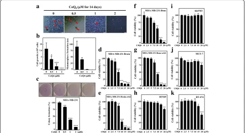
COQ0attenuates mammosphere formation in MDA-MB-231
cells
A previous research depicted the development of mam-mospheres, which are spherical clusters of nonadherent mammary stem/progenitor cells [33]. To investigate whether CoQ0affects tumor cell mammosphere
forma-tion, we exposed cells to various concentrations of CoQ0
for 14 d. Intriguingly, our results indicate that CoQ0
inhibited nonadherent spherical breast cancer clusters in vitro so that the cells became noncompetent in generat-ing secondary spheres and differentiatgenerat-ing along more than one lineage (Fig. 2a-b). As indicated in Fig. 2a, reduction in the size and formation of such spheres was observed for CoQ0 (0.5, 1, and 2μM) treatment.
proliferation capacity of these cells in the presence of CoQ0. A significant reduction in cell growth of
MDA-MB-231 was noted (Fig. 2b). The collective outcome of the aforementioned results suggests that CoQ0exhibits a
strong anti-mammosphere forming capability.
CoQ0attenuates colony formation
Subsequently, we determined whether CoQ0could affect soft
agar–cultured MDA-MB-231 cells anchorage-independent growth, a property that tumor cells demonstrate with in vivo tumorigenesis. Dose-dependent inhibition (5 d) of the growth of anchorage-independent MDA-MB-231 cells was noted (Fig.2c). Colony number inhibition increased with a reduc-tion in colony size. The decreased colony formareduc-tion ability with CoQ0indicates reduced MDA-MB-231-cell tumorigenic
ability. These results reveal that CoQ0 effectively hinders
MDA-MB-231-cell survival and growth.
CoQ0inhibits breast cancer cells viability
CoQ0 and its cytotoxic effects on the proliferation of
TNBC MDA-MB-231-Brain, MDA-MB-231-Brain-erb2,
MDA-MB-231-Bone, MDA-MB-231-Bone-erb2, BT549, Hs578T, BT474, and estrogen receptor-positive MCF-7 were explored. Cells underwent 24-h treatment with 0– 20μM concentrations of CoQ0. Significant cytotoxic
ef-fects were observed in the MB-231-Bone, 231-Bone-erb2, 231-Brain, and MDA-MB-231-Brain-erb2 cell lines dose dependently (Fig. 2d-g), whereas CoQ0 showed no to minimal effect against
BT549, Hs578T, MCF-7, and BT474 (Fig.2h-k). These re-sults indicated that CoQ0 was more potent against the
brain and bone metastatic variants of TNBC cells than against other tumorigenic cells.
CoQ0attenuates NFκB activation by suppressing I-κBα
degradation in MDA-MB-231 cells
The NFκB transcription factor family regulates myriad genes involved in MMP, VEGF, or uPA expression. Therefore, the effect of CoQ0on NFκB signaling and its
regulatory proteins in MDA-MB-231 cell were studied. Immunofluorescence assay shows that CoQ0 treatment
diminished nuclear p65 protein dose dependently (Fig.3a).
a
b
c
d
e
f
g
h
Fig. 1CoQ0inhibits proliferation, cell migration and invasion in human breast cancer cells.aStructure of CoQ0.bHuman tumorigenic breast cancer cell lines TNBC, MDA-MB-231, and non-tumorigenic line MCF-10A were treated with CoQ0(2.5–20μM) or vehicle control (0.1% DMSO) for 24 h. MTT colorimetric assay was used to determine cell viability.c-fCoQ0inhibits migration and invasion of MDA-MB-231 cells. Cells MDA-MB-231 were treated with corresponding concentration of CoQ0for 24 h.c-dCells were scratched, and migration was observed by an optical microscope (200 × magnification). The area closure was calculated by commercially available software.e-fInvasiveness was determined by counting per sample three microscopic fields. The inhibitory percentage of invading cells was quantified and expressed with untreated cells (control) representing 100%.
NFκB activity, which was ascertained through luciferase re-porter assays, was high in the control group but decreased dose dependently in cells treated using CoQ0(Fig.3b).
Next, we determined whether the inhibition of NFκB by CoQ0 is related to I-κBα protein degradation, which
regulates NFκB stability. CoQ0 treatment attenuated
I-κBα degradation, which in turn hindered NFκB (p65) activation (Fig.3c). To determine whether I-κBα degrad-ation suppression was the result of IKKα phosphoryl-ation inhibition, we used Western blotting to explore IKKα phosphorylation. CoQ0 pre-treatment suppressed
IKKαphosphorylation dose dependently (Fig.3c). These results indicate that CoQ0suppressed nuclear activation
of NFκB by inhibiting I-κBαdegradation.
CoQ0inhibits MMP-9 through the inhibition of PI3K/AKT/
NFκB pathways in MDA-MB-231
NFκB has a major role in managing the MMP expres-sion in numerous cancer cell lines. To more profoundly comprehend the inhibitory mechanisms of CoQ0on the
transcriptional regulation of MMP-9, we investigated NFκB and MMP-9 through Western blotting. The cells underwent celastrol (an NFκB inhibitor) pre-treatment
and then 24-h CoQ0treatment. The results revealed that
CoQ0 lowered p65 and MMP-9 protein expression.
However, inhibition was more pronounced in the case of celastrol pre-treatment (Fig.3d).
Based on the findings that CoQ0 inhibited NFκB and
MMP-9 expression and because PI3K/AKT is the major pathway involved in NFκB/MMP-9 activation, we hypothe-sized that CoQ0regulates the PI3K/AKT pathway. We
eval-uated this hypothesis by appraising the impact of CoQ0on
PI3K and AKT phosphorylation. As shown in Fig.3e, CoQ0
substantially decreased p-PI3K and p-AKT expression dose dependently. Subsequently, we investigated the impact of LY294002, a PI3K inhibitor, on nuclear p65 and MMP-9 expression in the absence or presence of CoQ0. Lower
nu-clear p65 and MMP-9 expression was recorded in the pres-ence of LY294002 (Fig.3f). These results suggest that CoQ0
suppressed MMP-9 by inhibiting the PI3K/AKT/NFκB pathways of MDA-MB-231 cells.
CoQ0suppresses EMT by restoring E-cadherin pathways
in MDA-MB-231
Incubating cells by using varying CoQ0 concentrations
(0.5–2μM) for 24 h resulted in abrupt morphological
a
b
c
d
e
f
g
h
i
j
k
a
b
c
e
d
f
Fig. 3CoQ0inhibits metastasis through the downregulation of PI3K/AKT/NFκB signaling pathways in MDA-MB-231 cells.aCells were grown in chamber slides and exposed to CoQ0(0.5–2μM) for 2 h, fixed and permeabilized. Cells were incubated with anti-p65 antibody followed by FITC-labeled secondary antibody. The subcellular localization of p65 was visualized using a confocal microscope of 40 × magnification.bNFκB activity was evaluated via luciferase reporter gene assay. Cells were transfected with the luciferase reporters and co-transfected with either NFκB or empty vector. Then, they were treated with CoQ0(0.5–2μM) for 4 h, and luciferase activity was determined and normalized withβ-gal activity, shown as relative luciferase activity.cCoQ0induces nuclear p65 and inhibits cytosolic I-κB and p-IKK degradation. Cells were treated withCoQ0
changes compared with the control group (Fig. 4a). E-cadherin, a protein that has a role in regulating EMT, was assayed in MDA-MB-231 cells. A rise in E-cadherin expression due to CoQ0 may be because E-cadherin
transcriptional activity was activated. Thus, we measured E-cadherin promoter activity on the basis of luciferase activity: the data are provided in Fig. 4b. We transfected an E-cadherin promoter construct in a pcDNA vector into MDA-MB-231 cells; we then ascertained cell lucif-erase activity. The findings indicated that luciflucif-erase ac-tivity from the E-cadherin promoter consistently and dose dependently improved following CoQ0 treatment
(0.5–2μM) (Fig.4b).
The immunofluorescence assay revealed that CoQ0 in-duced the expression of E-cadherin. E-cadherin anti-bodies were used for immunofluorescence staining, and the results indicated E-cadherin upregulation in the CoQ0-treated cells (Fig.4c). Western blotting as well as
RT-PCR affirmed E-cadherin upregulation by CoQ0.
Increased dose-dependent expression of Occludin, followed by reduced Vimentin, Slug, Twist, and Snail ex-pression, while an increase expression of E-cadherin was only observed at 0–1μM and not at 2μM was recorded
for CoQ0-treated MDA-MB-231 cells (Fig.4d).
Further-more, increased mRNA E-cadherin expression and re-duced Snail and Slug expression with dose-dependent 6-h CoQ0treatment were observed (Fig.4e). These
out-comes indicate that CoQ0 inhibited EMT through the
upregulation of E-cadherin pathways.
CoQ0attenuates EMT by inhibiting Wnt/β-catenin signaling
pathways in MDA-MB-231
E-cadherin/β-catenin protein complexes play an active role in EMT and are critical to cancer progression [34]. To evaluate the effects of CoQ0on the E-cadherin/β
-ca-tenin complex; we first measured the β-catenin expres-sion of MDA-MB-231 cells. The results demonstrated that CoQ0treatment suppressedβ-catenin expression in
the cytoplasm and nucleus dose dependently (Fig. 5a). Moreover, the mRNA gene expression of β-catenin was subdued by CoQ0 treatment in the MDA-MB-231 cells
(Fig. 5b). To prove if CoQ0 treatment in MDA-MB-231 cells modulated β-catenin transcriptional activity, a TOP/FOP luciferase reporter system was used. De-creased luciferase activity expression was observed in cells transfected with TOP reporter vector when dose
a
b
d
c
e
dependently treated with CoQ0. By contrast, CoQ0 did
not affect cells that were transfected with FOP reporter vector and used as negative control. Subsequently, en-hanced E-cadherin and β-catenin association was noted (Fig. 5d).
Next, we examined E-cadherin by using a luciferase re-porter construct. This construct was stably transfected into MDA-MB-231 cells. A luciferase reporter assay revealed that cells treated with CoQ0 (1μM) caused a
profound increase in E-cadherin promoter activity. Intri-guingly, cells that underwent pre-treatment with the NFκB inhibitor celastrol (0.3μM), β-catenin inhibitor iCRT5 (5μM), or both exhibited a significant increase in E-cadherin luciferase activity relative to the control cells (Fig.5e), suggesting that CoQ0induces the expression of
E-cadherin by suppressing the β-catenin and NFκB sig-naling pathways in MDA-MB-231 cells. We transfected MDA-MB-231 cells with β-catenin siRNA to confirm this phenomenon; consequently, we observed changes in E-cadherin, Snail, and Slug proteins following CoQ0
treatment. Through Western blotting, we determined that CoQ0treatment considerably increased the expression of
E-cadherin and suppressed that of Slug and Snail in MDA-MB-231 cells transfected with β-catenin siRNA (Fig. 5e). These results suggest that CoQ0 would act on E-cadherin expression thus modulating EMT by not only Wnt/β-catenin, but also NFκB pathway.
CoQ0inhibits metastasis and EMT induced by TNF-α/TGF-β
The non-toxic concentrations of CoQ0 (2μM) were
employed to assess the anti-EMT and antimetastatic ef-fects in TNF-α/TGF-β-stimulated non-tumorigenic MCF-10A breast cells. MCF-10A intrusion was ascer-tained using a Boyden chamber assay. This assay deter-mined cells’ ability to traverse a Matrigel-coated filter’s extracellular matrix layer. The findings revealed that TNF-α/TGF-β treatment enhanced MCF-10A cell inva-siveness significantly compared with that of untreated cells. Additionally, CoQ0pre-treatment resulted in
consid-erable inhibition of TNF-α/TGF-β-induced invasiveness of MCF-10A (Fig. 6a). Subsequently, we investigated the effect of CoQ0 on TNF-α/TGF-β-induced stimulation of
MMPs and uPA levels. Zymography and Western blotting analysis showed that CoQ0pre-treatment attenuated the
a
c
d
b
e
f
Fig. 5CoQ0inhibits EMT through downregulation ofβ-catenin signaling pathways in MDA-MB-231 cells.aCoQ0inhibitedβ-catenin nuclear translocation and transcriptional activation.β-catenin levels in total nuclear and cytoplasmic fractions were determined by Western blotting. The cells were incubated with or without CoQ0(0.5–2μM) for 24 h. Histone H3 andβ-actin were used as internal loading controls for nuclear and cytoplasmic fractions, respectively.bβ-catenin mRNA expression was determined by RT-PCR analyses after 6 h treatment with CoQ0(0.5–2μM).
MMP-2 and MMP-9 activity (Fig.6b) and uPA protein ex-pression in MCF-10A cells induced by TNF-α/TGF-β (Fig.6c).
To ascertain the impact of CoQ0on TNF-α/TGF-β
-in-duced cell morphology, MCF-10A cells underwent 24-h TNF-α/TGF-β treatment. Subsequently, their F-actin distribution was analyzed. As shown in Fig.6d, cells that underwent TNF-α/TGF-β treatment were redistributed from the epithelial to fibroblastic phenotype, whereas CoQ0pre-treatment reversed the TNF-α/TGF-β-induced
morphological changes. This result was, as verified through DAPI nuclear staining, independent of apop-tosis (Fig. 6d). Notably, our findings confirmed that TNF-α/TGF-βstimulation decreased E-cadherin expres-sion and increased β-catenin expression in MCF-10A cells, a hallmark of EMT (Fig. 6e). Nevertheless, CoQ0
pre-treatment increased the TNF-α/TGF-β-induced downregulation of E-cadherin while it downregulated β-catenin (Fig.6e). These findings confirmed that CoQ0
can attenuate EMT by upregulating the E-cadherin and downregulating β-catenin signaling pathways and sup-pressing metastasis by inhibiting invasion and downreg-ulating the expression of MMP-2/9 and uPA in TNF-α/ TGF-β-activated MCF-10A cells.
CoQ0induces apoptosis through generation of ROS in
MDA-MB-231 cells
Studies have implicated ROS generation as a cellular apoptosis inducer [35]. To ascertain whether the gener-ation of ROS is associated with CoQ0-engendered
apop-tosis, the intracellular ROS level in CoQ0-treated
MDA-MB-231 cells was determined. CoQ0dose
depend-ently induced apoptosis in breast cancer cells; its effect was observed through death Annexin V-FITC/PI flow cytometry and staining (Additional file 1 a-d). Incuba-tion of cells with CoQ0 (15μM) for 5–60 min caused
DCF fluorescence to increase time dependently, which was directly proportionate to the amount of ROS gener-ated maximum at 15 min (Fig. 7a-b). ROS levels dose-dependently increased in CoQ0-treated (5–15μM
for 15 min) MDA-MB-231 cells (Fig. 7c-d). However, cells subjected to ROS inhibitor treatment (1 mM NAC for 60 min) before CoQ0 treatment exhibited
signifi-cantly reduced ROS generation (Fig. 7c-d). Another line of evidence revealed that CoQ0-induced MDA-MB-231
cell death did not occur in NAC-pre-treated cells (Fig.7e). Western blot results revealed that NAC preincubation re-sulted in a gradual decrease in CoQ0-induced apoptotic
Bax and p53 protein expression (Fig. 7f ). These results
a
d
b
c
e
Fig. 6CoQ0inhibits TGF-β/TNF-α-induced metastasis and EMT in MCF-10A cells. Cells were pretreated with 2μM CoQ0for 1 h and then stimulated with TGF-β/TNF-α(10 ng/mL) for 24 h. (A-C) CoQ0inhibits TNF-α/TGF-β-induced metastasis.aCells invasiveness determined by counting cells in three microscopic fields per sample.bCoQ0inhibits TNF-α/TGF-β-induced MMP-2/−9 and uPA. Inhibition of MMP-2 and MMP-9 activity in conditioned medium from MCF-10A cells was evaluated using gelatin zymography.c CoQ0inhibits TNF-α/TGF-β-induced uPA. uPA protein expression was monitored by using Western blot analyses. (D-E) CoQ0 inhibits TNF-α/TGF-β-induced EMT.dCytoskeletal pattern of F-actin was measured by immunofluorescence analyses (100 × magnification).eCoQ0-induced TNF-α/TGF-β decreased E-cadherin and inhibited TNF-α/TGF-β-induced
evince that CoQ0 triggered the production of ROS in
MDA-MB-231 cells that could engender apoptotic cell death.
In vivo growth inhibition in xenografted mouse model by CoQ0
To determine the in vivo impact of CoQ0 on tumor
growth, xenografted nude mice were used. We xeno-grafted MDA-MB-231 cells into them. Observations in-dicated that all mice were healthy and that their body weights were unaffected during CoQ0 treatment (FiA).
The MDA-MB-231 xenografted animals were treated with CoQ0 (0.75 mg/kg three times/week) or with only
vehicle. A significant time-dependent inhibition of tumor volume was observed for CoQ0treatment (Fig.8a).
Add-itionally, a reduction in tumor weight in the CoQ0-treated
xenografted mice was observed (Fig.8b). After 12 weeks, the animals were killed and the xenografted tumor was ex-tracted. Concomitantly, excised tumor sections were ob-served under the microscope to discern the differences in nuclei and cytoplasmic morphology. The cancer cells in the xenografted mice used as controls appeared large and oval or round in form with myriad nucleoli, and expressed substantial mitotic figure and cellular activity levels (Fig. 8c). By contrast, the CoQ0-treated tumor-xenografted
mice demonstrated less angiogenesis, and their cells
appeared shrunken and condensed. Furthermore, their nu-clei exhibited karyopyknosis, implying carcinoma regression or activity (Fig.8c). These results suggest that CoQ0 pro-motes antitumor activity in xenografted mouse models.
Fragmentation of apoptotic DNA induced by CoQ0in
tumor-xenografted mouse model
CoQ0’s role in apoptosis in MDA-MB-231-xenografted
mice was ascertained using the TUNEL assay on cancer-ous sections. The presence of a greater number of TUNEL-positive cells in mouse tumors treated with CoQ0compared with those of the untreated controls (p<
0.001) suggested that CoQ0treatment was affiliated with
reduced cell proliferation and increased apoptosis (Fig. 8d). In addition, immunohistochemical analysis revealed that CoQ0-treated mice significantly decreased
Cyclin A, Cyclin B, and CDK1 expression and increased Bax, p53, and p21 expression in MDA-MB-231 xeno-grafted tumor tissues (Fig. 8e). By contrast, CoQ0
treat-ment in MDA-MB-231 cells resulted in G2/M arrest and downregulation of the expression of Cyclin B, Cyclin A, Cdc2, and Cdc25C (Additional file 2 a-b). Subsequently, Western blotting was employed to determine the effect of CoQ0on Cyclin A and B proteins in MDA-MB-231
xeno-grafted tumor tissues. Cyclin A and B expression was sup-pressed relative to that the controls (Fig.8f ). In summary,
a
b
e
c
d
f
a
b
c
d
e
f
the inhibition of tumor development by induction of apoptosis in TNBCs during CoQ0 treatment was
observed.
CoQ0attenuates in vivo lung metastasis
To validate the potential effect of CoQ0 on metastasis,
we treated mice with CoQ0 (1.5 or 2 mg/kg) and then
intravenously injected them with MDA-MB-231-lucifer-ase cells (1 × 10⁶ cells/well). The luciferase-labeled MDA-MB-231 cancer cells metastasized to the lungs after 28 d in the control mice (Fig. 9a). CoQ0treatment
blocked MDA-MB-231 cell lung metastasis to a statisti-cally significant level; photon flux in the mouse lungs fell by > 90% in the CoQ0treatment groups (1.5 and 2 mg/
kg) (Fig. 9a). These findings imply that CoQ0 entirely
suppresses highly metastatic breast cancer cell lung me-tastasis at concentrations of 1.5 or 2 mg/kg.
To validate the mechanism by which CoQ0attenuates
tumor metastasis, the metastasis-related proteins from control and CoQ0-treated (0.75 mg/kg) mouse tumors
were studied. Immunohistochemical and Western blot results revealed that CoQ0 inhibited MMP-9, MMP-2,
p-AKT, p65, and β-catenin expression and increased E-cadherin expression relative to the controls (Fig.9b-c). Comprehensive results confirmed that CoQ0suppresses
metastasis by downregulation of MMP-2, MMP-9, p65, β-catenin, and p-AKT and upregulation of E-cadherin proteins. Furthermore, the gene expression patterns of MMP-2 and MMP-9 mRNA were substantially suppressed by CoQ0 (Additional file 3 a). In addition,
immunohistochemical analysis revealed significant de-creases in uPA, uPAR, Vimentin, COX-2, Twist, VEGF, and p-mTOR in CoQ0-treated mice (Additional file3b).
Subsequently, we investigated the effects of CoQ0 on
EMT regulatory proteins. CoQ0 substantially reduced
uPA, uPAR, COX-2, and Vimentin expression and pro-moted Occludin protein levels compared with those of the control group as discerned through Western blot analysis (Additional file 3 c). These results indicate that CoQ0 attenuated metastasis and EMT in
MDA-MB-231-xenografted nude mice.
Discussion
EMT is a physiological process that is usually activated during wound healing and embryonic development. It is a crucial step in cancerous metastatic progression [36]. During EMT, the epithelial-derived tumor cells stimulate intercellular and intracellular changes that contribute to mesenchymal cell phenotypes, including cytoskeleton reorganization, polarity alteration, extracellular matrix remodeling, and migratory ability acquisition [12]. Numerous researchers have investigated EMT’s role in breast cancer. Mesenchymal EMT molecular marker overexpression in biopsies of breast cancer is correlated
with increased recurrence, adverse clinicopathological characteristics, reduced survival, and tumor aggressive-ness [37]. Therefore, efficacious therapeutic strategies must be established to reduce breast cancer cell tumor aggressiveness and prevent malignant growth. In our previous study, we reported that CoQ0exerts
antimeta-static effects in melanoma carcinomas. This action may be because of the modulation of the Wnt/β-catenin sig-naling pathway in B16F10 melanoma cells [24]. In the current study, CoQ0’s antimetastatic and anti-EMT
abil-ities were characterized, and mechanisms responsible for its effects in MDA-MB-231 were studied. Additionally, E-cadherin downregulation and alterations of the EMT-linked signaling regulator indicate that MDA-MB-231 cells can commence and propagate the EMT process in cancer cells. The salubrious impact of pre-treatment with CoQ0
was proven by the renewal of E-cadherin protein and tran-scriptional activity. The renewal of E-cadherin was linked to β-catenin, NFκB, and MMP-9 inhibition, a key molecular event in EMT inhibition. Increased cancer cell migration and invasion, mammosphere formation, colony formation ability, and tumor growth were effectively suppressed through CoQ0treatment. These findings indicate that CoQ0
is an antimetastatic and anti-EMT substance, and the poten-tial molecular signaling pathways that are involved in this process can be inferred.
E-cadherin is an adherens junction protein expressed in normal breast tissue; it is a useful phenotypic marker in cases of breast cancer [38]. In this study, the tran-scriptional activity and protein levels of E-cadherin were investigated to ascertain the manifestation of EMT with TNF-α/TGF-β-stimulation in TNBC cells. The results of immunofluorescence and luciferase activity and Western blotting revealed that TNF-α/TGF-β could undermine E-cadherin junctions by governing the organization of actin in MDA-MB-231 cells. These results were supported by evidence from previous studies [33]. E-cadherin loss stimulates EMT, which plays a major role in the develop-ment of carcinomas to a metastatic state. Although the mechanism that is involved in E-cadherin inactivation in cancer cells remains vague, alterations of transcriptional levels may explain its downregulation [39]. Therefore, an effective strategy for controlling metastasis and EMT pro-gression may be restoring or preventing E-cadherin down-regulation by using TNF-α/TGF-β. In this work, the restoration of E-cadherin protein levels and transcriptional activity through CoQ0 treatment inhibited EMT and
the associated carcinoma metastasis. Restoration of E-cadherin expression at the transcription and protein level by Withaferin A was linked to metastasis and cell proliferation inhibition in breast cancer cells [33]. Furthermore, our in vivo study proved that CoQ0
cell lung metastasis, implying that CoQ0 arrests EMT
programming because of its antimetastatic properties in breast cancer cells.
EMT is a crucial mechanism in cancer development and in the first phase of metastasis. Retardation of E-cadherin/β-catenin may facilitate tumor invasion and
metastasis [40]. Increasingly, evidence indicates that E-cadherin has a vital function inβ-catenin function and stabilization. When E-cadherin expression decreased, β-catenin was able to be separated from the E-cadherin/ β-catenin complexes and could translocate to the nu-cleus freely. Moreover, β-catenin bound with the TCF/
a
b
c
Fig. 9In vivo anti-metastatic activity of CoQ0.aCoQ0inhibited lung metastasis in living MDA-MB-231-luciferase-injected mice by bioluminescence imaging. Mice were treated with CoQ0(1.5 or 2 mg/kg) and then the MDA-MB-231-luciferase cells (1 × 10
6
LEF-1 element after which it activated certain promigra-tory genes required for EMT combined with related transcription factors [41]. Some transcription factors, in-cluding Slug and Snail, which are both among the tran-scriptional targets of β-catenin, may be associated with E-cadherin and EMT repression [42]. In this work, CoQ0 induced E-cadherin and significantly decreased
nuclearβ-catenin, Snail, and Slug protein association, as shown by Western blot analysis. Furthermore, immuno-precipitation assays revealed that CoQ0 increased
E-cadherin and β-catenin expression relative to that of the untreated group. This result indicates that CoQ0
may restore the formation of E-cadherin/β-catenin complexes in MDA-MB-231 cells, impeding nuclear transport of β-catenin to a greater extent, which sub-sequently enhances the expression of E-cadherin by inhibiting Slug. Our results are a strong indication notion that the anti-EMT impact of CoQ0 is
corre-lated with the governance of the formation of E-cadherin/β-catenin complexes.
Matrix metalloproteases (MMPs) have a key func-tion in extracellular matrix (ECM) remodeling and degradation [43]. MMPs play roles in all stages of breast carcinogenesis, from tumor initiation to me-tastasis. Among the several MMP family members, MMP-2 and MMP-9 were highly expressed in inva-sive breast cancer cells [44]. The present study de-termined that CoQ0 pre-treatment abrogated the
TNF-α/TGF-β-induced MMP-9 and MMP-2 expres-sion levels in MDA-MB-231 cells. Therefore, MMP-9 and MMP-2 could be CoQ0-responsive mediators
whose ECM degradation could result in ensuing can-cer invasion and migration.
The MMP-9 promoter region possesses cis-regulatory elements, such as two AP-1 and one NFκB binding sites. These sites are not present in MMP-2’s promoter region [45]. Therefore; we investigated the effects of CoQ0 on
NFκB, which plays a major role in the transcription of MMP-9. NFκB activation results in cell invasion, metas-tasis, and survival advantages and drug resistance to sev-eral cancer types [46]. Nuclear translocation and transcriptional activation of NFκB subunits are strictly governed by NFκB’s inhibitory protein, I-κBα, whose phosphorylation releases NFκB subunits [47]. The data from our experiments clearly demonstrate that treat-ment with CoQ0 suppressed the transcriptional
activa-tion and nuclear translocaactiva-tion of NFκB. This effect may have been caused by the inhibition of I-κB kinase phos-phorylation and I-κBα degradation. Furthermore, pre-treatment with celastrol remarkably reduced the ex-pression of MMP-9 and NFκB proteins. This result indi-cates that the NFκB pathway is the principal regulatory pathway in the suppression of MMP-9 expression by treatment with CoQ0.
PI3K/AKT is the major pathway for tumor invasion [48]. Therefore, we sought to determine whether CoQ0
suppresses the phosphorylation of PI3K/AKT because its signaling cascade is the main component upstream of NFκB and plays a key role in cellular adhesion, differen-tiation, and growth. The PI3K/AKT axis plays a principal role in metastasis and tumor invasion through activation of NFκB-mediated MMP-9 [49]. Our findings clearly demonstrate that CoQ0treatment suppressed PI3K/AKT
phosphorylation substantially. Furthermore, NFκB acti-vation and MMP-9 were significantly reduced by block-age of the PI3K/AKT pathway with LY249002 treatment. These findings reveal that CoQ0 lowers the expression
of MMP-9 by blocking NFκB activation through PI3K/ AKT and thus suppresses MMP-9-mediated cell intru-sion in MDA-MB-231 human breast cancer cells. Our findings are consistent with those of a report that sug-gested that LFG-500 extracted from flavonoid inhibits cancer cell intrusion by suppressing the PI3K/AKT/ NFκB/MMP-9 signaling pathways [49].
The present paper documents the EMT and anti-metastatic capabilities of CoQ0and lists the mechanisms
that may cause its effects in non-tumorigenic MCF-10A cells under stimulation induced by TNF-α/TGF-β. TGF-βenhances tumor development by activating EMT. TGF-β-induced EMT exhibited the following attributes: the loss of junctional E-cadherin localization, acquisition of fibroblastic morphology, and increased cellular motil-ity [50]. TNF-α is a proinflammatory cytokine and plays a vital role in tumor malignancy, including motility, tumor cell invasion, and metastasis [51]. TNF-αinduced EMT in renal cell carcinoma by suppressing E-cadherin expression and promoting Vimentin and MMP-9 protein expression [52]. Stimulation of TGF-β, TNF-α, or both may cause an EMT-like phenomenon, E-cadherin ex-pression reduction, and morphological changes in Madin–Darby canine kidney cells [53]. In the present study, downregulation of E-cadherin, upregulation of β-catenin, and changes in EMT-linked signaling regula-tors initiated and propagated EMT in MCF-10A cells stimulated using TNF-α/TGF-β. The advantageous im-pact of pre-treatment with CoQ0 was based on the
re-newal of transcriptional and E-cadherin promoter activity against losses induced by TNF-α/TGF-β. Add-itionally, the renewed E-cadherin promoter activity was linked to β-catenin, NFκB, and MMP-2/−9 inhibition, which is a vital molecular event in the inhibition of EMT induced by TNF-α/TGF-β.
many cancers, including breast cancer, are guided by a cellular subpopulation, designated as cancer stem cells (CSCs), that mediates tumor metastasis and resistance to conventional therapies. Thus, preventing CSC growth in breast cancer is the optimal strategy for inhibiting tumor development and metastasis [55]. Therefore, re-search on CoQ0-induced molecular mechanisms that
mediate CSC proliferation is vital to clarify CoQ0’s
anti-metastatic and anticancer activities. Our study revealed that CoQ0 treatment considerably lowered
mammo-sphere formation and mammo-sphere size. These results suggest that CoQ0inhibits mammosphere formation.
Apoptosis induction, restriction of cell proliferation by chemical or biological agents, and cell-cycle arrest are intended to be effective strategies in cancer manage-ment, particularly of TNBCs. Apoptosis-inducing agents are under investigation as alternative tools for cancer treatment management. A study reported that CoQ0
treatment caused the proportion of late apoptotic MDA-MB-231 cells to rise when Annexin V/PI staining and then flow cytometry were employed [56]. In the present study, the treatment of MDA-MB-231 cells with CoQ0, successfully inhibited anchorage-independent
growth and cell proliferation. Examples of the character-istic features of apoptosis are chromatin condensation, internucleosomal DNA cleavage, caspase activation, and cellular morphological changes [57]. In the current study, we demonstrated that, by treating MDA-MB-231 cells with CoQ0, apoptotic cell death linked to DNA
fragmentation increased considerably. In a study, treat-ing human lung cancer cells with CoQ0 increased the
number of early and late apoptotic cells and reduced apoptotic cell death through antioxidant treatment [19]. Other studies have demonstrated that methoxy-contain-ing analogs of CoQ0 and quinones that have similar
structures to CoQ0have a cytotoxic influence on human
cancer cells because they induce apoptosis [58]. Re-searchers employed various CoQ analogs and recorded enhanced DNA fragmentation, caspase-3 activation, and apoptosis for CoQ4and CoQ2in HL 60 human leukemia
cells. However, these effects were not observed for CoQ10 or CoQ6. These results suggest that CoQ0
ana-logs pro-apoptotic and anticancer attributes vary de-pending on the location of the methoxy-substitutions on the quinone nucleus and the length of the isoprenyl side chain. No matter the cell line, CoQ0, which possesses no
isoprenoid units, suppresses cancer cell growth and trig-gers early and late apoptosis.
The excessive generation of ROS can induce cell-cycle arrest, oxidative stress, damaged DNA in cancer cells, cell function loss, and cellular apoptosis [59]. CoQ0’s
favorable impact on breast cancer cell lines is linked to mitochondrial dysfunction and the overproduction of intracellular ROS. ROS causes the mitochondrial
permeability transition pore to open, mitochondrial proapoptotic factors to be released, and the mitochondrial membrane to depolarize during mitochondria-mediated apoptosis [60]. The present study determined that CoQ0
treatment leads to a notable increased in intracellular ROS production in MDA-MB-231 cells. By contrast, the antioxidant, NAC, inhibited ROS production, which re-duced apoptosis significantly, indicated that MDA-MB-231-cell apoptosis induced by CoQ0had a close link with
ROS production. CoQ0potentially play roles as upstream
signaling molecules to induce cell apoptosis mediated by mitochondria. Our findings are agreement with those of prior investigations indicating that natural compounds (e.g., celastrol and deltonin) induce MDA-MB-231 cell ROS-mediated mitochondrial apoptosis [61].
Disruption of the cancer cell cycle is a therapeutic ob-jective of research on novel cancer drugs. This is linked to lower Cyclin A, Cyclin B, Cdc2, and Cdc25C expres-sion and higher CDK inhibitor p21 expresexpres-sion. In eu-karyotes, cell-cycle progression included the resultant triggering of CDKs; their activation is cyclin associated. Among CDKs, Cdc2 and CDK2 kinases are mainly trig-gered with Cyclin B and Cyclin A during G2/M phase progression [62]. Cdc2/Cyclin A and Bi kinase complex activity was suppressed by phosphorylating Tyr15 of Cdc2. Cdc25C phosphatase catalyzed the dephosphory-lation of Tyr15 of Cdc2. This reaction was considered the rate-limiting step in their progression into mitosis [63]. P21 might facilitate G2/M cell-cycle arrest main-tenance through CyclinB1/Cdc2 complex inactivation, thereby disrupting the cell nuclear antigen–Cdc25C interaction [64]. The findings suggest that Cdc25C, Cdc2, Cyclin A, and Cyclin B expression is downregu-lated, and the CDK inhibitor p21 increased in MDA-MB-231 cells treated with CoQ0, which arrests G2/M
phase. The present study’s data suggest that the moni-tored suppression of MDA-MB-231 cell proliferation linked to CoQ0 treatment was because of G2/M-phase
cell-cycle arrest and not G1 arrest. Intriguingly, our re-sults differ from those of the previous report that indi-cated that treatment of MDA-MB-231 cells with CoQ0
led to G0/G1-phase cell-cycle arrest.
To enhance CoQ0’s antimetastatic and anticancer
attri-butes, an in vivo investigation of CoQ0-treated
MDA-MB-231-xenografted nude mice was executed. CoQ0
-treated xenografted nude mice resulted in a significant fall in tumor volume and significantly prevented lung metastasis. The observed anticancer action is seemingly related to mitotic cell inhibition and substantial prolifer-ation of apoptotic cells in tumors treated with CoQ0.
Furthermore, antimetastatic activity may be linked to the upregulation of E-cadherin and the downregulation of MMP-2, MMP-9, p-AKT, p65, andβ-catenin proteins. These in vivo results verify CoQ0’s effective
antimeta-static and antitumor attributes against TNBC that are in agreement with its in vitro anticancer attributes.
Conclusion
This work determined for the first time that non-cyto-toxic concentrations of CoQ0exhibit antimetastatic and
anti-EMT attributes in MDA-MB-231 breast cancer cell. EMT inhibition in MDA-MB-231 cells was linked to the renewal of transcriptional activity and E-cadherin pro-tein. CoQ0 inhibited mammosphere formation and
ex-hibited its anti-invasive activity by downregulating the PI3K/AKT/NFκB/MMP-9 signaling pathways. Further-more, CoQ0 induced ROS-mediated apoptosis and
sig-nificantly inhibited the growth of tumors in MDA-MB-231 xenografted nude mice. Our results provide new insight into the potential molecular mechanisms that underlie CoQ0’s promising anticancer attributes.
Our findings justify further preclinical and clinical as-sessments of CoQ0for metastatic breast cancer therapy.
Additional files
Additional file 1:CoQ0-induced apoptosis in MDA-MB-231 cells. The cells were exposed to CoQ0(5–15μM for 24 h). (a-b) The TUNEL assay was performed to determine apoptotic DNA fragmentation. The green florescence indicates the number of TUNEL positive cells in the microscopic fields (400 × magnification) from three separate samples. The percentage of apoptotic cells was calculated by measuring the florescence intensity of treated cells using commercially available software. (c) Annexin V-FITC and PI staining was used to identify the early/late apoptosis or necrosis, and the data were analyzed using flow cytometry. The results in each quadrant are labeled and interpreted as follows: (Q1) PI positive, Annexin V-FITC-negative stained cells/necrosis. (Q2) PI positive, Annexin V-FITC-positive stained cells/ late apoptosis. (Q3) Cells negative for both PI and Annexin V-FITC staining/ normal live cells. (Q4) PI-negative, Annexin V-FITC-positive stained cells/early apoptosis. (d) Effects of CoQ0on apoptotic-related proteins. Protein levels of mitochondria/cytosolic cytochrome c, caspases-9, caspase-3, and PARP, Bax, Bcl-2, and p53 were analyzed by Western blotting. The results are presented as the mean ± SD of three independent assays. ***p< 0.001 significant compared to control cells. (PPTX 51519 kb)
Additional file 2:CoQ0treatment induces G2/ M cell-cycle arrest in MDA-MB-231 cells. (a) Cells were treated with CoQ0(5–15μM) for 24 h, stained with PI and analyzed for cell-cycle phase using flow cytometry. The cellular distributions (%) in different phases of the cell cycle (sub-G1, G1,S and G2/M) were determined after treatment with AS. The flow cytometry graph shown here is from one representative experiment that was performed in triplicates. (b) The effects of CoQ0AS on cell-cycle regulatory proteins. HL-60 cells were treated with increasing concentrations of CoQ0(5–15μM) for 24 h. Cell-cycle regulatory proteins, including Cyclin A, Cyclin B, p21, Cdc2, Cdc25C, CDK2, and CDK4 were examined using Western blot analyses. The results are presented as the mean ± SD of three independent assays. **p< 0.05, ***p< 0.001 significant compared to control cells. (PPTX 4253 kb)
Additional file 3:Metastasis and EMT inhibition by CoQ0in MDA-MB-231 xenografted tumors. Tumor sections were from control animals and experimental analogues treated with CoQ0(0.75 mg/kg). (a) Cells positive for the indicated proteins were counted from 3 fields (200 × magnification)
for each tumor sample, and MMP-2 and MMP-9 were examined using RT-PCR. (b) uPA, uPAR, Vimentin, COX-2, Twist, VEGF, and p-mTOR were examined using immunohistochemical staining. (c) uPA, uPAR, Vimentin, COX-2, and Occludin were examined using Western blotting. Western blot on the effects of CoQ0on the total protein contents in the xenograft tumors.β-actin was used as the control. Relative changes in protein bands were measured by densitometric analysis with the control being 100%. The results are the mean (±SE) numbers of cells/microscope field (as percentage) for 3 animals per group. Significant at *p< 0.05; **p< 0.01; ***p< 0.001 compared to untreated control cells. (PPTX 12155 kb)
Abbreviations
EMT:Epithelial–mesenchymal transition; HER2: Human epidermal growth factor receptor 2; TNBC: Triple Negative Breast Cancer Cell
Acknowledgements
The authors would sincerely like to thank the reviewers and the editor of this manuscript for taking the time and effort in going through our manuscript.
Funding
This study was financially supported from grants MOST-106-2320-B-039-054-MY3, MOST-107-2320-B-039-013-MY3 and CMU 107-S-47 from the Ministry of Science and Technology, Taiwan as well as by the“Chinese Medicine Research Center, China Medical University”from the Featured Areas Research Center Program within the framework of the Higher Education Sprout Project by the Ministry of Education (MOE) in Taiwan (CMRC-CHM-8).
Availability of data and materials
The supplementary material for this research is available online.
Authors’contributions
Conceived and designed the experiments: YCH, HLY, and KYL conceived the idea. YCH, PCS, HLY, and KYL performed the experiments. PCS and DCM analyzed the data. YCH, DCM, VT, and HLY wrote the paper. All authors read and approved the final manuscript.
Ethics approval
All experiments involving animals were performed with the permission and under the strict guidance of the Institutional Animal Care and Treatment Committee of China Medical University. A written consent to participate for these studies was given by all authors.
Consent for publication
All authors have given their consent for the publication of this article.
Competing interests
The authors declare that they have no competing interests.
Publisher’s Note
Springer Nature remains neutral with regard to jurisdictional claims in published maps and institutional affiliations.
Author details
1Institute of Nutrition, College of Biopharmaceuticals and Food Sciences,
China Medical University, Taichung 40402, Taiwan.2Department of Cosmeceutics, College of Biopharmaceutical and Food Sciences, China Medical University, No. 91, Hsueh-Shih Road, Taichung 40402, Taiwan.3Department of Medical Research, Chi-Mei Medical Center, Tainan 710, Taiwan.4Graduate Institute of Veterinary Pathology, National Chung Hsing University, Taichung 40227, Taiwan. 5