Open Access
Research
Telomerase prevents accelerated senescence in
glucose-6-phosphate dehydrogenase (G6PD)-deficient human
fibroblasts
Yi-Hsuan Wu
1, Mei-Ling Cheng
2, Hung-Yao Ho
2, Daniel Tsun-Yee Chiu*
2and Tzu-Chien V Wang*
3Address: 1Graduate Institute of Basic Medical Sciences, Chang Gung University, Kwei-San, Tao-Yuan 333, Taiwan, 2School of Medical Biotechnology, Chang Gung University, Kwei-San, Tao-Yuan 333, Taiwan and 3Department of Molecular and Cellular Biology, Chang Gung University, Kwei-San, Tao-Yuan 333, Taiwan
Email: Yi-Hsuan Wu - d9101302@stmail.cgu.edu.tw; Mei-Ling Cheng - chengm@mail.cgu.edu.tw; Hung-Yao Ho - hoh01@mail.cgu.edu.tw; Daniel Tsun-Yee Chiu* - dtychiu@mail.cgu.edu.tw; Tzu-Chien V Wang* - tcvwg@mail.cgu.edu.tw
* Corresponding authors
Abstract
Fibroblasts derived from glucose-6-phosphate dehydrogenase (G6PD)-deficient patients display retarded growth and accelerated cellular senescence that is attributable to increased accumulation of oxidative DNA damage and increased sensitivity to oxidant-induced senescence, but not to accelerated telomere attrition. Here, we show that ectopic expression of hTERT stimulates telomerase activity and prevents accelerated senescence in G6PD-deficient cells. Stable clones derived from hTERT-expressing normal and G6PD-deficient fibroblasts have normal karyotypes, and display no sign of senescence beyond 145 and 105 passages, respectively. Activation of telomerase, however, does not prevent telomere attrition in earlier-passage cells, but does stabilize telomere lengths at later passages. In addition, we provide evidence that ectopic expression of hTERT attenuates the increased sensitivity of G6PD-deficient fibroblasts to oxidant-induced senescence. These results suggest that ectopic expression of hTERT, in addition to acting in telomere length maintenance by activating telomerase, also functions in regulating senescence induction.
Background
Normal human cells grown in vitro replicate for a limited period of time before entering senescence [1], a term that has been used primarily to describe a signal transduction pathway that leads to the irreversible growth arrest of cells in culture. Among the various stimuli that are known to trigger senescence [2,3], telomere attrition and oxidative damage (produced during normal cellular proliferation) are the basis for the "telomere hypothesis" [4,5] and "free
radical theory" [6,7], respectively, postulated to account for the aging process.
According to the "telomere hypothesis" of aging, dysfunc-tional telomeres caused by telomere attrition are postu-lated to initiate the senescent phenotypes. Telomere attrition occurs because the ends of linear chromosomal DNA cannot be completely replicated by normal DNA polymerases, and, therefore, telomere DNA becomes
Published: 5 February 2009
Journal of Biomedical Science 2009, 16:18 doi:10.1186/1423-0127-16-18
Received: 29 October 2008 Accepted: 5 February 2009
This article is available from: http://www.jbiomedsci.com/content/16/1/18
© 2009 Wu et al; licensee BioMed Central Ltd.
shortened with each round of DNA replication [8,9]. When telomeres reach a critical length, they become dys-functional and trigger so-called replicative senescence. Activation of telomere length-maintenance mechanisms, such as expression of telomerase, a specialized reverse transcriptase that synthesizes telomeric DNA repeats at chromosome ends, is thought to counteract replicative senescence [5,10,11]. In support of this postulate, normal human somatic cells express low or undetectable telomer-ase activity and are mortal. In contrast, a majority of immortal and cancer cells have an indefinite proliferative capacity and maintain their telomere length by upregulat-ing telomerase [12,13]. Ectopic expression of telomerase has been shown to extend the lifespan of many normal human cells cultured in vitro [14-16].
The free radical theory of aging postulates that the accu-mulation of oxidative damage is the central mediator of the aging process [6,7]. Reactive oxygen species produced during normal cellular metabolism or from exogenous sources, such as drugs and radiation, are known to react with biomolecules, including proteins and DNA. It is this accumulation of oxidative damage over time that is postu-lated to trigger senescence [6]. In support of this hypothe-sis, exposure to oxidative stress (e.g., tert-butyl hydroperoxide, hydrogen peroxide, or a hyperbaric atmosphere with high O2 partial pressure) triggers senes-cence, and oxidative DNA damage is known to accrue dur-ing senescence [17-23]. In addition, growth of cells under hypoxic conditions (3% [v/v] O2 instead of the normal atmospheric O2 level) is known to delay cellular
senes-cence of fibroblasts [24].
Despite supporting evidence for both of these theories, based on multiple experimental approaches, little is known about the relative roles of these two mechanisms in the induction of senescence or the potential interac-tions of the signaling pathways they trigger. Recently, we have shown that fibroblasts derived from glucose-6-phos-phate dehydrogenase (G6PD)-deficient patients dis-played retarded growth and accelerated cellular senescence [25]. Evidence indicates that the accelerated cellular senescence observed in G6PD-deficient cells arises because of increased accumulation of oxidative DNA damage and an increased sensitivity to oxidant-induced senescence, but not to accelerated telomere attrition [26]. These data indicate that G6PD status – and thus proper redox balance – is a determinant of cellular senescence. It is not yet known, however, whether increased oxidative DNA damage is the only important determinant of senes-cence induction in G6PD-deficient cells. To address this point, we asked whether activation of telomerase activity might be capable of overcoming the accelerated senes-cence observed in such cells. Here, we present evidence that ectopic expression of hTERT, the key regulator of
tel-omerase, activates telomerase activity and prevents pre-mature senescence in G6PD-deficient cells.
Materials and methods
Chemicals, enzymes and oligonucleotides
Dulbecco's modified Eagle's medium (DMEM), trypsin, penicillin, streptomycin and amphotericin were pur-chased from Gibco (Karlsruhe, Germany). The anti-G6PD antibody was from Genesis Biotech (Taiwan). The anti-actin antibody was from Santa Cruz Biotechnologies (Santa Cruz, CA, USA). The antibiotic, G418 sulfate, was from Promega (Madison, WI, USA). The TeloTAGGG assay kit, alkaline phosphatase and protein kinase K were from Roche (Roche, Mannheim, Germany). Taq DNA polymerase was from Qiagen. The sequence and source of TS and CX oligonucleotides have been described [27].
Cell culture
Normal human fibroblasts (HFF3) and G6PD-deficient fibroblasts (HFF1) were routinely cultured in DMEM sup-plemented with 10% fetal bovine serum, 100 units/ml penicillin, 100 units/ml streptomycin and 0.25 mg/ml amphotericin at 37°C in a humidified atmosphere con-taining 5% CO2. Human fibrosarcoma cells (HT1080), used as a positive control in soft agar and telomerase assays, were from American Type Culture Collection (ATCC). BOSC23 and PT67 cells were used for prepara-tion of retroviral particles as previously described [26].
Plasmids, retroviral packaging and infection
The plasmid, pBABE-Puro-hTERT [28], a retroviral con-struct that expresses hTERT, was kindly provided by Dr. E. Blackburn. Retroviral packaging and infection were accomplished as previously described [26], with the exception that the retrovirus producer cells and hTERT-expressing clones were selected in a medium containing 2
μg/ml puromycin.
Telomerase activity assay
A PCR-based telomeric amplification protocol (TRAP) was used to assay telomerase activity. The preparation of cell extracts, the PCR amplification conditions, and the analysis of PCR products by electrophoresis on polyacry-lamide gel were as described [29].
Soft agar assay
To assay for contact-independent growth in soft agar, cells were trypsinized and resuspended at 3.3 × 10³ cells/ml in
Karyotyping of hTERT-expressing cells
Cytogenetic examination was performed with hTERT-expressing cells at passage 11–14. After incubating with 0.06 μg/ml colcemid for 16 h to arrest cells in mitosis, fibroblasts were collected by trypsinization and fixed with Carnoy's fixative (a 3:1 mixture of methanol and glacial acetic acid). Fixed fibroblasts were placed on a glass slide, air-dried and stained with a 6% Giemsa solution for 3.5 min. The number of chromosomes in at least 40 cells was analyzed from each fibroblast preparation.
Assay for telomere length
Cell pellets were lysed in 1 M Tris-EDTA buffer (pH 7.4) containing 0.5% SDS, and treated with 200 μg/ml protei-nase K for 18 h at room temperature. Genomic DNA was then isolated by phenol-chloroform extraction. Genomic DNA was digested with HinfI and RsaI, and DNA frag-ments were separated by electrophoresis in 0.8% agarose gels. The DNA fragments containing telomeric repeats were identified by Southern blotting using a TeloTAGGG telomere-length assay kit (Roche, Mannheim, Germany). Average telomere fragment length (TFR) was determined from measurements of the intensity of chemiluminescent signals at each molecular mass, obtained by scanning the radiogram using a Molecular Dynamics Personal Densito-meter (Sunnyvale, CA, USA). Signal densities were ana-lyzed with ImageQuaNT software (Molecular Dynamics), and mean TRF lengths were calculated according to man-ufacturer's recommendation.
Assay for H2O2-induced premature senescence
Premature senescence was tested in fibroblasts treated with different concentrations of H2O2 for 1.5 h using senescence-associated (beta)-galactosidase (SA-β-gal) as a biomarker of senescence. After H2O2 treatment, the medium was replaced with fresh complete medium, and cells were cultured for 72 h before staining for SA-β-Gal activity as previously described [26]. Quantification of premature senescence was determined by calculating the rate of conversion of 4-methylumbellliferyl-β -D-galacto-pyranoside (MUG) to the fluorescent product, 4-methyl-umbelliferone (4-MU), at pH 6.0 as previously described [30]. The relative increase in 4-MU fluorescence per mg protein was determined by subtracting the untreated con-trol values from the H2O2-treated values.
Results
Ectopic expression of hTERT immortalizes fibroblasts derived from normal and G6PD-deficient patients
To address the relative role of oxidative stress and tel-omere attrition in cellular senescence, we asked whether ectopic expression of hTERT might be capable of restoring telomerase activity and extending the lifespan of G6PD-deficient fibroblasts. Normal (HFF3) and G6PD-G6PD-deficient fibroblasts (HFF1) were infected with the retroviral
con-struct, pBABE-Puro-hTERT, which expresses hTERT under the control of the LTR promoter, and G418-resistant clones were screened for the expression of telomerase activity. G418-resistant, telomerase-positive, clones from infected normal fibroblasts (T5 and T9), or G6PD-defi-cient fibroblasts (GT3 and GT8), were randomly selected for further study. As shown in Figure 1, G418-resistant cells from pBABE-Puro-hTERT-transfected normal and G6PD-deficient fibroblasts expressed similarly high tel-omerase activity that was comparable to that of a telomer-ase-positive fibrosarcoma cancer cell line (HT1080), whereas no telomerase activity was detected in G418-resistant cells transfected with the pBABE-Puro vector.
To determine whether ectopic expression of telomerase might be capable of extending the limited population doublings previously observed for normal and G6PD-deficient fibroblasts, we examined the proliferative capac-ity of hTERT-expressing fibroblasts. As shown in Fig. 2A and 2B, cells transfected with vector alone ceased to pro-liferate after about 55 and 40 passages for V1 and GV1, respectively. In contrast, hTERT-expressing fibroblasts derived from normal or G6PD-deficient patients contin-ued to proliferate over 105 passages and showed no evi-dence of senescence, indicating that these cells were immortal. Accompanying the acquisition of unlimited growth potential in these fibroblasts was stable expression of telomerase activity, which persisted over the entire time of cell propagation (Fig. 2C).
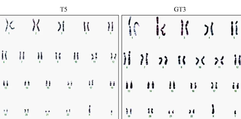
hTERT-immortalized G6PD-deficient fibroblasts do not exhibit a transformed phenotype
To address the possibility that hTERT-immortalization of G6PD-deficient fibroblasts could induce changes associ-ated with a transformed phenotype, we examined karyo-type and anchorage-independent growth. Representative results of a karyotype analysis are shown for T5 and GT3 in Figure 3A. Both the hTERT-expressing normal and G6PD-deficient fibroblasts had a normal complement of diploid chromosomes (i.e., 46 + XY), and no abnormal chromosomal structures (e.g., translocations) were detected from a total of 20 cells analyzed in each prepara-tion. To analyze anchorage-independent growth, we examined the ability of hTERT-expressing normal and G6PD-deficient fibroblasts to form colonies in soft agar (Fig. 3B). Whereas the fibrosarcoma HT1080 cells dis-played 60% colony-forming efficiency, colony formation was not detected in hTERT-expressing normal or G6PD-deficient fibroblasts from six independent experiments with 5 × 10³ cells plated in each experiment, indicating
Telomere length stabilizes after reaching a critical length in hTERT-expressing G6PD-deficient fibroblasts
Expression of telomerase is thought to counteract tel-omere attrition and thus provide escape from replicative senescence. To determine whether the ectopic expression of hTERT stabilizes telomere length in G6PD-deficient fibroblasts, we analyzed TRF lengths. As shown in Figure 4A, telomere length continued to decrease in hTERT-expressing normal fibroblasts, T5 and T9, in early-passage cells, despite the fact that these cells express high levels of telomerase activity. After passage 36, the TRF length in T5 cells appeared to stabilize at approximately 3.3 kb, whereas TRF length in T9 cells increased in later passages, increasing from ~3.3 kb to 6.2–8.5 kb. Both hTERT-expressing G6PD-deficient fibroblasts, GT3 and GT8, dis-played a pattern of telomere length reduction and stabili-zation that was similar to that of normal T5 cells, with TRF decreasing in earlier-passage cells but then stabilizing at 3–3.7 kb after passage 36 (Fig. 4B).
Resistance of hTERT-expressing G6PD-deficient fibroblasts to H2O2-induced premature senescence
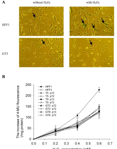
To address the mechanism by which ectopic expression of hTERT enables G6PD-deficient cells to overcome prema-ture senescence, we measured the ability of hTERT-expressing cells to cope with oxidative stress induced by treatment with exogenous H2O2. As shown in Figure 5A, under normal culture conditions there were very few cells among G6PD-deficient HFF1 fibroblasts (passage 22) and hTERT-expressing G6PD-deficient GT3 fibroblasts (pas-sage 72) that were positive for SA-β-Gal staining (less than 5%). Treatment with H2O2 increased the number of SA-β -Gal-positive cells in both the HFF1 and GT3 populations, indicating that H2O2induces premature senescence in both cells. Interestingly, we noted that there is a greater increase of SA-β-Gal-positive cells in the H2O2-treated HFF1 cells (55 ± 9%) than in the H2O2-treated GT3 cells (21 ± 1.4%). To confirm the significance of this observa-tion, we treated parental and hTERT-expressing fibroblasts
Telomerase activity of hTERT-transfected normal and G6PD-deficient fibroblasts
Figure 1
Proliferation capability of hTERT-transfected normal and G6PD-deficient fibroblasts
Figure 2
The karyotypes and anchorage-independent growth potential of hTERT-expressing normal and G6PD-deficient fibroblasts
Figure 3
Telomere lengths of hTERT-expressing normal and G6PD-deficient fibroblasts
Figure 4
H2O2-induced senescence in G6PD-deficient fibroblasts
Figure 5
H2O2-induced senescence in G6PD-deficient fibroblasts. A. Cells were treated with 350 μM H2O2 for 1.5 h and then cultured for another 72 h before staining for senescence-associated β-galactosidase (SA-β-Gal) activity. Representative results for HFF1 and GT3 cells with or without H2O2 treatment are shown in the right and left panels, respectively. Examples of cells
that are positive for SA-β-Gal staining are indicated by arrow. In the quantification, any cell that has detectable green staining was scored as positive for SA-β-Gal. B. Cells were treated with different concentrations of H2O2 for 1.5 h and then cultured
for another 72 h. Quantification of premature senescence was determined by the rate of conversion of
from both normal and G6PG-deficient patients with dif-ferent concentrations of H2O2 and quantified the extent of H2O2-induced premature senescence. As shown in Figure 5B, the levels of H2O2-induced premature senescence in the early passage (p13) and late passage (p72) cells of T5, T9, GT3 and GT8 were similar to HFF3, but were signifi-cantly greater in HFF1 cells. These results indicate that ectopic expression of hTERT in G6PD-deficient cells renders the cells more resistant to H2O2-induced prema-ture senescence.
Discussion
The accelerated cellular senescence characteristic of G6PD-deficient fibroblasts would seem to represent a clear case of a senescence mechanism based on accumu-lated oxidative DNA damage rather than one involving accelerated telomere attrition [26]. This mechanistic transparency makes these cells an ideal system for addressing the relative role of oxidative stress and tel-omere attrition in cellular senescence. Somewhat surpris-ingly, we found that ectopic expression of hTERT prevented the accelerated senescence of G6PD-deficient cells and led to their immortalization (Fig. 2). The growth rate of hTERT-expressing G6PD-deficient fibroblasts, however, remained slower than that of hTERT-expressing normal fibroblasts (Fig. 2A and 2B), as noted previously (25). This finding suggests that hTERT overexpression may attenuate senescence induction by oxidative stress, but does not suppress the growth defect caused by G6PD-deficiency. To test this hypothesis, we measured the abil-ity of hTERT-expressing G6PD-deficient cells to cope with H2O2-induced oxidative stress. Similar to results reported by others [31], we found no difference in stress-induced premature senescence (SIPS) between normal and expressing normal fibroblasts (Fig. 5). However, hTERT-expressing G6PD-deficient cells became more resistant to H2O2-induced premature senescence (Fig. 5), indicating that the increased sensitivity to oxidative stress in G6PD-deficient cells is prevented by the expression of hTERT.
Ectopic expression of hTERT has also been shown previ-ously to immortalize fibroblasts derived from individuals with Ataxia telangiectasia (A-T), Nijimegen breakage syn-drome (NBS), Hutchinson-Gilford progeria synsyn-drome (HGPS), and Werner Syndrome (WS) [32-36]. Although the genetic defects in A-T, NBS, HGPS, WS, and G6PD-deficiency patients are very different, fibroblasts derived from these individuals have one common phenotype: they all undergo accelerated senescence in vitro
[25,34,36,37]. The premature senescence of mitotic cells derived from A-T, HGPS, and NBS patients has been cor-related with an increased rate of telomere loss [31,34,38], whereas the mechanism responsible for the premature senescence of WS and G6PD-deficient fibroblasts appears to be different and has been postulated to reflect the
accu-mulation of DNA damage [26,38]. The fact that ectopic expression of hTERT immortalizes fibroblasts derived from individuals with any of these different defects indi-cates that telomerase may attenuate senescence induction triggered by either telomere attrition or genome-wide DNA damage. Ectopic expression of hTERT has also been shown to increase radioresistance of adult human mesen-chymal stem cells [39], to circumvent hyperglycemia-induced premature senescence [40], and to prevent apop-tosis induced by tumor necrosis factor [41]. These obser-vations suggest that expression of hTERT not only activates telomere maintenance but may also affect signal-ing that protects cells from oxidative stress and other stim-uli. Indeed, increasing evidence is emerging to implicate that hTERT has functions beyond telomere maintenance [42].
hTERT-immortalization of cells is generally attributed to the activation of telomerase activity and the subsequent counteraction of telomere attrition. Surprisingly, we observed that telomere lengths continued to decrease in earlier-passage cells from both hTERT-expressing normal and G6PD-deficient fibroblasts, despite the fact that these cells expressed high levels of telomerase activity (Fig. 1). In fact, we observed that TRF lengths from one hTERT-expressing normal cell line (T5) and two hTERT-express-ing G6PD-deficient cell lines (GT3 and GT8) were short-ened to ~3–3.7 kb before stabilization (Fig. 4). The parental fibroblasts used for the derivation of these hTERT-expressing cells undergo senescence when TRF lengths decrease to approximately 5–6 kb [25], suggesting that dysfunctional telomeres may already have formed when the TRF length was reduced to 5–6 kb in normal fibroblasts. However, senescence was not induced in these hTERT-expressing cells even when the TRF lengths were reduced to less than 5 kb, suggesting that hTERT, in addi-tion to serving as a subunit for telomerase, acts by some other mechanism to prevent senescence induction. Short-ening of telomere lengths in the presence of telomerase activation has also been observed by others [28,43], and a protective role for hTERT in telomere capping has been suggested [28,44]. In addition, hTERT has been found to associate with human telomeres, and ectopic expression of hTERT causes transcriptional alterations in a subset of genes; this may lead to increased genomic stability and enhanced DNA repair activity [45]. At this time, the molecular details of hTERT involvement in senescence induction remain obscure.
and Pot1, are known to participate in telomere-length reg-ulation and chromosome-end protection [46]. It is likely that some of these factors prevent telomerase from func-tioning at the telomere until telomeres are shortened to a critical length. Our finding that the lengths of telomeres in hTERT-expressing cells were reduced to approximately 3– 3.7 kb before stabilization (Fig. 4) provides support for this notion.
In conclusion, we have shown that ectopic expression of hTERT immortalizes fibroblasts derived from normal and G6PD-deficient patients. The accelerated cellular senes-cence observed in G6PD-deficient cells has been shown to be attributable to increased accumulation of oxidative DNA damage [26]. As this oxidant-induced senescence was overridden by the ectopic expression of hTERT, we suggest that hTERT, in addition to providing a subunit for telomerase, may also function in regulating senescence induction.
Competing interests
The authors declare that they have no competing interests.
Authors' contributions
MC and HH participated in the preparation of retroviral particles and isolation of hTERT-expressing clones, YW carried out the rest of research, DTC and TVW conceived of the study and design research, YW and TVW wrote the paper. All authors read and approved the final manu-script.
Acknowledgements
This work was supported by grants from Chang Gung University (CMRP160231) and the National Science Council of Taiwan (NSC94-2320-B182-041) to DTYC; and from Chang Gung University (CMRPD140012) and the National Science Council of Taiwan (NSC95-2311-B182-001) to TCVW. This work was also supported by a grant from the Ministry of Edu-cation (EMRPD170581) to DTYC and TCVW.
References
1. Hayflick L, Moorhead PS: The serial cultivation of human diploid cell strains. Exp Cell Res 1961, 25:585-621.
2. Campisi J, Kim SH, Lim CS, Rubio M: Cellular senescence, cancer and aging: the telomere connection. Exp Gerontol 2001, 36:607-618.
3. Ben-Porath I, Weinberg RA: The signals and pathways activating cellular senescence. Int J Biochem Cell Biol 2005, 37:961-976. 4. Harley CB, Vaziri H, Counter CM, Allsopp RC: The telomere
hypothesis of cellular aging. Exp Gerontol 1992, 27:375-382. 5. Harley CB, Kim NW, Prowse KR, Weinrich SL, Hirsch KS, West MD,
Bacchetti S, Hirte HW, Counter CM, Greider CW, et al.: Telomer-ase, cell immortality, and cancer. Cold Spring Harb Symp Quant Biol 1994, 59:307-315.
6. Harman D: Aging: a theory based on free radical and radiation chemistry. J Gerontol 1956, 11:298-300.
7. Sohal RS, Weindruch R: Oxidative stress, caloric restriction, and aging. Science 1996, 273:59-63.
8. Harley CB, Futcher AB, Greider CW: Telomeres shorten during ageing of human fibroblasts. Nature 1990, 345:458-460. 9. Lingner J, Cooper JP, Cech TR: Telomerase and DNA end
repli-cation: no longer a lagging strand problem? Science 1995, 269:1533-1534.
10. Greider CW, Blackburn EH: Telomeres, telomerase and cancer.
Sci Am 1996, 274:92-97.
11. Stewart SA, Weinberg RA: Telomerase and human tumorigen-esis. Semin Cancer Biol 2000, 10:399-406.
12. Kim NW, Piatyszek MA, Prowse KR, Harley CB, West MD, Ho PL, Coviello GM, Wright WE, Weinrich SL, Shay JW: Specific associa-tion of human telomerase activity with immortal cells and cancer. Science 1994, 266:2011-2015.
13. Shay JW, Bacchetti S: A survey of telomerase activity in human cancer. Eur J Cancer 1997, 33:787-791.
14. Bodnar AG, Ouellette M, Frolkis M, Holt SE, Chiu CP, Morin GB, Har-ley CB, Shay JW, Lichtsteiner S, Wright WE: Extension of life-span by introduction of telomerase into normal human cells. Sci-ence 1998, 279:349-352.
15. Vaziri H, Benchimol S: Reconstitution of telomerase activity in normal human cells leads to elongation of telomeres and extended replicative life span. Curr Biol 1998, 8:279-282. 16. Nakayama J, Tahara H, Tahara E, Saito M, Ito K, Nakamura H,
Nakan-ishi T, Tahara E, Ide T, Ishikawa F: Telomerase activation by hTRT in human normal fibroblasts and hepatocellular carci-nomas. Nat Genet 1998, 18:65-68.
17. Toussaint O, Houbion A, Remacle J: Aging as a multi-step proc-ess characterized by a lowering of entropy production lead-ing the cell to a sequence of defined stages. II. Testlead-ing some predictions on aging human fibroblasts in culture. Mech Ageing Dev 1992, 65:65-83.
18. Dumont P, Burton M, Chen QM, Gonos ES, Frippiat C, Mazarati JB, Eliaers F, Remacle J, Toussaint O: Induction of replicative senes-cence biomarkers by sublethal oxidative stresses in normal human fibroblast. Free Radic Biol Med 2000, 28:361-373. 19. Chen Q, Ames BN: Senescence-like growth arrest induced by
hydrogen peroxide in human diploid fibroblast F65 cells. Proc Natl Acad Sci USA 1994, 91:4130-4134.
20. Honda S, Matsuo M: Shortening of the in vitro lifespan of human diploid fibroblasts exposed to hyperbaric oxygen. Exp Gerontol 1983, 18:339-345.
21. von Zglinicki T, Saretzki G, Docke W, Lotze C: Mild hyperoxia shortens telomeres and inhibits proliferation of fibroblasts: a model for senescence? Exp Cell Res 1995, 220:186-193. 22. Chen Q, Fischer A, Reagan JD, Yan LJ, Ames BN: Oxidative DNA
damage and senescence of human diploid fibroblast cells.
Proc Natl Acad Sci USA 1995, 92:4337-4341.
23. Gaubatz S, Lees JA, Lindeman GJ, Livingston DM: E2F4 is exported from the nucleus in a CRM1-dependent manner. Mol Cell Biol
2001, 21:1384-1392.
24. Ho HY, Cheng ML, Cheng PF, Chiu DT: Low oxygen tension alle-viates oxidative damage and delays cellular senescence in G6PD-deficient cells. Free Radic Res 2007, 41:571-579.
25. Ho HY, Cheng ML, Lu FJ, Chou YH, Stern A, Liang CM, Chiu DT: Enhanced oxidative stress and accelerated cellular senes-cence in glucose-6-phosphate dehydrogenase (G6PD)-defi-cient human fibroblasts. Free Radic Biol Med 2000, 29:156-169. 26. Cheng ML, Ho HY, Wu YH, Chiu DT: Glucose-6-phosphate
dehy-drogenase-deficient cells show an increased propensity for oxidant-induced senescence. Free Radic Biol Med 2004, 36:580-591.
27. Yu CC, Lo SC, Wang TC: Telomerase is regulated by protein kinase C-zeta in human nasopharyngeal cancer cells. Biochem J 2001, 355:459-464.
28. Zhu J, Wang H, Bishop JM, Blackburn EH: Telomerase extends the lifespan of virus-transformed human cells without net tel-omere lengthening. Proc Natl Acad Sci USA 1999, 96:3723-3728. 29. Sheng WY, Chien YL, Wang TC: The dual role of protein kinase
C in the regulation of telomerase activity in human lym-phocytes. FEBS Lett 2003, 540:91-95.
30. Gary RK, Kindell SM: Quantitative assay of senescence-associ-ated beta-galactosidase activity in mammalian cell extracts.
Anal Biochem 2005, 343:329-334.
31. Naka K, Tachibana A, Ikeda K, Motoyama N: Stress-induced pre-mature senescence in hTERT-expressing ataxia telangiecta-sia fibroblasts. J Biol Chem 2004, 279:2030-2037.
Publish with BioMed Central and every scientist can read your work free of charge "BioMed Central will be the most significant development for disseminating the results of biomedical researc h in our lifetime."
Sir Paul Nurse, Cancer Research UK
Your research papers will be:
available free of charge to the entire biomedical community
peer reviewed and published immediately upon acceptance
cited in PubMed and archived on PubMed Central
yours — you keep the copyright
Submit your manuscript here:
http://www.biomedcentral.com/info/publishing_adv.asp
BioMedcentral 33. Nakamura H, Fukami H, Hayashi Y, Kiyono T, Nakatsugawa S,
Hamaguchi M, Ishizaki K: Establishment of immortal normal and ataxia telangiectasia fibroblast cell lines by introduction of the hTERT gene. J Radiat Res (Tokyo) 2002, 43:167-174. 34. Ranganathan V, Heine WF, Ciccone DN, Rudolph KL, Wu X, Chang
S, Hai H, Ahearn IM, Livingston DM, Resnick I, Rosen Fred, See-manova E, Jarolim P, DePinho RA, Weaver DT: Rescue of a tel-omere length defect of Nijmegen breakage syndrome cells requires NBS and telomerase catalytic subunit. Curr Biol 2001, 11:962-6.
35. Wyllie FS, Jones CJ, Skinner JW, Haughton MF, Wallis C, Wynford-Thomas D, Faragher RG, Kipling D: Telomerase prevents the accelerated cell ageing of Werner syndrome fibroblasts. Nat Genet 2000, 24:16-17.
36. Ouellette MM, McDaniel LD, Wright WE, Shay JW, Schultz RA: The establishment of telomerase-immortalized cell lines repre-senting human chromosome instability syndromes. Hum Mol Genet 2000, 9:403-411.
37. Faragher RG, Kill IR, Hunter JA, Pope FM, Tannock C, Shall S: The gene responsible for Werner syndrome may be a cell divi-sion "counting" gene. Proc Natl Acad Sci USA 1993, 90:12030-12034.
38. Allsopp RC, Vaziri H, Patterson C, Goldstein S, Younglai EV, Futcher AB, Greider CW, Harley CB: Telomere length predicts replica-tive capacity of human fibroblasts. Proc Natl Acad Sci USA 1992, 89:10114-10118.
39. Serakinci N, Christensen R, Graakjaer J, Cairney CJ, Keith WN, Alsner J, Saretzki G, Kolvraa S: Ectopically hTERT expressing adult human mesenchymal stem cells are less radiosensitive than their telomerase negative counterpart. Exp Cell Res 2007, 313:1056-1067.
40. Blazer S, Khankin E, Segev Y, Ofir R, Yalon-Hacohen M, Kra-Oz Z, Gottfried Y, Larisch S, Skorecki KL: High glucose-induced repli-cative senescence: point of no return and effect of telomer-ase. Biochem Biophys Res Commun 2002, 296:93-101.
41. Dudognon C, Pendino F, Hillion J, Saumet A, Lanotte M, Segal-Bend-irdjian E: Death receptor signaling regulatory function for tel-omerase: hTERT abolishes TRAIL-induced apoptosis, independently of telomere maintenance. Oncogene 2004, 23:7469-7474.
42. Cong Y, Shay JW: Actions of human telomerase beyond telom-eres. Cell Res 2008, 18:725-732.
43. Yang J, Chang E, Cherry AM, Bangs CD, Oei Y, Bodnar A, Bronstein A, Chiu CP, Herron GS: Human endothelial cell life extension by telomerase expression. J Biol Chem 1999, 274:26141-26148. 44. Chan SW, Blackburn EH: Telomerase and ATM/Tel1p protect
telomeres from nonhomologous end joining. Mol Cell 2003, 11:1379-1387.
45. Sharma GG, Gupta A, Wang H, Scherthan H, Dhar S, Gandhi V, Iliakis G, Shay JW, Young CS, Pandita TK: hTERT associates with human telomeres and enhances genomic stability and DNA repair. Oncogene 2003, 22:131-146.