C A S E R E P O R T
Open Access
Autosomal dominant polycystic kidney disease
with ectopic unilateral multicystic dysplastic
kidney
Jing Xu
1, Dong-Ping Chen
1, Zhi-Guo Mao
1, He-Feng Huang
2, Chen-Ming Xu
2, Cong-Rong Wang
3,
Wei-Ping Jia
3and Chang-Lin Mei
1*Abstract
Background:Autosomal dominant polycystic kidney disease (ADPKD) is the most common hereditary renal disorder. In most cases, ADPKD similarly affects bilateral kidneys.
Case presentation:Among the 605 ADPKD patients that were followed up by our center, we identified two male patients with unilateral ADPKD. The cases were remarkable because the patients also had ectopia and multicystic dysplasia in the contralateral kidney, which are generally sporadic disease conditions. Both patients tested positive for polycystic kidney disease 1 mutation, but negative for hepatocyte nuclear factor 1 beta mutation. Moreover, the deterioration of their kidney function seemed to be quicker than their age- and sex-matched controls and siblings. Both patients had started a long-term hemodialysis in their 40s.
Conclusion:Anatomical and genetic abnormality in patients with ADPKD may be more frequent and complex than previously believed. The compensatory capacity in patients with ADPKD is fragile, and missing one kidney could accelerate the deterioration of renal function.
Keywords:Autosomal dominant polycystic kidney disease, Ectopia, Multicystic dysplasia, Unilateral
Background
Autosomal dominant polycystic kidney disease (ADPKD) is the most common hereditary renal disorder and the fourth cause of death of end-stage renal disease, ac-counting for 10% of patients on dialysis [1]. The disease is caused by mutations in thePKD1(85% of cases) orPKD2 (15% of cases) [2]. In most ADPKD patients, bilateral kidneys are similarly affected, with numerous fluid-filled cysts arising from different nephron segments. Only a few cases of ADPKD with unilateral renal agenesis or severe hypoplasia have been reported [2].
Among the 605 ADPKD patients followed by our cen-ter, we report two cases of ADPKD patients with pelvic ectopic unilateral multicystic dysplastic kidney (MCDK), which refers to a sporadic disease condition and is related totranscription factor 2 (TCF2) mutations [3]. The clinical
characteristics of these two rare cases were compared with their siblings and other male ADPKD patients with similar ages; these characteristics were then summarized and analyzed. The PKD1 and PKD2 mutations and the rele-vant MCDK (TCF2) mutations were also measured.
Case presentation
Case 1
A 48-year-old man with ADPKD was admitted for long-term hemodialysis because his estimated glomerular filtration rate (eGFR) dropped to 14 ml/min/1.73 m2. His blood pressure was within the normal range. The patient was diagnosed with ADPKD 10 years earlier with a positive family history (Figure 1). He and his relatives had no history of intracranial aneurysm. He had no albu-minuria and did not complain of renal pain. During diagnosis, an enlarged polycystic left kidney was found, but the right kidney was absent. An ultrasonography after admission detected a fist-sized, multi-cystic mass located before his right iliac artery. An enlarged polycystic liver
* Correspondence:chlmei@hotmail.com
1Division of Nephrology, Kidney Institute of CPLA, Changzheng Hospital,
Second Military Medical University, 415 Fengyang Rd, Shanghai 200003, China
Full list of author information is available at the end of the article
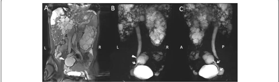
was also detected. The magnetic resonance imaging (MRI) scan confirmed a 6 cm diameter polycystic mass in the right pelvis (Figure 2, Panels A and B, triangles), and the left kidney was significantly enlarged, measuring 18 cm × 12 cm × 8 cm. A renal volume assessment was then performed. The renal volume for the left kidney was 1127.6 cm3 and that for the right was only 74.6 cm3. Further MR urography revealed a tubular structure con-necting the pelvic polycystic mass to the bladder with a fluid signal along the tubular lumen area (Figure 2, Panels B and C, arrows). This ADPKD patient had a right pelvic multicystic kidney with congenital aplasia, and the tubular structure was his right ureter.
Case 2
A 45-year-old male with fatigue and elevated creatinine (542 μmol/l, eGFR = 14 ml/min/1.73 m2) was admitted to set up long-term vascular access. The patient had hypertension controlled with nifedipine. No history of intracranial aneurysm was reported. He had a positive family history (Figure 3) and was diagnosed with ADPKD through an ultrasonography 16 years prior this study. He
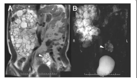
had no albuminuria and did not complain of renal pain. During diagnosis, an exaggerated right kidney was found. However, the left kidney was absent. After admission, MR urography detected a 22.8 cm × 12.2 cm × 10.5 cm right kidney with a typical polycystic phenotype, and a 5.8 cm × 4.2 cm × 3.9 cm multicystic space-occupying lesion before the left psoas (Figure 4, Panels A and B, triangles), with signals similar to the right polycystic kidney, indicating a small left pelvic kidney with multicystic aplasia. Based on renal volume assessment, the left kidney measured 2290.7 cm3, whereas the right one only measured 44.0 cm3. A polycystic liver was also detected. The CT scan result agreed with that of MR urography.
Screening and genotyping
No ectopic aplastic cystic kidney was detected among the family members of these two patients, both affected and non-affected, who were screened using abdominal ultrasonography. These two patients were the only ones with this presentation among their families. A genetic analysis of PKD1 and PKD2 was performed by Athena Diagnostics (MA, U.S.A.) and verified at the Zhejiang
? ?
Figure 1Family tree of Case 1 patient.(Arrow): affected proband with unilateral ADPKD; (filled square, filled circle): male/female affected by ADPKD with two kidneys; (blank square, blank circle): male/female did not have ADPKD; (?): unsure for ADPKD; (box with a stripe): family member passed away.
University affiliated Gynecology and Obstetrics Hospital (Hangzhou, Zhejiang, China).
Genotyping of the two cases indicated that each had different mutations in thePKD1gene. Case 1 had a pro-line in place of leucine at amino acid 55 and a premature termination signal in codon 60 in exon 1 [NM_000296.3 P.L56PfsX60 (168_174dupCGCGGGC,novel,Het)]. Case 2 had a 7 bp duplication mutation (11650_11651) in exon 41, resulting in a frameshift at amino acid 3814, which is a predicted disease-associated mutation. He also had two nonsense mutations in exon 15 (an A in place of a G at nucleotide position of 6625) and exon 23 (an A in place of a G at nucleotide position of 8851). None of these po-lymorphisms had been previously reported.
Both patients tested negative for TCF2 mutation, also known ashepatocyte nuclear factor 1 beta(HNF1β).
Comparison of the reported cases to age- and sex-matched controls with bilateral ADPKD or siblings A matched control group for each case was defined among the remaining 603 patients with ADPKD followed up by our center. All patients with the same sex and age (with no more than a five-year difference) from the respective ages of the cases were recruited. Three of their age- and sex-matched siblings were also chosen as another control group (Table 1). The
single-kidney volume (SKV) and eGFR were expressed as mean and 95% confidence interval (CI). Compared with the matched bilateral ADPKD controls, the median SKV of the ectopic MCDK kidneys of the two reported cases were significantly smaller [Case 1: 74.6 vs. 692.4 95% CI (611.3, 781.3) cm3; Case 2: 44.0 vs. 657.1 95% CI (566.9, 743.4) cm3]. However, the SKV of their normal contralat-eral kidneys seemed to be significantly larger [Case 1: 1127.6 vs. 692.4 95% CI (611.3, 781.3) cm3; Case 2: 2290.7 vs. 657.1 95% CI (566.9, 743.4) cm3]. The mean eGFR of the two cases were significantly lower than their age- and sex-matched siblings (14.0 vs. 68.3, 39.6, 65.0 ml/min/ 1.73 m2) and bilateral ADPKD controls [Case 1: 14.0 vs. 84.7 95% CI (77.7, 91.7) ml/min/1.73 m2; Case 2: 14.0 vs. 88.8 95% CI (81.7, 96.4) ml/min/1.73 m2].
Discussion
In most cases, bilateral kidneys are involved in ADPKD patients. No previous report has been conducted on uni-lateral ADPKD with contrauni-lateral ectopic MCDK.
Among the 605 patients followed up by our center, we identified two subjects with unilateral ADPKD. No kid-ney tissue on the contralateral location was found on both patients’ kidneys. MRI revealed ectopic dysplasia kidney remnants with multiple cysts in the pelvis. Both patients had advanced disease during presentation. Compared with their matched bilateral ADPKD controls, the median SKV of the ectopic MCDK kidneys of the two reported cases were significantly smaller (Table 1), whereas their contralateral kidneys were significantly lar-ger. The mean eGFR of the two cases (Table 1), which indicated a more advanced stage of disease progression with the unilateral ADPKD, were significantly lower than their age- and sex-matched siblings and bilateral ADPKD controls.
In a cohort of 182 patients with ADPKD, Poster et al. [2] identified three patients with unilateral renal agenesis or severe hypoplasia. These three cases had different truncating mutations in theirPKD1gene. Although their kidney volumes and volume progression rates were greater than the mean values of their two polycystic kid-ney controls, which is normally associated with an
? ?
?
?
Figure 3Family tree of Case 2 patient.(Arrow): affected proband with unilateral ADPKD; (filled square, filled circle): male/female affected by ADPKD with two kidneys; (blank square, blank circle): male/female not had ADPKD; (?): not sure for ADPKD; (box with a stripe): deceased family member.
accelerated decrease in renal function, the eGFR in these patients was remarkably well-preserved [4]. This charac-teristic may be partly due to compensatory parenchymal hypertrophy. However, renal parenchyma is unlikely to make a large contribution to the total kidney volume because the enlarged single cystic kidneys were grossly cystic, as shown in the MRI images [2]. Moreover, all their patients were much younger than ours (23 years old, 38 years old, and 40 years old), and the disease pro-gressions of the three subjects after long-term follow-up remain unknown. The two patients in the present study had different mutations on thePKD1gene. The relation-ship between the PKD1 mutation sites and the disease progression was difficult to explain because of the small sample size limitation of the reported cases.
Apart from unilateral ADPKD, our patients also had concurrent ectopic MCDK. MCDK generally refers to a sporadic disease condition of abnormal metanephric differentiation. The incidence is approximately 1 in 4300 in the general population [3]. We identified two cases of MCDK among the 605 ADPKD patients, indicating an incidence rate of 3.3%. Generally, the total kidney func-tion of many subjects with MCDK could be well-maintained [5]. However, both our patients prepared for long-term hemodialysis when they were in their 40s, mainly in consideration of the kidney structure destruc-tion by the continuous cyst growth in their contralateral kidneys.
TCF2 (HNF1β) abnormalities cause congenital anom-alies of the kidney and urinary tract [6]. Some studies have reported that MCDK is related to TCF2 mutation [7,8]. However, both of our patients tested negative for
TCF2mutation. Mutations in genes such asEYA1,SIX1, andPAX2are related to the occurrence of MCDK. How-ever, neither of our patients had concomitant ear and eye structural abnormalities or dysfunction (data not shown). Their MCDK could be related to other genetic abnormalities.
Conclusions
We conclude that the anatomical and genetic abnormal-ity in patients with ADPKD could be more frequent and complex than previously believed. Patients with ADPKD have fragile compensatory capacities, and missing one kidney could accelerate the deterioration of the renal function.
Consent
The clinical information of the two patients and their relatives were provided by the index patients or their relatives after obtaining their consents. The study was conducted following the Declaration of Helsinki. Written informed consents were obtained from the patients for publication of this case report and any accompanying images. Copies of the written consents are available for review by the editor of this journal.
Abbreviations
ADPKD: Autosomal dominant polycystic kidney disease; CI: Confidence interval; eGFR: Estimated glomerular filtration rate; HNF1β: Hepatocyte nuclear factor 1 beta; MCDK: Multicystic dysplastic kidney; MRI: Magnetic resonance imaging; SKV: Single-kidney volume; TCF2: Transcription factor 2.
Competing interests
The authors declare that they have no competing interests.
Authors’contributions
XJ participated in the conception and design of the study, data acquisition, and data analysis and interpretation, performed the statistical analysis, and drafted the manuscript; CDP participated in the data acquisition and statistical analysis; MZG participated in the conception and design, as well as in data acquisition; HHF, XCM, WCR, and JWP carried out the genetic studies on mutations; MCL participated in the design and coordination and helped revise the study critically for important intellectual content. All authors read and approved the final manuscript.
Acknowledgements
This work was funded in part by the National Nature Science Fund of China (Nos. 30971368, 81000281, and 81200499) and the Outstanding Young Investigator Fund of the Second Military Medical University, Shanghai, China.
Author details
1
Division of Nephrology, Kidney Institute of CPLA, Changzheng Hospital, Second Military Medical University, 415 Fengyang Rd, Shanghai 200003,
Table 1 Kidney volumes and renal function in two patients with ectopic unilateral MCDK compared with their age- and sex-matched siblings and bilateral ADPKD controls
Parameter Case 1 Age- and sex-matched bilateral ADPKD for case 1
Case 2 Age- and sex-matched bilateral ADPKD for case 2
Age- and sex-matched siblings
No. of Patients 1 66 1 60 3
Age 48.0 47.9 45.0 45.6 41, 47, 49
Mean (range) (43.2, 53.0) (40.3, 49.7)
SKV (cm3) Left: 1127.6 692.4 Right: 2290.7 657.1 NA
Mean (95%CI) Right: 74.6 (611.3, 781.3) Left: 44.0 (566.9, 743.4)
eGFR (ml/min/1.73 m2) 14.0 84.7 14.0 88.8 68.3, 39.6, 65.0
Mean (95%CI) (77.7, 91.7) (81.7, 96.4)
China.2Zhejiang University affiliated Gynecology and Obstetrics Hospital, Key
Laboratory of Reproductive Genetics, Zhejiang University, Hangzhou, China.
3Division of Endocrinology, Shanghai Diabetes Institute, Shanghai Sixth
People’s Hospital, School of Medicine, Shanghai Jiao Tong University, Shanghai, China.
Received: 4 November 2012 Accepted: 13 February 2013 Published: 17 February 2013
References
1. Alam A, Perrone RD:Management of ESRD in patients with autosomal dominant polycystic kidney disease.Adv Chronic Kidney Dis2010,17:164–172. 2. Poster D, Kistler AD, Krauer F, Blumenfeld JD, Rennert H, Weishaupt D,
Wüthrich RP, Serra AL:Kidney function and volume progression in unilateral autosomal dominant polycystic kidney disease with contralateral renal agenesis or hypoplasia: a case series.Am J Kidney Dis
2009,54:450–458.
3. Schreuder MF, Westland R, van Wijk JA:Unilateral multicystic dysplastic kidney: a meta-analysis of observational studies on the incidence, associated urinary tract malformations and the contralateral kidney.
Nephrol Dial Transplant2009,24:1810–1818.
4. Grantham JJ, Torres VE, Chapman AB, Guay-Woodford LM, Bae KT, King BF Jr, Wetzel LH, Baumgarten DA, Kenney PJ, Harris PC, Klahr S, Bennett WM, Hirschman GN, Meyers CM, Zhang X, Zhu F, Miller JP, CRISP Investigators:
Volume progression in polycystic kidney disease.N Engl J Med2006,
354:2122–2130.
5. Aslam M, Watson AR, Trent & Anglia MCDK Study Group:Unilateral multicystic dysplastic kidney: long term outcomes.Arch Dis Child2006,
91:820–823.
6. Nakayama M, Nozu K, Goto Y, Kamei K, Ito S, Sato H, Emi M, Nakanishi K, Tsuchiya S, Iijima K:HNF1B alterations associated with congenital anomalies of the kidney and urinary tract.Pediatr Nephrol2010,25:1073–1079. 7. Weber S, Moriniere V, Knüppel T, Charbit M, Dusek J, Ghiggeri GM,
Jankauskiené A, Mir S, Montini G, Peco-Antic A, Wühl E, Zurowska AM, Mehls O, Antignac C, Schaefer F, Salomon R:Prevalence of mutations in renal developmental genes in children with renal hypodysplasia: results of the ESCAPE study.J Am Soc Nephrol2006,17:2864–2870.
8. Hains DS, Bates CM, Ingraham S, Schwaderer AL:Management and etiology of the unilateral multicystic dysplastic kidney: a review.
Pediatr Nephrol2009,24:233–241.
doi:10.1186/1471-2369-14-38
Cite this article as:Xuet al.:Autosomal dominant polycystic kidney disease with ectopic unilateral multicystic dysplastic kidney.BMC Nephrology201314:38.
Submit your next manuscript to BioMed Central and take full advantage of:
• Convenient online submission
• Thorough peer review
• No space constraints or color figure charges
• Immediate publication on acceptance
• Inclusion in PubMed, CAS, Scopus and Google Scholar
• Research which is freely available for redistribution