TECHNICAL NOTES
Implementation of the
agmatine-controlled expression system
for inducible gene expression
in
Lactococcus lactis
Daniel M. Linares, Patricia Alvarez‑Sieiro, Beatriz del Rio, Victor Ladero, Begoña Redruello, Mª Cruz Martin,
Maria Fernandez
*and Miguel A. Alvarez
Abstract
Background: Lactococcus lactis has been safely consumed in fermented foods for millennia. This Gram‑positive bac‑ terium has now become of industrial importance as an expression host for the overproduction of lipopolysaccharide‑ free recombinant proteins used as food ingredients, therapeutic proteins and biotechnological enzymes.
Results: This paper reports an agmatine‑controlled expression (ACE) system for L. lactis, comprising the lactococcal agmatine‑sensor/transcriptional activator AguR and its target promoter PaguB. The usefulness and efficiency of this system was checked via the reporter gene gfp and by producing PEP (Myxococcus xanthus prolyl‑endopeptidase), an enzyme of biomedical interest able to degrade the immunotoxic peptides produced during the gastrointestinal breakdown of gluten.
Conclusion: The ACE system developed in this work was suitable for the efficient expression of the functional recom‑ binant proteins GFP and PEP. The expression system was tightly regulated by the agmatine concentration and allowed high protein production without leakiness.
Keywords: Lactococcus lactis, Expression vector, Agmatine induction, Myxococcus xanthus, Prolyl‑endopeptidase
© 2015 Linares et al. This article is distributed under the terms of the Creative Commons Attribution 4.0 International License (http://creativecommons.org/licenses/by/4.0/), which permits unrestricted use, distribution, and reproduction in any medium, provided you give appropriate credit to the original author(s) and the source, provide a link to the Creative Commons license, and indicate if changes were made. The Creative Commons Public Domain Dedication waiver (http://creativecommons.org/ publicdomain/zero/1.0/) applies to the data made available in this article, unless otherwise stated.
Background
Heterologous protein production is a multi-billion dol-lar market of particudol-lar importance to manufacturers of biopharmaceuticals and enzymes for industrial use. Microbial production systems are often the best option for making such products given their ease of handling and high synthesis rates [1]. At present, Escherichia coli
remains the first choice of host system given its high overexpression yields, ease of genetic handling, and the wealth of information available on this microorgan-ism [2]. However, it is not without its drawbacks, such as the formation of inclusion bodies, the presence of an outer membrane that hampers secretion, its relatively
complicated aerobic fermentation system, and the forma-tion of endotoxins such as cell wall lipopolysaccharides [3]. The presence of bacterial endotoxins is one of the major concerns of regulatory agencies, and the need to add downstream steps to ensure their removal can make otherwise simple processes quite costly [4].
The Gram-positive bacterium Lactococcus lactis has emerged as an attractive alternative for the overproduc-tion of recombinant proteins. Due to its long, safe his-tory of use in dairy fermentations, this bacterium has been classified as a food grade microorganism ‘Generally Recognized As Safe’ (GRAS) by the US Food and Drug Administration (FDA), and has led it to receive ‘Qualified Presumption of Safety’ (QPS) status from the European Food Safety Authority (EFSA) [5]. In addition, it is an effi-cient secretor of extracellular recombinant proteins, has low protease activity (allowing for simplified purification
Open Access
*Correspondence: mfernandez@ipla.csic.es
processes), and a very simple metabolism that allows for rapid growth without aeration—all properties that facili-tate scaling-up [4]. Moreover, L. lactis is likely to provide a good membrane environment for the overproduction of eukaryotic proteins [6]. Indeed, a number of eukaryotic membrane transporters, yeast mitochondrial proteins and human proteins have been heterologously expressed in this host [7]. Further, L. lactis is an efficient cellular factory able to turn out recombinant viral antigens, inter-leukins, allergens, virulence factors, bacteriocins and enzymes [8, 9]. It can even be genetically engineered to produce proteins from pathogenic species on its cell sur-face, and thus be used as a vector for the production and delivery of oral vaccines against HIV, cholera, malaria, human papillomavirus, stomach ulcers, tetanus and bru-cellosis [10–18].
For most of these applications, the availability of vec-tors that allow the cloning and expression of foreign genes is of paramount importance. Although the genetic accessibility and ease of handling of L. lactis lags far behind that of E. coli, the molecular biology techniques and genetic tools available for use with this bacterium have increased over recent years [9, 19]. So far, a num-ber of inducible expression systems regulated by envi-ronmental factors have been documented, including the chloride-inducible expression cassette [20], the zinc-inducible expression system [21], the lactate-inducible P170 system [22], the heat shock-inducible system [23], systems based on sugar or peptide concentration-regu-lated promoters, and bacteriophage-derived promoters [24, 25]. However, some of these systems are less useful since they are controllable only to a limited extent, show low efficiency, or are associated with some degree of basal expression [21, 25, 26]. These limitations may be due to (1) the corresponding inducer being an essential nutrient or metabolite, the concentration of which in the culture cannot be fully controlled, (2) by being strongly sensi-tive to catabolite repression (i.e., certain sugar-inducible systems) [21, 25, 26], or (3) the promoter showing leaky activity. To date, the most widely used and potent gene expression system in L. lactis has been the nisin-induc-ible controlled expression (NICE) system. When added to the medium as an inducer, nisin binds to the mem-brane receptor NisK, which subsequently activates NisR by phosphorylation, and the activated NisR induces the
nisin A promoter [27–30].
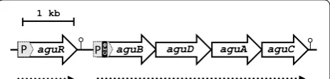
It has been shown that the agmatine deiminase (AGDI) cluster of L. lactis subsp. cremoris CECT 8666 (formerly GE2-14) encodes the enzymatic activities responsible for the catabolism of agmatine to putrescine [31]. Briefly,
aguD codes for the agmatine/putrescine antiporter, aguA
encodes agmatine deiminase, aguB encodes putres-cine transcarbamylase, and aguC encodes a specific
carbamate kinase [32, 33]. Transcriptional analyses of these genes has shown the expression of aguB, aguD,
aguA and aguC to be driven by the aguB promoter, and has confirmed the four genes to be co-transcribed as a single polycistronic mRNA [32, 33]. A cre site exists in the promoter of aguB which is transcriptionally regulated by carbon catabolite repression (CCR) mediated by the catabolite control protein CcpA [32, 33]. Also included in the AGDI cluster, upstream of the aguBDAC genes, is the aguR gene, which is transcribed constitutively under its own promoter (PaguR) (Fig. 1). We previously showed
that aguR is a regulatory gene encoding a transmem-brane protein (AguR) that acts as a one-component sig-nal transduction system able to sense the extracellular agmatine concentration and trigger transcriptional acti-vation of the aguB promoter (which drives expression of the aguBDAC operon) [34].
The present work reports the adaptation of the ACE system—an inducible gene expression system that involves aguR and the PaguB promoter (the latter with
its natural ribosome binding site)—to L. lactis. This was successfully tested via the production of green fluores-cent protein (GFP) and Myxococcus xanthus prolyl-endo-peptidase (PEP) [35, 36].
Results
Site‑directed mutagenesis of the cre site of PaguB
In previous work [34] we showed that PaguB of L. lactis
CECT 8666 drives expression of the aguBDAC operon in response to agmatine supplementation. Moreover, aguB-DAC expression is regulated by CCR, mediated by the catabolite control protein A (CcpA) [33]. To develop an efficient gene expression system, CCR had to be inacti-vated so that glucose could be used as a carbon source. This allowed high bacterial cell densities to be obtained, and in turn high recombinant protein yields. For this we introduced arbitrarily directed mutations into the cre site (sequence 5′-TGAAATCGTTCCCA-3′; the nucleotides
aguR aguB aguA
P
1 kb
cre aguD aguC
P
Fig. 1 Genetic organization of the AGDI cluster of L. lactis CECT 8666. Physical map showing the cluster to be composed of five genes: aguR which encodes a transcription regulator, followed by
aguB, aguD, aguA and aguC, which encode the proteins involved in the putrescine biosynthesis pathway (GenBank: AZSI00000000.1). The PaguR and PaguB promoters are shaded in grey, and the termina‑
matching the cre consensus sequence are underlined) within PaguB (see “Methods”). The pAGDI plasmid [33],
containing the PaguR-aguR-PaguB cassette fused to the
reporter gene gfp (encoding green fluorescent protein [GFP]), was used to construct new plasmids to assess the effect of cre mutation on PaguB activity. Using this
plas-mid as a template, and the primers indicated in Table 1, three new plasmids were generated which contained spe-cific mutations in the cre site: pAGDIcre1 (containing 10 nucleotide mutations from positions 5–14 of the cre site), pAGDIcre2 (containing 3 nucleotide mutations from positions 6–8 of the cre site) and pAGDIcre3 (containing 1 nucleotide mutation at position 5) (Fig. 2a). The 10 and 3 nucleotide mutations (plasmids pAGDIcre1 and pAG-DIcre2 respectively) both had a dramatic effect on PaguB:
no activity was detected with either construct at any glu-cose concentration (Fig. 2c, d; compare with Fig. 2b [wild type]). The introduction of a single mutation (A > T) in the cre site (pAGDIcre3 construct) gave the expected result, i.e., expression was not repressed at 120 mM glucose (Fig. 2e). This mutated promoter was therefore selected for the construction of the expression vector.
Agmatine‑induced heterologous expression of gfp
To verify the usefulness of the ACE system in the expression of heterologous genes, the reporter gene gfp
was cloned into the pACE vector under the control of PaguB, thus generating the plasmid pACE-gfp. L. lactis
NZ9000—a strain without the AGDI cluster—was trans-formed with pACE-gfp, thus resulting in L. lactis
pACE-gfp. The presence of 10 mM agmatine in a culture of L. lactis pACE-gfp induced the expression of gfp, which was measured in terms of the fluorescence produced
(7.87 arbitrary units) (Fig. 3). It should be noted that the expression of gfp was undetectable (<0.5 arbitrary units) in agmatine-uninduced cultures of L. lactis pACE-gfp. Fluorescence was also undetectable (<0.5 arbitrary units) in parallel agmatine-induced cultures of L. lactis NZ9000 harbouring the pACE vector (L. lactis pACE).
Sensitivity of the ACE system to the inducer: dose– response curve
The production of GFP in cultures of L. lactis
pACE-gfp induced using a range of agmatine concentrations (between 0 and 60 mM) was analysed by whole-cell fluo-rescence. The ACE expression system showed great sen-sitivity to very low agmatine levels; a significant increase in fluorescence was seen after induction with agmatine at concentrations as low as 10−5 mM (Fig. 3). Above this concentration, the induction levels increased in line with the agmatine concentration until a maximum induction level (fluorescence ~8 arbitrary units) was reached at 0.5 mM agmatine (no significant increases in induction were seen with concentrations of >0.5 mM). The absence of any leaky activity of the promoter PaguB, as verified by
the absence of fluorescence in uninduced cultures (0 mM agmatine, Fig. 3), is remarkable.
Heterologous production of GFP using the ACE system The efficiency of the ACE system in overexpressing recombinant protein was tested with the reporter protein GFP. The expressed His-tagged GFP protein was puri-fied using immobilized metal affinity chromatography (IMAC). The eluted protein fractions were examined by SDS-PAGE (data not shown) and their GFP activity. Pure protein with GFP activity was obtained in fraction two
Table 1 Oligonucleotides used in this study
Primer Function Sequence (5′ to 3′)
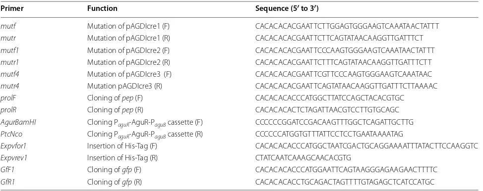
mutf Mutation of pAGDIcre1 (F) CACACACACGAATTCTTGGAGTGGGAAGTCAAATAACTATTT
mutr Mutation of pAGDIcre1 (R) CACACACACGAATTCTTCAGTATAACAAGGTTGATTTCT
mutf1 Mutation of pAGDIcre2 (F) CACACACACGAATTCCCAAGTGGGAAGTCAAATAACTATTT
mutr1 Mutation of pAGDIcre2 (R) CACACACACGAATTCTTTCAGTATAACAAGGTTGATTTCTT
mutf4 Mutation of pAGDIcre3 (F) CACACACACGAATTCGTTCCCAAGTGGGAAGTCAAATAAC
mutr4 Mutation pAGDIcre3 (R) CACACACACGAATTCAGTATAACAAGGTTGATTTCTTAAAAC
prolF Cloning of pep (F) CACACACACCCATGGCTTATCCAGCTACACGTGC
prolR Cloning of pep (R) CACACACACTCTAGATTAACGTCCTTGTGCAGC
AgurBamHI Cloning PaguR‑AguR‑PaguB cassette (F) CCCCCCGGATCCGACAAGTTTGGCTCAGATTGCTTG PtcNco Cloning PaguR‑AguR‑PaguB cassette (R) CCCCCCATGGTGTTTATTCCTCCTGAATAAAATAG
Expvfor1 Insertion of His‑Tag (F) CACACACACCCATGGCTAATCGACTGCAGGAAAATTTATACTTCCAAGGTC
Expvrev1 Insertion of His‑Tag (R) CTATCAATCAAAGCAACACGTG
GfF1 Cloning of gfp (F) CACACACACCCATGGAATTCAGTAAGGGAGAAGAACTTTTC
(of the four obtained); the yield was 47 % (Table 2). Fluo-rescence was found in the soluble fraction only (data not shown).
Heterologous overproduction of M. xanthus
prolyl‑endopeptidase and comparison with the NICE system
To confirm the usefulness and efficiency of the ACE system, the pep gene of M. xanthus, which encodes a prolyl-endopeptidase of biomedical interest, was cloned into appropriate plasmids for introduction into L. lactis NZ9000. The resulting L. lactis NZ9000 pACE-pep was induced with different agmatine con-centrations (0, 0.001, 0.01, 0.1, 0.5, 1, 2, 5, 10, 20, 40 and 60 mM) and the PEP activity assayed. No activity was detected in cultures without agmatine, but was observed even with the lowest agmatine concentra-tion tested (0.001 mM). Above this concentraconcentra-tion, the induction level increased with the agmatine concen-tration until 0.1 mM agmatine (21.04 mU mg−1) (no significant increase in PEP activity was obtained with concentrations of >0.1 mM) (Fig. 4). PEP activity was sought in the soluble and insoluble fractions, but was only seen in the former.
For comparison, PEP was also produced using the NICE system at different nisin concentrations (0, 0.05, 0.1, 0.25, 0.5, 1, 2.5, 5, 10 ng ml−1) (Fig. 4). The activity increased with the nisin concentration until 2.5 ng ml−1. The highest specific activity obtained (15.2 mU mg−1) was lower than that obtained with the ACE system pAGDIcre1
pAGDI
pAGDIcre2 pAGDIcre3
TTATACTGAAATCGTTCCCAAGTG
TTATACTGAA
GAATTCTTGG
AGTG
TTATACTGAAA
GAA
TTCCCAAGTG
TTATACTGAA
T
TCGTTCCCAAGTG
cre site -10
gfp gfp gfp gfp aguR
aguR aguR aguR
RBS
P
aguBa
b
c
d
e
2.5 5.0
0 7.5 10
2.5 5.0
0 7.5 10
2.5 5.0
0 7.5 10
2.5 5.0
0 7.5 10
30 120 Glucose (mM)
Fluorescence
(a.u.
)
30 120 Glucose (mM)
Fluorescence
(a.u.
)
30 120 Glucose (mM)
Fluorescence
(a.u.
)
30 120 Glucose (mM)
Fluorescence
(a.u.
)
pAGDI pAGDIcre1 pAGDIcre2 pAGDIcre3
*
Fig. 2 Generation of mutations in the cre site of PaguB in the AGDI cluster of L. lactis CECT 8666, and their effect on promoter activity. a Genetic
detail of the different gfp fusions made with the wild type PaguB region (fusion pAGDI) and the derived promoters carrying different mutations in
the cre site (fusions pAGDIcre1, pAGDIcre2 and pAGDIcre3). The introduced mutations are highlighted in black. Dashed lines indicate sequence dis‑ continuities. b–e Promoter activity reported by GFP fluorescence (arbitrary units) for the wild type PaguB and mutants assayed in the presence of 30
and 120 mM glucose under induction by 20 mM agmatine. Data represent the average of three independent experiments. Bars indicate standard deviations (*p < 0.05)
0 1 2 3 4 5 6 7 8 9 10
1 2 3 4 5 6 7 8 9 10 11 12 13 14 15
Fluo
re
scence (a.u.)
0.1 0.5 10 20 40 60
10-6 10-510-410-310-2 1 2 5
*
*
*
*
*
0
(21.04 mU mg−1). Again, all PEP activity was observed in the soluble fraction.
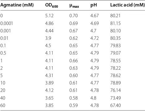
Influence of agmatine on Lactococcus lactis growth
Since high induction concentrations of agmatine were tested in the present work, assays were performed to see whether these affected bacterial fitness. L. lactis cul-tures were grown in liquid GM17 supplemented with 0, 0.001, 0.01, 0.1, 0.5, 1, 2, 5, 10, 20, 40 or 60 mM agmatine. Similar growth curves were obtained (data not shown). Table 3 shows the OD600, μmax and pH values and lac-tate concentrations reached after 24 h of incubation. Growth slightly decreased with increasing concentra-tion of agmatine. Although the 0.5 and 0.1 mM agmatine concentrations led to the highest GFP and PEP activi-ties, no differences in OD600 were observed compared to the uninduced cultures. Organic acids and sugars were analyzed by HPLC, and no differences observed in the presence or absence of agmatine (data not show). Agmatine had a weak effect on L. lactis cell viability after 24 h of exposure; optical density values suitable for pro-tein production were obtained even at high agmatine concentrations.
Discussion
Lactococcus lactis has long been used in the food indus-try and has emerged as a cost-effective cellular factory for the production of proteins of interest [4]. At present, the genome sequences of several strains have been elu-cidated, and the genetic tools available for use with lac-tic acid bacteria (LAB) are ever increasing the number of fully lipopolysaccharide-free recombinant proteins that can be produced [3, 37].
The control of gene expression is critical in achieving high protein yields. For example, it is essential for ensur-ing the conservation of energy for the production of bio-mass prior to the directed overproduction of the target protein, and in controlling the production of products that might be toxic to the host cell (i.e., membrane pro-teins, autolysins, lysis-related proteins from phages) [3, 38]. Systems that allow for well controlled induc-ible expression, and that allow no basal expression, are essential in setting production rates. A number of induc-ible expression systems for L. lactis are available [20–22, 25, 39], however, the use of some of them, especially in large-scale fermentations, may be hampered by the low-level induction achieved, high background expression
Table 2 Purification of GFP protein using the ACE system
a Recovery of fluorescence activity relative to the total activity of the soluble extract
Step Total protein (µg) Protein (µg ml−1) Total activity (U mg−1) Protein yield (%)a
Lysate 63,927 15,981 80
Flow through 53,463 13,365 0
Wash 1 3244 811 0
Wash 2 4220 1055 0
Elution 1 3049 6098 0
Elution 2 235 471 171 47
Elution 3 55 111 0
Elution 4 488 977 0
0 5 10 15 20 25 30
0 0.001 0.01 0.1 0.5 1 2 5 10 20 40 60
PEP
-1)
*
*
*
0 5 10 15 20 25 30
0 0.05 0.1 0.25 0.5 1 2.5 5 10
PEP
-1)
Nisin (ng ml-1)
*
*
*
* *
b
a
Fig. 4 Comparison of PEP activity. a In the NICE system, PEP activity was monitored in L. lactis NZ9000 pNZ8048‑pep cells induced at different nisin concentrations for 3 h after reaching OD600, while b in the ACE system, PEP activity was monitored in L. lactis NZ9000 pACE‑pep cells induced with
before induction (leaky activity), and/or a lack of con-trol over the inducer [21, 25, 26, 39]. Such drawbacks are not suffered with the NICE system, probably the most commonly used regulated expression system for Gram-positive bacteria [27–29, 40]. However, when using a host strain other than L. lactis NZ9000, the NICE system requires the regulatory genes nisR and nisK to be sup-plied in trans. Thus, although the NICE system has been optimised to be used as a single-plasmid expression vec-tor [41], its exploitation nearly always requires the inte-gration of nisR and nisK, either in the chromosome of the host strain or via their cloning into a separate plasmid (the dual-plasmid strategy).
The present work describes an alternative system based on the aguR/PaguB cassette (the regulatory part of
the AGDI cluster of L. lactis CECT 8666) for agmatine-controlled gene expression in L. lactis using a vector that includes all the elements required. It should be noted that this system requires the expression of no additional genes supplied in trans. The developed vec-tor relies on the regulavec-tory transmembrane protein AguR, which responds to extracellular agmatine, and in so doing triggers the induction of gene expression via PaguB [34]. It was previously shown that PaguB of L. lactis
CECT 8666 contains a cre site involved in CCR and that this is repressed by glucose concentrations of >30 mM [33]. Since higher concentrations of glucose may be required in culture media to obtain the densities of bac-terial cells needed to provide high recombinant protein yields, this repression mechanism was eliminated in the present work through the introduction of a single A > T mutation at position 5 in the cre site. Larger muta-tions of either 3 or 10 nucleotides completely impaired the activity of the promoter, most likely by preventing
some additional regulatory signal. Other authors have also shown CCR to be relieved when single mutations occur in the cre site of CCR-controlled genes in L. lactis. For example, a single mutation in the cre site of the celB
promoter allows fully active transcription of the cryp-tic cel cluster involved in lactose utilization in L. lactis
MG1363 [42]. Similarly, two single mutations in the
cre site of the ptcC promoter do away with the glucose-repressor effect and allow cells to constitutively metabo-lize cellobiose [43].
An expression vector combining the one-component signal transduction system, i.e., aguR and the aguB pro-moter, followed by convenient cloning sites for introduc-ing the gene of interest, was constructed. An important feature of the developed pACE vector is the possibility of fusing a His-tag to the protein of interest by cloning the encoding gene in frame into the NcoI-PstI sites. His-tags have been used in other L. lactis expression vectors previously shown to perform efficiently in the immuno-detection and purification of proteins [6, 44]. We here confirm the functionality of the His-tag in purifying the GFP (protein yield 47 %).
The control of the ACE system was assessed via the expression of gfp, the reporter gene coding for GFP. Strong fluorescence was seen in the presence of ≥0.5 mM agmatine (8 arbitrary units compared to 0 in uninduced cells). Interestingly, the system was associated with no basal expression, indicating PaguB to have no leaky
activ-ity. Further, agmatine is not present in common culture media, thus allowing for tight control over the gene to be expressed. Neither is it found in milk nor any deriva-tive dairy environment where L. lactis occurs [45]. The optimal moment of induction, which can change from one overexpressed gene to another, needs to be evalu-ated. In the ACE system, the inducer agmatine can be added when the culture medium is prepared. The AguR/ PaguB cassette, on which this expression system is based,
is the regulatory part of the AGDI cluster of L. lactis [33] and is not active until the transition between the expo-nential and stationary phases is reached (5–6 h of cul-ture) [46]. Thus, even when agmatine is supplied to the culture medium, the time of net expression would lie between 5 and 7 h after culturing began. This induction time is comparable to that associated with the NICE system (2–3 h after adding nisin). The addition of the inducer at the beginning of culturing avoids the problem of monitoring the culture’s optical density to determine the optimal moment for induction. Moreover, the aboli-tion of sampling and inducer addiaboli-tion steps may prevent contamination, which could have serious economic con-sequences in industrial protein production. As seen for nisin, agmatine affected cell viability and caused a 20 % reduction in bacterial yield. However, the final OD (>3.3)
Table 3 Effect of agmatine on growth, μmax, pH and
pro-duction of lactic acid
Agmatine (mM) OD600 μmax pH Lactic acid (mM)
0 5.12 0.70 4.67 80.21
0.0001 4.86 0.69 4.69 81.15 0.001 4.44 0.67 4.7 80.10
0.01 3.9 0.62 4.72 80.35
0.1 4.5 0.65 4.77 79.83
0.5 4.11 0.65 4.79 79.07
1 4.11 0.66 4.79 78.55
2 4.11 0.63 4.79 78.22
5 4.31 0.60 4.77 78.62
10 3.89 0.61 4.77 78.89
20 4.12 0.61 4.78 76.14
40 3.65 0.58 4.8 73.49
of agmatine-induced cultures was still optimal for indus-trial protein production.
The price of the inducer is important in large scale fer-mentations. That of agmatine varies widely, depending on the supplier, but even the cheapest found (marketed as dietary supplement) worked properly as an inducer (data not shown). Certainly, it was much cheaper than nisin.
An agmatine induction system combining AguR and PaguB of Enterococcus faecalis was earlier used to develop
an expression vector suitable for the latter species [47]. However, the proposed lactococcal ACE system shows some differences to the E. faecalis system: (1) the aguR
gene is in the same orientation as PaguB (reflecting the
corresponding organization of the AGDI cluster in each system), (2) the highest expression rate is reached at ≥0.5 mM agmatine in the ACE system but at ≥60 mM in the E. faecalis system, and (3) the level of induction (as determined by fluorescence) is less than that achieved with the E. faecalis system.
In the present work, the performance of the ACE sys-tem for the heterologous production of the M. xanthus
PEP was compared to that obtained using the NICE sys-tem, the most widely used and potent gene expression system in L. lactis [40]. The proposed system achieved higher PEP activity (circa 38 %) under similar laboratory conditions. The observed differences might be related to the characteristics of each promoter, or to the effect on signal transduction of the two-component NICE system compared to the one-component ACE system. More studies are required to understand how the agmatine sig-nal is transduced.
In summary, the present results confirm the ACE sys-tem as an attractive candidate for high level recombinant protein production. The lactococcal aguR/PaguB system
can effectively control the expression of genes in response to agmatine in L. lactis without any basal expression, and combines both the expression cassette and regulatory gene in one plasmid. This vector expands the genetic toolbox available for this species, and could be a powerful and straightforward alternative system for overexpressing proteins in lactococcal strains lacking nisR and nisK. It might also be used to complement the NICE system and be used in co-expression.
Conclusions
The present work describes the construction of a L. lac-tis agmatine-controlled expression system based on the
aguR/PaguB cassette of the putrescine biosynthesis gene
cluster. A single mutation of the cre site in PaguB abolished
the CCR of this promoter. This system was assessed by expressing the reporter gene gfp, and fluorescence was found strictly dependent on the agmatine concentration added to the culture medium, with maximum induction
occurring at 0.5 mM agmatine (7 arbitrary units com-pared to 0 in uninduced cells). An important potential benefit of this system is the lack of leaky activity asso-ciated with it, and the fact that gene expression can be tightly controlled via the addition of the appropriate concentration of agmatine. The pACE vector allowed the agmatine-inducible expression of the gene encoding
M. xanthus PEP, an enzyme that can degrade the immu-notoxic peptides of gluten breakdown. Moreover, enzy-matic activity was greater than that obtained with the NICE expression system. The addition of a His-tag to the pACE vector renders the system suitable for protein purification and immunodetection purposes. Together, these findings suggest that the ACE expression system could be a very valuable addition to the L. lactis genetic toolbox, and offers a straightforward, alternative induc-ible gene expression system that to be used in functional studies and in the large-scale production of recombinant proteins.
Methods
Bacterial strains and growth conditions
L. lactis CECT 8666 (formerly GE2-14) and L. lactis
NZ9000 were grown at 30 °C in M17 medium (Oxoid, Basingstoke, United Kingdom) supplemented with 30 mM glucose (GM17). When required, agmatine (Sigma-Aldrich, St. Louis, MO, USA) was added to the medium. Chloramphenicol (5 μg ml−1) was added as required for plasmid maintenance. For overexpression using the NICE system, cultures of L. lactis NZ9000 in exponential phase (OD600 = 0.4–0.5), grown in GM17, were induced for 3 h with various nisin concentrations (0, 0.05, 0.1, 0.25, 0.5, 1, 2.5, 5 and 10 ng ml−1) (Sigma-Aldrich). Solid media were prepared by adding 2 % (w/v) agar (Merck, Darmstad, Germany). Microbial growth was examined in all cultures by measuring absorbance at 600 nm (OD600) using a spectrophotometer (Eppendorf, NY, USA). The pH of the samples was measured using a CRISON miCropH 2001 pH meter (Crison Instruments S.A., Barcelona, Spain). The maximum specific growth rate (μmax) was determined experimentally in the expo-nential growth phase, as described by O´Sullivan and Condon [48].
DNA manipulation
of Korea). All enzymes for DNA technology were used according to the manufacturer’s specifications.
Construction of plasmids
The core of the lactococcal vector pNZ8048 [29]—which includes the replication cassette and the chlorampheni-col resistance marker—was used as a starting point for the construction of the pACE vector. First, a fragment of the AGDI cluster from L. lactis CECT 8666 (Table 4), including the aguR promoter (PaguR), the aguR gene, and
the aguB promoter (PaguB) carrying the mutation in the
cre site, was PCR-amplified (using pAGDIcre3 as a tem-plate) and cloned into the BglII-NcoI sites of pNZ8048. Subsequently, a fragment including the multicloning site and a histidine tag encoding 10 consecutives histidines (His-tag) was amplified from plasmid pNZErmC [6] and cloned into the NcoI-XbaI sites of the previous construct, thus yielding vector pACE (Fig. 5). This vector offers the option to fuse in-frame the gene encoding the protein of interest to a C-terminal His-Tag by cloning the target gene into the NcoI-PstI sites. Thus, this vector could be used for immunodetection or purification of the proteins encoded by overexpressed genes. The target gene could also be cloned without the His-tag for use in functional studies.
For the heterologous expression of GFP, the gfp
gene (amplified from pAG2 [34]) was inserted into the
NcoI-PstI sites of pACE, thus generating the plasmid pACE-gfp. To produce PEP using the ACE system, the
pep gene was PCR-amplified from pNZ8048-pep and cloned into the NcoI-XbaI sites in the pACE vector,
resulting in the pACE-pep plasmid. To produce PEP using the NICE system, the pep gene was released from plasmid pUC57-pep [36] as a NcoI-XbaI fragment and cloned into the same sites in pNZ8048 under the con-trol of the nisA promoter [29], thus generating plasmid pNZ8048-pep.
Directed mutagenesis of the cre site of PaguB
Modification of the cre site was achieved by in vitro site-directed mutagenesis. Mutation(s) were introduced by PCR using two divergent primers (Table 1) spanning the
cre site of PaguB and containing the desired mutation(s).
Each primer was complementary to the opposite strand of the pAGDI vector, which was used as template to gen-erate plasmids pAGDIcre1, pAGDIcre2 and pAGDIcre3 containing the specific mutations in the cre site. The pAGDI plasmid was first methylated with Dam methyl-ase and S-adenosyl methionine following the manufac-turer’s instructions (New England Biolabs, Hertfordshire, UK). Phusion High-Fidelity DNA Polymerase (New Eng-land Biolabs) was used to amplify both plasmid strands with high fidelity. The PCR thermoycling conditions were as follows: initial denaturation (98 °C for 30 s), 32 cycles of amplification (98 °C for 10 s; 55 °C for 30 s; and 72 °C for 2.5 min) plus a final extension step (72 °C for 7 min). An EcoRI target site was included in the primers so that the obtained amplicons could be digested with EcoRI and self-ligated. Before transformation in L. lactis NZ9000, the ligation mixture was treated with DpnI order to digest the original, Dam methylated pAGDI plasmid used as a template.
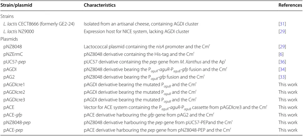
Table 4 Strains and plasmids
PaguR, aguR promoter; PaguB, aguB promoter; pep, prolyl endopeptidase gene; Cmr, chloramphenicol resistance marker; Apr, ampicillin resistance marker
Strain/plasmid Characteristics References
Strains
L. lactis CECT8666 (formerly GE2‑24) Isolated from an artisanal cheese, containing AGDI cluster [31]
L. lactis NZ9000 Expression host for NICE system, lacking AGDI cluster [29]
Plasmids
pNZ8048 Lactococcal plasmid containing the nisA promoter and the Cmr [29]
pNZErmC pNZ8048 derivative containing the His‑tag and the Cmr [6]
pUC57‑pep pUC57 derivative containing the pep gene from M. Xanthus and the Apr [36]
pAGDI pNZ8048 derivative bearing the PaguR‑aguR‑PaguB‑gfp fusion and the Cmr [34]
pAG2 pNZ8048 derivative bearing the PaguB‑gfp fusion and the Cmr [33]
pAGDIcre1 pAGDI derivative bearing the mutated PaguB and the Cmr This work
pAGDIcre2 pAGDI derivative bearing the mutated PaguB and the Cmr This work
pAGDIcre3 pAGDI derivative bearing the mutated PaguB and the Cmr This work
pACE Vector for ACE system containing the PaguR‑aguR‑PaguB cassette from pAGDIcre3 and the Cmr This work
pACE‑gfp pACE derivative harbouring the gfp gene from pAG2 and the Cmr This work
pNZ8048‑pep pNZ8048 derivative harbouring the pep gene from pUC57‑PEPand the Cmr This work
Measurement of green fluorescence
For whole-cell fluorescence measurements, overnight cultures of L. lactis NZ9000 harbouring either pACE or pACE-gfp were transferred (1 %) to fresh medium (GM17) supplemented with different agmatine concentrations (0–60 mM) and grown for 7 h. Equal amounts of cells were harvested, washed, and then resuspended in 50 mM potas-sium phosphate buffer, pH 7.2, as previously described [6]. GFP emission was measured in a volume of 200 μl of cells, using a Cary Eclipse fluorescence spectrophotometer (Varian Inc., Palo Alto, CA, USA) at an excitation wave-length of 485 nm and an emission wavewave-length 530 nm. To facilitate direct comparisons, the bacterial cultures used for GFP fluorescence measurements contained similar amounts of cells (estimation was made based on OD600). Background fluorescence levels were assessed using non-fluorescent control cells (lacking the gfp gene), and these values subtracted from the experimental results.
Prolyl‑endopeptidase assay
PEP activity was determined using a synthetic substrate, succinyl-Ala-Pro-p-NA (NA, nitroanilide) (Bachem, Bubendorf, Switzerland), as previously described [36] with slight modifications. Bacterial cultures (10 ml) of
L.lactis pNZ8048-pep (induced with nisin for 3 h after the cells reached an OD600 of 0.6) and L. lactis pACE-pep
(induced with 20 mM agmatine [added when the cul-ture medium was prepared] and grown for 7 h) were harvested by centrifugation (8000g for 10 min), washed twice, and resuspended in 2 ml of 50 mM phosphate buffer, 0.2 M NaCl, pH 7.5. The samples were then dis-rupted using 200 mg glass beads (<106 µm) (Sigma-Aldrich) in a Fast-Prep FP120 Instrument (Thermo Savant-BIO101/Q-Biogen, CA, USA) for 6 × 30 s at power setting 4.5 (with intermittent cooling). Cell debris was removed by centrifugation (10,000g for 30 min at 4 °C) and the supernatant used in activity assays. The assay mixture contained 625 μl of 50 mM phosphate buffer (pH 7.5), 0.2 M NaCl, 125 μl substrate (1.2 mM), and 250 μl of cell extracts. The reaction was stopped by adding 500 μl of 20 % trichloroacetic acid. The sam-ples were then centrifuged (10,000g for 10 min) and the release of the p-NA spectrophotometrically detected at 410 nm in a U-2800 Digilab Hitachi spectrophotometer (HitachiHigh-Technologies Corporation, Tokyo, Japan). One activity unit was defined as the amount of enzyme required to release 1 μmol of p-NA per min under the assay conditions. Assays were performed in triplicate. Specific enzyme activity was expressed as milliunits per milligram of protein. The protein concentration was measured using a Pierce BCA Assay Kit (Thermo Fisher scientific) following the manufacturer’s indications. AguR
PaguB*
Target gene
Recombinant protein
b
MCS
T1 T2
aguR
PaguRPaguB
*
HisT
ag
Nc
o
I
RBS PstI
T3
pACE
repA
repC
cat
BglII
a
4784 bp
Nc
o
I
Ps
t
I
HisT
ag
Sp
h
I
Kp
n
I
Sp
e
I
Xb
a
I
Sa
c
I
PaguB*
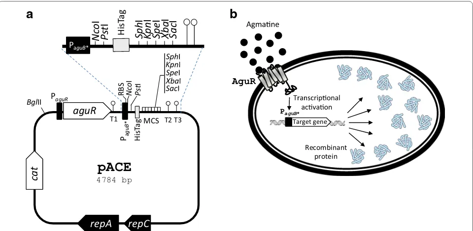
Fig. 5 a Genetic map of the pACE expression vector. repC and repA, replication genes; cat, chloramphenicol resistance gene; aguR, gene encoding the regulatory agmatine‑sensor‑regulator protein AguR; PaguR, aguR promoter; RBS, ribosome binding site; T1, T2 and T3, transcription terminators
(ΔG =−10.3, −9.7 and −8.3 kcal/mol respectively); PaguB*, aguB agmatine‑inducible promoter carrying the A > T mutation in the fifth nucleotide
of the cre site; MCS, multicloning site; HisTag; C‑terminal histidine tag. Representative restriction sites are indicated. b Overview of the AguR/PaguB
Purification His‑tagged protein
Purification of the His-tagged GFP protein was per-formed by immobilized metal ion affinity chromatog-raphy (IMAC). Cells (200 ml) induced with 10 mM agmatine were harvested by centrifugation at 8000g, at 4 °C for 10 min after 7 h of growth. The supernatant was discarded and cells washed twice and resuspended in 4 ml phosphate buffer (50 mM pH 7.5). They were then disrupted using a French Press operating at 2.3 kbar [Constant Cell Disruption Systems (Low March, Dav-entry, Norttants, UK)]. Cell debris was removed by cen-trifugation (10,000g for 30 min at 4 °C). Imidazole was then added to a concentration of 10 mM, and the result-ing samples employed in protein purification usresult-ing the QIAexpressionist kit (Quiagen, Madrid, Spain) follow-ing the manufacturer’s instructions. Aliquots of collected fractions were analyzed by SDS-PAGE using 12 % acryla-mide gels to determine the purity of the His6-taggeted proteins. Their GFP activities and protein concentrations were determined using the protocols mentioned above to determine the protein yield.
Soluble and insoluble protein fractions
Soluble and insoluble proteins fractions were prepared following the protocol described by Cano-Garrido et al. [51]. Samples (10 ml) of bacterial cultures grown at dif-ferent agmatine concentrations were pelleted by cen-trifugation at 8000g at 4 °C for 10 min and the sediment resuspended in 1 ml of the appropriate buffer depend-ing on the protein expressed (GFP—phosphate buffer 50 mM, pH 7.5; PEP—phosphate buffer 50 mM, pH 7.5, 0.2 M NaCl). The samples were then disrupted with 200 mg glass beads (<106 µm) (Sigma-Aldrich) in a Fast-Prep FP120 Instrument (Thermo Savant-BIO101/Q-Bio-gen, CA, USA) for 6 × 30 s at a power setting of 4.5 (with
intermittent cooling). Total cell extracts were centrifuged at 15,000g at 4 °C for 15 min. Finally, the insoluble frac-tions were resuspended in 1 ml of the appropriate buffer and fluorescence and PEP activity monitored in both the soluble and insoluble-resuspended fractions.
Determination of organic acids and sugars by HPLC
Sugar and organic acid concentrations were determined using a chromatographic system composed of an Alli-ance 2690 module injector, a Photodiode Array PDA 996 detector, and a 410 Differential Refractometer detec-tor, all controlled with Millennium 32 software (Waters, Milford, MA, USA). Supernatants (50 μl) were isocrati-cally separated in a 300 × 7.8 mm HPX-87H Aminex
ion-exchange column (Hewlett Packard, Palo Alto, CA, USA) protected by a cation H+ Microguard cartridge
(BioRad, Laboratories, Richmond, CA, USA), at a flow rate of 0.7 ml/min and a temperature of 65 °C. Sulphuric
acid (3 mM) was used as the mobile phase. A PDA 996 detector at 210 nm was used to identify and quantify the organic acids detected, whereas the sugars were analyzed with a 410 Refractometer. Solutions of lactic and acetic acids, glucose, galactose, lactose, and sucrose were used as standards in the identification and quantification procedure.
Statistical analysis
The Student t test was used to examine differences between groups. Significance was set at p < 0.05.
Abbreviations
ACE: agmatine‑controlled expression; AGDI: agmatine deiminase; CCR: carbon catabolite repression; GFP: green fluorescence protein; NICE: nisin‑inducible controlled expression; PEP: prolyl‑endopeptidase.
Authors’ contributions
DML designed and performed some experiments and drafted the manuscript; PAS and BR performed some experiments; VL, BdR, MC and MF participated in the design of the study and helped to write the manuscript; MAG provided the general concept and supervised the work and the manuscript. All authors contributed to the discussion of the research. All authors read and approved the final manuscript.
Acknowledgements
This work was performed with the financial support of the Spanish Ministry of Economy and Competitiveness (AGL2013‑45431‑R), the Principality of Asturias Plan for Science, Technology and Innovation 2013‑2017, and FEDER funds (GRUPIN14‑137). We are grateful to Bert Poolman for providing the GFP‑based cloning vectors and Adrian Burton for linguistic assistance. L. lactis NZ9000 and plasmid pNZ8048 were kindly provided by NIZO Food Research. We acknowledge support of the publication fee by the CSIC Open Access Publica‑ tion Support Initiative through its Unit of Information Resources for Research (URICI). P.A.S. was the beneficiary of a fellowship from the FICYT, Principality of Asturias, Spain (BP09093). D.M.L. and B.d.R. were beneficiaries of JAE DOC contracts (CSIC).
Competing interests
The authors declare that they have no competing interests.
Received: 17 July 2015 Accepted: 16 December 2015
References
1. Nocon J, Steiger MG, Pfeffer M, Sohn SB, Kim TY, Maurer M, Rußmayer H, Pflügl S, Ask M, Haberhauer‑Troyer C, Ortmayr K, Hann S, Koellensperger G, Gasser B, Lee SY, Mattanovich D. Model based engineering of Pichia pastoris central metabolism enhances recombinant protein production. Metab Eng. 2014;24:129–38.
2. Rosano GL, Ceccarelli EA. Recombinant protein expression in Escherichia coli: advances and challenges. Front Microbiol. 2014;5:172.
3. Mierau I, Olieman K, Mond J, Smid EJ. Optimization of the Lactococcus lactis nisin‑controlled gene expression system NICE for industrial applica‑ tions. Microb Cell Fact. 2005;4:16.
4. Cano‑Garrido O, Rueda FL, Sànchez‑García L, Ruiz‑Ávila L, Bosser R, Vil‑ laverde A, García‑Fruitós E. Expanding the recombinant protein quality in
Lactococcus lactis. Microb Cell Fact. 2014;13:167.
6. Linares DM, Geertsma ER, Poolman B. Evolved Lactococcus lactis strains for enhanced expression of recombinant membrane proteins. J Mol Biol. 2010;401:45–55.
7. Kunji ER, Chan KW, Slotboom DJ, Floyd S, O’Connor R, Monné M. Eukary‑ otic membrane protein overproduction in Lactococcus lactis. Curr Opin Biotechnol. 2005;16:546–51.
8. Bermúdez‑Humarán LG, Kharrat P, Chatel JM, Langella P. Lactococci and lactobacilli as mucosal delivery vectors for therapeutic proteins and DNA vaccines. Microb Cell Fact. 2011;10:S4.
9. D’Souza R, Pandeya DR, Hong ST. Lactococcus lactis: an efficient Gram positive cell factory for the production and secretion of recombinant protein. Biomed Res. 2012;23:1–7.
10. Lee MH, Roussel Y, Wilks M, Tabaqchali S. Expression of Helicobacter pylori urease subunit B gene in Lactococcus lactis MG1363 and its use as a vaccine delivery system against H. pylori infection in mice. Vaccine. 2001;19:3927–31.
11. Ribeiro LA, Azevedo V, Le Loir Y, Oliveira SC, Dieye Y, Piard JC, Gruss A, Lan‑ gella P. Production and targeting of the Brucella abortus antigen L7/L12 in Lactococcus lactis: a first step towards food‑grade live vaccines against brucellosis. Appl Environ Microbiol. 2002;68:910–6.
12. Xin KQ, Hoshino Y, Toda Y, Igimi S, Kojima Y, Jounai N, Ohba K, Kushiro A, Kiwaki M, Hamajima K, Klinman D, Okuda K. Immunogenicity and protective efficacy of orally administered recombinant Lactococcus lactis
expressing surface‑bound HIV Env. Blood. 2003;10:223–8.
13. Robinson K, Chamberlain LM, Lopez MC, Rush CM, Marcotte H, Le Page RW, Wells JM. Mucosal and cellular immune responses elicited by recom‑ binant strains of Lactococcus lactis expressing tetanus toxin fragment C. Infect Immun. 2004;72:2753–6.
14. Bermúdez‑Humarán LG, Cortes‑Perez NG, Lefèvre F, Guimarães V, Rabot S, Alcocer‑Gonzalez JM, Gratadoux JJ, Rodriguez‑Padilla C, Tamez‑Guerra RS, Corthier G, Gruss A, Langella P. A novel mucosal vaccine based on live lactococci expressing E7 antigen and IL‑12 Induces systemic and mucosal immune responses and protects mice against human papillomavirus type 16‑Induced tumors. J Immunol. 2005;175:7297–302.
15. Ramasamy R, Yasawardena S, Zomer A, Venema G, Kok J, Leenhouts K. Immunogenicity of a malaria parasite antigen displayed by Lactococcus lactis in oral immunisations. Vaccine. 2006;24:3900–8.
16. Van Roosmalen ML, Kanninga R, El Khattabi M, Neef J, Audouy S, Bosma T, Kuipers A, Post E, Steen A, Kok J, Buist G, Kuipers OP, Robillard G, Leen‑ houts K. Mucosal vaccine delivery of antigens tightly bound to an adju‑ vant particle made from food‑grade bacteria. Methods. 2006;38:144–9. 17. Mercenier A, Müller‑Alouf H, Grangette C. Lactic acid bacteria as live vac‑
cines. Curr Issues Mol Biol. 2000;2:17–25.
18. Zamri HF, Shamsudin MN, Rahim RA, Neela V. Oral vaccination with
Lactococcus lactis expressing the Vibrio cholerae Wzm protein to enhance mucosal and systemic immunity. Vaccine. 2012;30:3231–8.
19. Douillard FP, O’Connell‑Motherway M, Cambillau C, van Sinderen D. Expanding the molecular toolbox for Lactococcus lactis: construction of an inducible thioredoxin gene fusion expression system. Microb Cell Fact. 2011;10:66.
20. Sanders JW, Venema G, Kok J. A chloride‑inducible gene expression cassette and its use in induced lysis of Lactococcus lactis. Appl Environ Microbiol. 1997;63:4877–82.
21. Mu D, Montalbán‑López M, Masuda Y, Kuipers OP. Zirex: a novel zinc‑regu‑ lated expression system for Lactococcus lactis. Appl Environ Microbiol. 2013;79:4503–8.
22. Jorgensen CM, Vrang A, Madsen SM. Recombinant protein expression in
Lactococcus lactis using the P170 expression system. FEMS Microbiol Lett. 2014;351:170–8.
23. van Asseldonk M, Simons A, Visser H, de Vos WM, Simons G. Cloning, nucleotide sequence, and regulatory analysis of the Lactococcuslactis dnaJ gene. J Bacteriol. 1993;175:1637–44.
24. Marugg JD, Meijer W, van Kranenburg R, Laverman P, Bruinenberg PG, de Vos WM. Medium‑dependent regulation of proteinase gene expression in
Lactococcus lactis: control of transcription initiation by specific dipeptides. J Bacteriol. 1995;177:2982–9.
25. de Vos WM. Gene expression systems for lactic acid bacteria. Curr Opin Microbiol. 1999;2:289–95.
26. de Ruyter PG, Kuipers OP, de Vos WM. Controlled gene expression systems for Lactococcus lactis with the food‑grade inducer nisin. Appl Environ Microbiol. 1996;62:3662–7.
27. Kuipers OP, Beerthuyzen MM, de Ruyter PG, Luesink EJ, de Vos WM. Autoregulation of nisin biosynthesis in Lactococcus lactis by signal trans‑ duction. J Biol Chem. 1995;270:27299–304.
28. Kuipers OP, Rollema HS, Beerthuyzen MM, Siezen RJ, de Vos WM. Protein engineering and biosynthesis of nisin and regulation of transcription of the structural nisA gene. Int Dairy J. 1995;5:785–95.
29. Kuipers OP, de Ruyter PG, Kleerebezem M, de Vos WM. Quorum sensing‑controlled gene expression in lactic acid bacteria. J Biotechnol. 1998;64:15–21.
30. Mierau I, Kleerebezem M. 10 years of the nisin‑controlled gene expres‑ sion system (NICE) in Lactococcus lactis. Appl Microbiol Biotechnol. 2005;68:705–17.
31. Ladero V, Del Rio B, Linares DM, Fernandez M, Mayo B, Martin MC, Alvarez MA. Genome sequence analysis of the biogenic amine‑producing strain
Lactococcus lactis subsp. cremoris CECT 8666 (Formerly GE2‑14). Genome Announc. 2014;2:e01088–114.
32. Ladero V, Rattray FP, Mayo B, Martín MC, Fernández M, Alvarez MA. Sequencing and transcriptional analysis of the biosynthesis gene cluster of putrescine‑producing Lactococcus lactis. Appl Environ Microbiol. 2011;77:6409–18.
33. Linares DM, del Río B, Ladero V, Redruello B, Martín MC, Fernández M, Alvarez MA. The putrescine biosynthesis pathway in Lactococcus lactis is transcriptionally regulated by carbon catabolic repression, mediated by CcpA. Int J Food Microbiol. 2013;165:43–50.
34. Linares DM, del Rio B, Redruello B, Ladero V, Martin MC, de Jong A, Kuipers OP, Fernandez M, Alvarez MA. AguR, a transmembrane transcrip‑ tion activator of the putrescine biosynthesis operon in Lactococcus lactis, acts in response to agmatine concentration. Appl Environ Microbiol. 2015;81(18). doi:10.1128/AEM.00959‑15.
35. Plugis NM, Khosla C. Therapeutic approaches for celiac disease. Best Pract Res Clin Gastroenterol. 2015;29:503–21.
36. Alvarez‑Sieiro P, Martin MC, Redruello B, Del Rio B, Ladero V, Palanski BA, Khosla C, Fernandez M, Alvarez MA. Generation of food‑grade recombi‑ nant Lactobacillus casei delivering Myxococcus xanthus prolyl endopepti‑ dase. Appl Microbiol Biotechnol. 2014;98:6689–700.
37. Morello E, Bermúdez‑Humarán LG, Llull D, Solé V, Miraglio N, Langella P, Poquet I. Lactococcus lactis, an efficient cell factory for recombinant pro‑ tein production and secretion. J Mol Microbiol Biotechnol. 2008;14:48–58. 38. Kunji ER, Slotboom DJ, Poolman B. Lactococcus lactis as host for over‑
production of functional membrane proteins. Biochim Biophys Acta. 2003;1610:97–108.
39. Llull D, Poquet I. New expression system tightly controlled by zinc avail‑ ability in Lactococcus lactis. Appl Environ Microbiol. 2004;70:5398–406. 40. Kuipers OP, de Ruyter PG, Kleerebezem M, de Vos WM. Controlled
overproduction of proteins by lactic acid bacteria. Trends Biotechnol. 1997;15:135–40.
41. Bryan EM, Bae T, Kleerebezem M, Dunny GM. Improved vectors for nisin‑ controlled expression in gram‑positive bacteria. Plasmid. 2000;44:183–90. 42. Solopova A, Bachmann H, Teusink B, Kok J, Neves AR, Kuipers OP. A
specific mutation in the promoter region of the silent cel cluster accounts for the appearance of lactose‑utilizing Lactococcus lactis MG1363. Appl Environ Microbiol. 2012;78:5612–21.
43. Linares DM, Kok J, Poolman B. Genome sequences of Lactococcus lactis
MG1363 (revised) and NZ9000 and comparative physiological studies. J Bacteriol. 2010;192:5806–12.
44. Geertsma ER, Groeneveld M, Slotboom DJ, Poolman B. Quality con‑ trol of overexpressed membrane proteins. Proc Natl Acad Sci USA. 2008;105:5722–7.
45. Galgano F, Caruso M, Condelli N, Favati F. Focused review: agmatine in fermented foods. Front Microbiol. 2012;3:199.
46. del Rio B, Ladero V, Redruello B, Linares DM, Fernández M, Martín MC, Alvarez MA. Lactose‑mediated carbon catabolite repression of putrescine production in dairy Lactococcus lactis is strain dependent. Food Microbiol. 2015;48:163–70.
47. Linares DM, Perez M, Ladero V, Del Rio B, Redruello B, Martin MC, Fer‑ nandez M, Alvarez MA. An agmatine‑inducible system for the expres‑ sion of recombinant proteins in Enterococcus faecalis. Microb Cell Fact. 2014;13:169.
48. O’Sullivan E. Condon S Relationship between acid 499 tolerance, cytoplasmic pH, and ATP and H+ ‑ATPase levels in chemostat cultures of
49. Sambrook JD, Russell D. Molecular cloning a laboratory manual. Cold Spring Harbor: Cold Spring Harbor Laboratory; 2001.
50. de Vos WM, Vos P, Dehaard H, Boerritger I. Cloning and expression of the
Lactococcus lactis ssp cremoris SK11 gene encoding an extracellular serine proteinase. Gene. 1989;85:169–76.
51. Cano‑Garrido O, Rueda FL, Sànchez‑García L, Ruiz‑Ávila L, Bosser R, Vil‑ laverde A, García‑Fruitós E. Expanding the recombinant protein quality in
Lactococcus lactis. Microb Cell Fact. 2014;13:167.
• We accept pre-submission inquiries
• Our selector tool helps you to find the most relevant journal
• We provide round the clock customer support
• Convenient online submission
• Thorough peer review
• Inclusion in PubMed and all major indexing services
• Maximum visibility for your research
Submit your manuscript at www.biomedcentral.com/submit