Open Access
Research article
Cardiovascular responses to cognitive stress in patients with
migraine and tension-type headache
Rune B Leistad*
1,2, Trond Sand
1,2, Kristian B Nilsen
1,2, Rolf H Westgaard
3and Lars Jacob Stovner
1,2Address: 1Department of Neurosciences, Norwegian University of Science and Technology, Trondheim, Norway, 2Department of Neurology and
Clinical Neurophysiology, St. Olavs Hospital, Trondheim, Norway and 3Department of Industrial Economics and Technology Management,
Norwegian University of Science and Technology, Trondheim, Norway
Email: Rune B Leistad* - rune.leistad@ntnu.no; Trond Sand - trond.sand@ntnu.no; Kristian B Nilsen - kristian.b.nilsen@ntnu.no; Rolf H Westgaard - rolf.westgaard@iot.ntnu.no; Lars Jacob Stovner - lars.stovner@ntnu.no
* Corresponding author
Abstract
Background: The purpose of this study was to investigate the temporal relationship between autonomic changes and pain activation in migraine and tension-type headache induced by stress in a model relevant for everyday office-work.
Methods: We measured pain, blood pressure (BP), heart rate (HR) and skin blood flow (BF) during and after controlled low-grade cognitive stress in 22 migraineurs during headache-free periods, 18 patients with tension-type headache (TTH) and 44 healthy controls. The stress lasted for one hour and was followed by 30 minutes of relaxation.
Results: Cardiovascular responses to cognitive stress in migraine did not differ from those in control subjects. In TTH patients HR was maintained during stress, whereas it decreased for migraineurs and controls. A trend towards a delayed systolic BP response during stress was also observed in TTH. Finger BF recovery was delayed after stress and stress-induced pain was associated with less vasoconstriction in TTH during recovery.
Conclusion: It is hypothesized that TTH patients have different stress adaptive mechanisms than controls and migraineurs, involving delayed cardiovascular adaptation and reduced pain control system inhibition.
Background
Prolonged physiologic (e.g. autonomic) responses to a stressor, or insufficient recovery from stress, may cause disease, chronic pain or other subjective complaints [1-3]. Stress may also trigger headache in both migraine and ten-sion-type headache (TTH) patients [4-7]. In other head-ache syndromes (e.g. "trigeminal autonomic cephalalgias") there seems to be a clear association
between headache attacks and autonomic hyperactivity [8,9], and migraine symptoms in the prodromal phase and during attacks (e.g. nausea and vomiting) suggest autonomic imbalance also in these patients. Trigeminal and brainstem dysfunction is also implicated in migraine during attacks [10-12], and pain sensitivity is increased in TTH suggesting abnormal pain processing [13,14]. Because of the known interactions between autonomic Published: 7 August 2007
BMC Neurology 2007, 7:23 doi:10.1186/1471-2377-7-23
Received: 22 January 2007 Accepted: 7 August 2007
This article is available from: http://www.biomedcentral.com/1471-2377/7/23
© 2007 Leistad et al; licensee BioMed Central Ltd.
and pain control centres in the brainstem (e.g. for the baroreceptor reflex [15]), and because autonomic hyper-activity may sensitize peripheral nociceptors [16], it makes sense to study if autonomic activation to stress is abnormal in migraine and TTH compared to healthy sub-jects, and if autonomic activation is related to the pain responses in these patients [17].
Cardiovascular responses to short-lasting acute stress have been measured in migraine but no clear pattern emerges [18-24], and data about responses and adaptation to more long-lasting cognitive stress are lacking. For TTH most studies concerning physiological responses to stres-sors have focused on muscular activity [25], and studies investigating cardiovascular responses to stressors report inconsistent findings [18,21,23,26-29]. Low-grade long-lasting cognitive stress may be more relevant to daily (e.g. work-related) stress than short-lasting stressors used in previously published studies, such as deep breathing tests, orthostatic tests, the cold pressor test and mental arithme-tic tests. In addition, data about physiologic recovery after stress, which may be of particular importance as disease promoting factors [2,30], are insufficient in the headache research literature. Since migraine and TTH patients in some cases can have rather similar symptoms [31-33], although they are considered as clearly different syn-dromes, it was reasonable to include both entities in one study.
We have recently found that migraine and TTH patients have more stress-induced muscle pain and slower muscle pain recovery after long-lasting cognitive stress than con-trols [34,35]. This experimental task induces muscular activity and pain in the shoulders, neck and head of patients with migraine [34], TTH [34,36], cervicogenic headache [37], fibromyalgia [38,39] as well as in healthy controls [40]. However, muscular activation did not cor-relate with pain responses and no muscular response dif-ferences were found between migraine, TTH and controls [34]. Measuring cardiovascular and skin blood flow (BF) responses to stress and recovery after stress in parallel with pain in migraine and TTH may give insight into other potentially pain-inducing or contributing mechanisms in primary headache disorders.
The main questions were: do the early and the late auto-nomic activation pattern during stress and the recovery pattern differ in migraine, TTH and headache-free con-trols? Do the autonomic activation and recovery pattern correlate with increased pain during stress and recovery in migraine and TTH patients? We accordingly recorded blood pressure (BP), heart rate (HR) and skin BF develop-ment as well as head and shoulder/neck pain in these sub-jects during low-grade cognitive stress for one hour followed by 30 minutes of relaxation.
Methods
Subjects
Forty-four healthy control subjects, 35 women (mean age 39.7 years) and 9 men (36.6 years) and 40 patients with headache participated in this study. Twenty-two patients had migraine, 20 women (39.8 years) and 2 men (45.0 years), and 13 of these patients had aura. Eighteen patients had TTH, 9 women (33.8 years) and 9 men (35.7 years). Twelve of the TTH patients had chronic TTH. Detailed subject and headache history data are shown in Table 1. Patients were diagnosed after interview and phys-ical examination by a neurologist according to the Inter-national Headache Society classification of headache from 1988 [41]. Control subjects did not suffer from headache or musculoskeletal pain for more than one day per month. Exclusion criteria were: neoplastic disease, hypertension, infectious disease, metabolic, endocrine or neuromuscular diseases, significant psychiatric disorders, connective tissue disorder, tendinitis, recent significant accident or injury, pregnancy, daily medication with neu-roleptics, antiepileptics, Ca2+-blockers, β-blockers,
antide-pressants, and significant associated diseases affecting either the heart, lungs, cerebrovascular system, central or peripheral nervous system. Migraineurs with TTH more than 7 days per month were also excluded. The project was approved by the Regional Ethics Committee. All par-ticipants gave written informed consent and received NOK 500 (USD 75) for transport expenses and inconven-ience. The participants were provided with written infor-mation concerning the aim of the study prior to the day of the stress test. The aim of studying pain and headache was mentioned, but the information focused on the practical details of the procedure.
Questionnaire and interview
A structured interview concerning headaches and muscu-loskeletal complaints (distribution, severity, and dura-tion) was performed. One of these questions was: "Please state the level of general tension you have felt during the last 2–3 months", and the response was scored on a visual analogue scale (VAS) with endpoints: not tense – very tense. Participants also kept a headache diary for 7 days before and after the stress test. All subjects answered a questionnaire on marital status, weight, stimulant use, exercise habits, and sleep problems (data not shown). With the exception that migraineurs had lower alcohol consumption than controls (Chi-Square test, p = 0.034), there were no statistically significant differences in these parameters.
Physiological recordings
Muscular activity was recorded with surface electromyog-raphy (EMG) bilaterally in the trapezius, splenius, tempo-ralis and frontalis muscles, as described in a previous paper [34]. Autonomic activity was measured indirectly by continuous recording of non-invasive finger BP (Port-apres, TNO Biomedical Instrumentation, Amsterdam, The Netherlands) [42] and skin BF in the thumbs (Moorlab, time constant 0.02 s, low-pass filter 22 kHz; Moor Instru-ments Ltd, Devon, England). The BP cuffs were mounted on the intermediate phalanx on the left middle and ring fingers. Finger skin BF was measured bilaterally with the electrodes (fiber separation 0.5 mm) placed on the volar side of the distal phalanx (pulp) of the thumbs. The aver-age from the left and right thumb was used for analysis, because a significant side difference was not found. Sig-nals were sampled at 200 Hz. HR and BP was calculated with the Beatscope 1.0 software (TNO, Amsterdam, The Netherlands). Respiration was recorded with a thermistor (Embla S-AF-010, Flaga, Reykjavik, Iceland) below the nose with active elements in each nostril and in front of the mouth, but respiration frequency was not analysed in this study due to technical difficulties (Seven controls, eight migraineurs and two patients with TTH had cor-rupted respiration rate data).
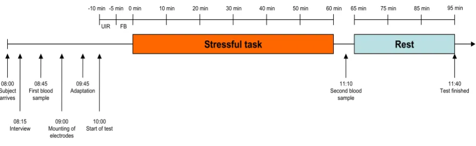
Procedure
The subjects were seated in an ordinary office chair with-out armrests and performed a two-choice reaction-time test presented on a PC monitor for 60 minutes [40]. The test involved a grid (7 columns by 5 rows) in which a large and a small square were placed randomly [43]. The sub-ject was then presented with a suggestion on how to move the small square to superimpose it on the large square (for instance, "two up, four right"), and the
subjects responded by pressing either "right" or "wrong" on a panel before them with their right index or ring fin-gers, respectively. Then the positions of the squares were changed, and a new suggestion was displayed. The sub-jects were instructed to carry out the assignment as fast and correctly as possible, and the computer provided feedback on performance by informing whether the answer was correct or not, and how fast the trial was per-formed (very slow, slow, normal, fast or very fast) [44]. The "normal" response for each subject was determined as the mean response time during a 5-minute trial period. The subjects were acclimated to the lab environment for 30 minutes, during which the procedure was explained and the recording equipment were mounted. The record-ing started with 5 minutes uninstructed rest (UIR) fol-lowed by 5 minutes active, instructed rest with visual EMG feedback (FB). FB-data are shown in figures but were not included in the statistical analysis because it was decided that UIR probably is a more realistic "real-life" baseline. The cognitive task was then performed for one hour (800– 1500 trials), followed by 30 minutes recording during rest (recovery period). The subjects were asked to relax while seated and to move as little as possible during the recovery period. After the UIR and FB periods, at 10-minute inter-vals during the cognitive task, and at 10- minute interinter-vals during the recovery period, the subjects were asked to mark on a VAS scale their level of pain (no pain – worst bearable pain), tension, fatigue and sleepiness. The differ-ent locations of pain corresponded with the positions of the EMG electrodes. Figure 1 shows an overview of the test day procedure. No patient had to be excluded because of headache attacks during the test. Venous blood was sam-pled before the test (immediately after the interview was concluded) and immediately after the stress period (after Table 1: Background data on subjects included in the study. Pain/tension responses and recoveries are given in group means.
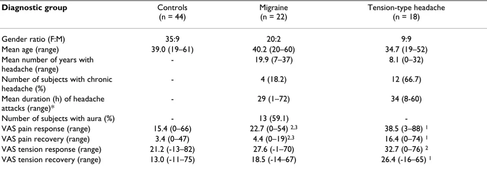
Diagnostic group Controls (n = 44)
Migraine (n = 22)
Tension-type headache (n = 18)
Gender ratio (F:M) 35:9 20:2 9:9
Mean age (range) 39.0 (19–61) 40.2 (20–60) 34.7 (19–52)
Mean number of years with headache (range)
- 19.9 (7–37) 8.1 (0–32)
Number of subjects with chronic headache (%)
- 4 (18.2) 12 (66.7)
Mean duration (h) of headache attacks (range)*
- 29 (1–72) 34 (8-60)
Number of subjects with aura (%) - 13 (59.1)
-VAS pain response (range) 15.4 (0–66) 22.7 (0–54) 2,3 38.5 (3–88) 1
VAS pain recovery (range) 3.4 (0–47) 4.4 (0–19)2,3 16.4 (0–74) 1
VAS tension response (range) 21.2 (-13–82) 27.6 (-1–70) 32.7 (0–76) 2
VAS tension recovery (range) 13.0 (-11–75) 18.5 (-14–67) 26.4 (-16–65) 1
* One migraine patient had some attacks of short duration.
60 minutes). Blood sample data will not be reported in this paper.
Some subjects had partly missing data due to technical difficulties: Two controls and two migraineurs had cor-rupted BP and HR data during the test and recovery period. One control was missing pain data at t95min, while one patient with TTH had corrupted BP, HR, BF, pain and tension data during the recovery period.
Data analysis
Mean values for systolic blood pressure (SBP), diastolic blood pressure (DBP), HR and finger BF were calculated for the UIR and FB period, and for each 10-minute inter-val throughout the stress test and recovery period. These data were used in statistical ANOVA models (see below).
In order to minimize the number of correlations we also defined summery variables for autonomic response and recovery, and for pain response and pain recovery. Two summary variables were used for each autonomic variable (SBP, DBP, HR and finger BF) in correlation analyses: mean response (average of 60 minutes during stress – UIR) and mean recovery (average of 30 minutes recovery – UIR). The pain response was defined as the highest pain response (max pain at t10–60min – pain at t0min) among the
eight location- and side-specific responses (trapezius, splenius, temporalis and frontalis muscles, left and right side). The muscle-specific pain data have been published in a previous paper [34]. The minimal pain during recov-ery was used first to calculate eight location- and side-spe-cific pain recoveries (minimal pain at t75–95min – pain at t0min). Thereafter, the highest among these eight location-and side-specific pain recoveries was defined as pain
recov-ery. These definitions were chosen because the highest (worst) pain during test (and the least complete recovery) was considered to most clinically relevant. Tension response and recovery were defined identically to the pain variables. Pain and tension variables are shown in Table 1.
Statistical analysis
Baseline values were compared with univariate ANOVA
(F1 models). Repeated measures ANOVA time × group
interaction was used to explore differences in response patterns between groups. We do not report group-factor statistics in the present exploratory study because baseline values did not differ between groups (see results). Three different models with selected dependent variables were applied to explore different parts of the stress response and recovery curve. To examine how the novelty of the stressor influenced the subjects, the first 10 min and the baseline was compared in a F2-model (y = (baseline, 0–10
min)). This was described as the early (acute) stress response. After the first 10 min it was assumed that the novelty aspect of the stressor were gone, and we used a model named F6 with six repeated dependent variables (y = (0–10 min, 10–20 min, 20–30 min, 30–40 min, 40–50 min, 50–60 min)) to examine how the subjects adapted to the stressor. This was described as the late stress response. A F3-model with three dependent variables (y = (65–75
min, 75–85 min, 85–95 min)) was used to examine how fast and complete the subjects recovered from the stressor. The ANOVA models were corrected for non-sphericity by reduced degrees of freedom with Huyhn-Feldts method. Three-group ANOVA models were used as the primary analysis, followed by three two-group ANOVA models for the differences between controls and migraine, controls and TTH, and migraine and TTH respectively. Intra-group Overview of the test-day procedure
Figure 1
Overview of the test-day procedure. The subjects arrived at 08:00 and started with a structured interview, followed by the first blood sample. At approximately 09:00 the electrodes were mounted, and after a short adaptation period, the stress test started at 10:00. The stress test (incl. UIR and FB rest periods, stress period and recovery period) lasted for approximately 1 h 40 min.
Stressful task Rest
08:00 Subject arrives
11:10 Second blood
sample UIR FB
0 min 10 min 20 min 30 min 40 min 50 min 60 min 65 min 75 min 85 min 95 min
08:15 Interview
09:00 Mounting of
electrodes 09:45 Adaptation
10:00 Start of test
11:40 Test finished 08:45
First blood sample
contrasts were explored by poshoc Student's paired t-test. Group differences in pain and tension response and recovery (summary variables) were explored using Mann-Whitneys U-test. Univariate Spearman's rank order corre-lation analyses were used to explore associations between pain, tension and cardiovascular responses and recovery (summary variables). As our general statistical strategy involves a large number of comparisons, some might argue that there is a need for a multiple-comparison adjustment to control for type I errors [45]. We chose not to do this, as this would create other problems, such as an increase in type II errors [46,47]. Also, as the studies were considered to be mainly hypothesis-generating and not so much hypothesis-controlling, we believe that findings
wor-thy of further research might be missed by applying too rigid criteria to the statistical analyses. A two-tailed signif-icance level of <0.05 was considered significant in the sta-tistical analyses. P-values within a range of 0.05–0.10 were defined as trends.
Results
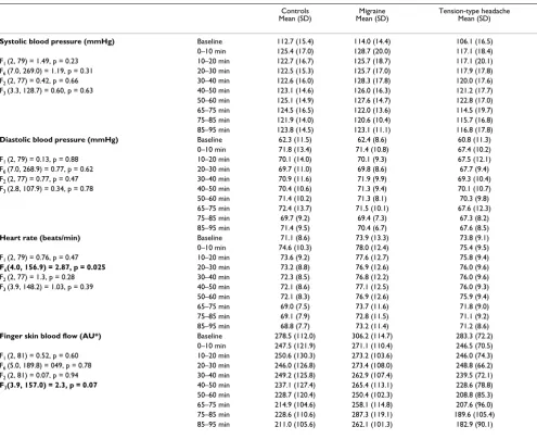
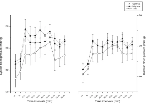
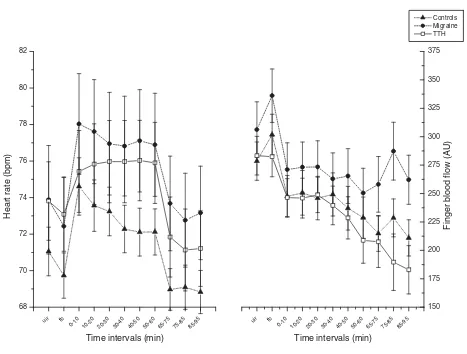
There were no statistically significant differences between the three subject groups when comparing physiological baseline values (see F1 values in Table 2). Inspecting Fig-ures 2 and 3, it appears that SBP, DBP and HR increased more abruptly and then decreased (i.e a "spiked" shape in Figures 2 and 3) at the start of the stressor in controls and migraineurs, but not in patients with TTH.
Table 2: Physiological mean values (SD) measured at baseline, during mental stress (0–60 min) and during the recovery period (65–95 min).
Controls
Mean (SD) Mean (SD)Migraine Tension-type headache Mean (SD)
Systolic blood pressure (mmHg) Baseline 112.7 (15.4) 114.0 (14.4) 106.1 (16.5)
0–10 min 125.4 (17.0) 128.7 (20.0) 117.1 (18.4)
F1 (2, 79) = 1.49, p = 0.23 10–20 min 122.7 (16.7) 125.7 (18.7) 117.1 (20.1)
F6 (7.0, 269.0) = 1.19, p = 0.31 20–30 min 122.5 (15.3) 125.7 (17.0) 117.9 (17.8)
F2 (2, 77) = 0.42, p = 0.66 30–40 min 122.6 (16.0) 128.3 (17.8) 120.0 (17.6)
F3 (3.3, 128.7) = 0.60, p = 0.63 40–50 min 123.1 (14.6) 126.0 (16.3) 121.2 (17.7)
50–60 min 125.1 (14.9) 127.6 (14.7) 122.8 (17.0)
65–75 min 124.5 (16.5) 122.0 (13.6) 114.5 (19.7)
75–85 min 121.9 (14.0) 120.6 (10.4) 115.7 (16.8)
85–95 min 123.8 (14.5) 123.1 (11.1) 116.8 (17.8)
Diastolic blood pressure (mmHg) Baseline 62.3 (11.5) 62.4 (8.6) 60.8 (11.3)
0–10 min 71.8 (13.4) 71.4 (10.8) 67.4 (10.2)
F1 (2, 79) = 0.13, p = 0.88 10–20 min 70.1 (14.0) 70.1 (9.3) 67.5 (12.1)
F6 (7.0, 268.9) = 0.77, p = 0.62 20–30 min 69.7 (11.0) 69.8 (8.6) 67.7 (9.4)
F2 (2, 77) = 0.77, p = 0.47 30–40 min 70.9 (11.6) 71.9 (9.9) 69.3 (10.4)
F3 (2.8, 107.9) = 0.34, p = 0.78 40–50 min 70.4 (10.6) 71.3 (9.4) 70.1 (10.7)
50–60 min 71.4 (10.2) 71.3 (8.1) 70.3 (9.8)
65–75 min 72.4 (13.7) 71.5 (10.1) 67.6 (12.3)
75–85 min 69.7 (9.2) 69.4 (7.3) 67.3 (8.2)
85–95 min 71.4 (9.5) 70.4 (6.7) 67.6 (8.5)
Heart rate (beats/min) Baseline 71.1 (8.6) 73.9 (13.3) 73.8 (9.1)
0–10 min 74.6 (10.3) 78.0 (12.4) 75.4 (9.5)
F1 (2, 79) = 0.76, p = 0.47 10–20 min 73.6 (9.2) 77.6 (12.7) 75.8 (9.4)
F6(4.0, 156.9) = 2.87, p = 0.025 20–30 min 73.2 (8.8) 76.9 (12.6) 76.0 (9.6)
F2 (2, 77) = 1.3, p = 0.28 30–40 min 72.3 (8.5) 76.8 (12.2) 76.0 (9.6)
F3 (3.9, 148.2) = 1.03, p = 0.39 40–50 min 72.1 (8.6) 77.1 (12.5) 76.0 (9.3)
50–60 min 72.1 (8.3) 76.9 (12.6) 75.9 (9.4)
65–75 min 69.0 (7.5) 73.7 (11.6) 71.8 (9.0)
75–85 min 69.1 (7.9) 72.8 (11.5) 71.1 (9.2)
85–95 min 68.8 (7.7) 73.2 (11.4) 71.2 (8.6)
Finger skin blood flow (AU*) Baseline 278.5 (112.0) 306.2 (114.7) 283.3 (72.2)
0–10 min 247.5 (121.9) 271.1 (110.4) 246.5 (70.5)
F1 (2, 81) = 0.52, p = 0.60 10–20 min 250.6 (130.3) 273.2 (103.6) 246.0 (74.3)
F6 (5.0, 189.8) = 049, p = 0.78 20–30 min 246.0 (126.8) 273.4 (108.0) 248.8 (66.2)
F2 (2, 81) = 0.07, p = 0.94 30–40 min 249.2 (125.8) 262.9 (107.4) 239.5 (72.1)
F3(3.9, 157.0) = 2.3, p = 0.07 40–50 min 237.1 (127.4) 265.4 (113.1) 228.6 (78.8)
50–60 min 228.7 (120.4) 250.4 (102.3) 208.8 (85.3)
65–75 min 214.9 (104.6) 258.1 (114.8) 207.6 (96.0)
75–85 min 228.6 (110.6) 287.3 (119.1) 189.6 (105.4)
85–95 min 211.0 (105.6) 262.1 (101.3) 182.9 (90.1)
F1: Oneway ANOVA F-statistic comparing baseline values between groups. F-statistic for group × time interaction in repeated measures ANOVA models is also tabulated for three different models in the left column: F2: Model for the early response to stress, with two intervals during the early stage of the stress test (baseline and 0–10 min). F6: Model for adaptation or potentiation during ongoing long-lasting stress, with six intervals during the stressful task (from 0–10 to 50–60 min). F3: Model to detect fast versus slow recovery patterns with three intervals during recovery (65–75, 75–85 and 85–95 min). p: Probabilities (degrees of freedom in parentheses) was adjusted for non-sphericity with Huyhn-Feldt's method. Significant interactions and trends in bold.
Cardiovascular responses to cognitive stress
ANOVA F2 analyses did not reveal any significant time ×
group interactions between the groups with regard to the initial (early) BP, HR or BF stress responses.
The late HR response pattern during ongoing stress from 0–10 to 50–60 min was significantly different between the three groups (see F6 time × group interaction value in Table 2) since HR adaptation in TTH differed significantly from HR adaptation in controls (Table 3). HR levels were stable in TTH patients whereas HR decreased after the ini-tial response in controls (Figure 3).
The SBP response tended to increase from the early (0–10 min) to the latest (50–60 min) part of stress (Student's paired t-test, p = 0.051) in TTH, while responses were sta-ble in migraine and in controls (p > 0.66; Figure 2). SBP tended to decrease from 0–10 to 10–20 min in migraine
patients (Student's paired t-test, p = 0.050) while no dif-ference was found in TTH (p = 0.97). Significant ANOVA time × group differences were not found in SBP and DBP adaptation during the stress test however (F6 models in Table 2 and 3),
Cardiovascular recovery after cognitive stress
TTH patients had a significant F3 time × group interaction for finger blood flow during the recovery period, com-pared to controls and migraine patients (Table 3). Figure 3 shows that finger blood flow in TTH patients continued to decrease throughout the recovery period, whereas this did not happen in the other groups.
Relationship between pain, tension and cardiovascular responses and recovery
In patients with TTH, mean finger BF recovery were related to the maximal pain response (rs = 0.49, p = 0.047),
mean-Systolic and diastolic BP development throughout the stress test and recovery period
Figure 2
Systolic and diastolic BP development throughout the stress test and recovery period. Values are given as group means (SEM). UIR: Uninstructed rest period (baseline EMG). FB: EMG feedback aided rest period. 0 – 60: During the cognitive stress test. 65 – 95: Relaxation period after the test.
uir fb
0-10 10-20 20-30 30-40 40-50 50-60 65-75 75-85 85-95
100 110 120 130
uir fb
0-10 10-20 20-30 30-40 40-50 50-60 65-75 75-85 85-95
60 70 80
Controls Migraine TTH
Time intervals (min)
Systolic blood pressure (mmHg) Diastolic blood pressure (mmHg)
Heart rate and finger blood flow development throughout the stress test and recovery period
Figure 3
Heart rate and finger blood flow development throughout the stress test and recovery period. Values are given as group means (SEM). UIR: Uninstructed rest period (baseline EMG). FB: EMG feedback aided rest period. T = 0 – 60: During the cog-nitive stress test. T = 65 – 95: Relaxation period after the test.
uir fb
0-10 10-20 20-30 30-40 40-50 50-60 65-75 75-85 85-95
68 70 72 74 76 78 80 82
Time intervals (min)
Heart rate (bpm)
Controls Migraine TTH
uir fb
0-10 10-20 20-30 30-40 40-50 50-60 65-75 75-85 85-95
150 175 200 225 250 275 300 325 350 375
Time intervals (min)
Finger blood flow (AU)
Table 3: F-statistic for group × time interaction in two-group repeated measures ANOVA models.
Controls vs Migraine Controls vs TTH Migraine vs TTH
Systolic blood pressure F2 (1, 60) = 0.33, p = 0.57 F2 (1, 58) = 0.22, p = 0.64 F2 (1, 36) = 0.97,. p = 0.33 F6 (3.3, 198.9) = 0.44, p = 0.74 F6 (3.5, 200.2) = 1.54, p = 0.20 F6 (3.5, 125.8) = 1.99, p = 0.11 F3 (1.68, 102.3) = 0.24, p = 0.75 F3 (1.6, 89.7) = 1.06, p = 0.34 F3 (1.9, 67.4) = 0.47, p = 0.62
Diastolic blood pressure F2 (1, 60) = 0.04, p = 0.85 F2 (1, 58) = 1.70, p = 0.20 F2 (1, 36) = 0.76, p = 0.39 F6 (3.2, 190.9) = 0.22, p = 0.89 F6 (3.5, 202.7) = 1.27, p = 0.29 F6 (3.4, 121.7) = 0.82, p = 0.50
F3 (1.4, 84.5) = 0.06, p = 0.88 F3 (1.3, 76.8) = 0.59, p = 0.50 F3 (1.6, 57.0) = 0.47, p = 0.59
Heart rate F2 (1, 60) = 0.06, p = 0.80 F2 (1, 58) = 1.98, p = 0.17 F2 (1, 36) = 2.85, p = 0.10 F6 (2.0, 122.2) = 1.46, p = 0.24 F6(2.0, 115.8) = 5.06, p = 0.008 F6 (2.1, 75.0) = 1.48, p = 0.23 F3 (1.9, 117.0) = 1.83, p = 0.17 F3 (2.0, 113.4) = 0.83, p = 0.44 F3 (1.9, 65.1) = 0.12, p = 0.88
Finger skin blood flow F2 (1, 64) = 0.06, p = 0.81 F2 (1, 60) = 0.15, p = 0.70 F2 (1, 38) = 0.01, p = 0.94 F6 (2.5, 161.1) = 0.31, p = 0.79 F6 (2.2, 133.1) = 066, p = 0.53 F6 (2.7, 100.6) = 0.57, p = 0.61 F3 (1.9, 124.3) = 0.66, p = 0.52 F3(2.0, 119.5) = 3.21, p = 0.04 F3(1.9, 72.7) = 3.47, p = 0.04
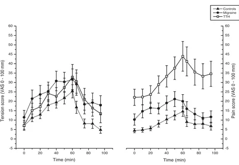
ing that a high pain response was related to less finger BF reduction in recovery. There were no correlations between maximal pain responses and BP or HR responses, or between pain recovery and mean cardiovascular recovery, in any of the diagnostic groups. Pain responses were abnormally large while pain recovery were delayed in TTH patients compared to controls while perceived tension responses did not significantly differ between groups (Table 1, Figure 4). TTH patients also had significantly less recovery from tension compared to controls. There were no correlations between tension and cardiovascular responses and recovery for any of the three groups.
Discussion
Controls, and to a certain degree also migraineurs, responded to the stressor in the present study with a rapid increase followed by a relatively fast decrease in BP and HR, giving the curve a spike-like shape. However, in TTH patients, the SBP, DBP and HR profiles increased slowly and did not decrease during the stress test. A trend towards a different SBP profile was found when compar-ing the first and last 10-min interval in controls and TTH. The possible lack of HR-adaptation during stress reflects the lack of a HR-spike (followed by a decrease in HR) in TTH. A reduced early cardiovascular response to mental stress, with the heart rate response inversely correlated to the pain response, was found for fibromyalgia patients in a study with a similar design [48]. Cardiac (HR) adapta-tion to mental stress has previously been reported in healthy students [49], while deficient cardiac adaptation to calculative mental stress has been found in migraine patients [50]. The migraine patients in our study did not show signs of deficient HR adaptation to stress. One may interpret the lack of an acute spike at the start of the cog-nitive task and the lack of HR adaptation as evidence of a deficient adaptive mechanism (or decreased autonomic excitability) to low-grade cognitive stress in TTH patients. It should be noted that due to a low sample size, espe-cially in the TTH group, these results are tentative and are considered to be hypothesis-generating and not hypothe-sis-controlling.
HR in migraineurs recovered as much during the relaxa-tion phase as controls. This is in accordance with another study [19] which did not show a difference in HR recovery between students with migraine and controls after three minutes of mental arithmetic, although the authors reported faster recovery in peripheral resistance in migraine compared to controls. On the other hand, Holm et al. [20] found that migraineurs had delayed HR recov-ery after four minutes of stressful speech-preparation. Methodological differences make it difficult to compare short-lasting cognitive stress with the one-hour test we applied.
The observed skin blood flow reduction during test is probably related to a gradually increasing sympathetic vasoconstrictor tone to skin arterioles and AV-shunts dur-ing cognitive stress [51]. However, we did not find any dif-ferences in finger BF development during the test between the three groups. This is in accordance with previous stud-ies that have utilized finger temperature and pulse ampli-tude as indirect measures of finger blood flow during short-duration stress with generally negative results in TTH [25] and migraine [19].
We did find a delayed finger BF recovery profile after stress in TTH compared to controls and migraineurs. Another study has previously reported prolonged skin vasocon-striction in TTH (earlobe pulse volume and finger temper-ature) [29], which is in accordance with our findings. In addition, TTH patients had delayed pain recovery (Table 1) and delayed EMG recovery in the trapezius area [34]. Our findings in general fit well with the theoretical mod-els of Eriksen & Ursin [1] and McEwen [2]. Our lack of HR adaptation in TTH is in accordance with McEwens concept of "allostatic load" which causes lack of adaptation to stress. Furthermore, the lack of skin BF recovery in TTH fits well both with the concept of "sustained arousal" in the model of Eriksen & Ursin, and with the concept of a pro-longed response to a stressor in McEwens model.
The role of the autonomic nervous subsystems in TTH is not clear [25]. Because muscular blood flow in tender points is decreased in TTH [52], and because we observed increased skin vasoconstriction (reduced BF) during recovery after stress, which was correlated to low pain response during stress, it is possible that sympathetic dys-regulation is involved, for instance as hyperactivity or hypersensitivity in the central autonomic network which again may be linked to increased central pain inhibition. It is also possible to explain this effect through pain-induced inhibition of sympathetic vasoconstriction in the skin however [53].
Recently, decreased muscle blood flow during muscle exercise was found in fibromyalgia patients, suggesting that muscle ischemia contributes to pain in these patients [54]. However, we were not able to measure intramuscu-lar blood flow in the present study. Muscle blood flow is regulated differently from skin blood flow [55] and the direct relevance of observed skin blood flow changes to the relationship between muscle blood flow and pain per-ceived as muscular is accordingly uncertain.
but not all studies report such autonomic dysfunction [64-66]. Many past studies have used procedures such as deep breathing tests, orthostatic tests, cold pressor tests and isometric work tests (sustained handgrip) and these responses are not directly comparable with autonomic response to cognitive stress of long duration used in the present study.
Cephalic and intracranial vessels may be regulated differ-ently from peripheral vessels. Painful stimuli to tooth pulp induce a blood flow increase in orofacial areas [67]. In chronic TTH patients, previously published data indi-cate cranial vasodilatation [68]. In migraine, cephalic pulse amplitude may increase during a mental task in migraine [18] but results are not consistent across studies [19], and both deficient and normal vasoactivity has gen-erally been reported in migraine [66]. Our results support the view that dysfunctional peripheral blood flow regula-tion is not a substantial part of migraine pathophysiology.
Although we did not measure perceived stress in this study, we believe that the measured perceived tension is an indirect measure of the level of stress. The Norwegian word "anspenthet" describe a feeling of general psycho-logical and muscular tension perceived in stressful situa-tions [69]. Tension responses did not differ, thus the level of stress seemed to be comparable between groups. How-ever, TTH patients had a significantly less recovery from tension, indicating an inability to unwind after the stres-sor is removed [70].
As to what is perceived as stressful, TTH-patients may be more likely to appraise daily situations as stressful, with a tendency towards passive coping, compared to non-head-ache controls [25]. Because cognitive processing involving the prefrontal cortex can change the activity in the ent parts of the periaqueductal grey matter (PAG), a differ-ence in stress adaptive mechanisms may infludiffer-ence both the autonomic nervous system and pain control system in Tension and pain development throughout the stress test and recovery period
Figure 4
Tension and pain development throughout the stress test and recovery period. Values given as group means (SEM), where maximal reported pain (from the trapezius, splenius, temporalis and frontalis areas, irrespective of side) for each subject was used in the calculations. T = 0 – 60: During the cognitive stress test. T = 65 – 95: Relaxation period after the test.
0 20 40 60 80 100
-5 0 5 10 15 20 25 30 35 40 45 50 55 60
Controls Migraine TTH
Tension score (VAS 0 - 100 mm)
Time (min)
0 20 40 60 80 100
-5 0 5 10 15 20 25 30 35 40 45 50 55 60
Pain score (VAS 0 - 100 mm)
several ways, for instance by delaying sympathetic cardio-vascular activation [71]. PAG is also important in pain control and in central sensitization, possibly explaining allodynia and hyperalgesia to pressure stimuli [72] and the increased stress-induced pain in TTH (Table 1, Figure 4).
Conclusion
In conclusion, we report a possible lack of HR adaptation to stress in TTH patients, as well as a delayed finger skin BF recovery after stress and a correlation between finger skin BF recovery and the pain response. Also, TTH had an increase in SBP from the first 10 min to the last 10 min of the stress test, whereas controls and migraineurs did not. Autonomic responses to cognitive stress were not abnor-mal in migraine. We hypothesize that TTH patients have different stress adaptive mechanisms compared to con-trols and migraine patients, involving both cardiovascular activation and the pain control system. The motor system is also involved in responses to stress [73-75], and low-threshold motor unit activity may contribute to local met-abolic changes and muscle pain [76,77]. However, because no associations between muscle activity and pain activation was found in a previous study [34], the present results suggest that cardiovascular responses are more closely linked to pain control than reflexes regulating muscle activity in TTH patients.
Abbreviations
BF Blood flowBP Blood pressure
DBP Diastolic blood pressure
EMG Electromyography
FB Feedback period
HR Heart rate
PAG Periaqueductal grey matter
SBP Systolic blood pressure
TTH Tension-type headache
UIR Uninstructed rest period
VAS Visual analogue scale
Competing interests
The author(s) declare that they have no competing inter-ests.
Authors' contributions
RBL participated in acquiring data from the stress test, per-formed the statistical analyses and drafted the manuscript. TS participated in the design of the study, assisted in the statistical analyses and helped draft the manuscript. KBN participated in the design of the study, acquired data from the stress test and helped draft the manuscript. RHW par-ticipated in the design of the study. LJS parpar-ticipated in the design of the study and helped draft the manuscript. All authors read and approved the final manuscript.
References
1. Eriksen HR, Ursin H: Sensitization and subjective health com-plaints. Scandinavian Journal of Psychology 2002, 43(2):189-196. 2. McEwen BS: Protective and damaging effects of stress
media-tors. New England Journal of Medicine 1998, 338(3):171-179. 3. McEwen BS, Stellar E: Stress and the individual. Mechanisms
leading to disease. Arch Intern Med 1993, 153(18):2093-2101. 4. Martin PR, Soon K: The relationship between perceived stress,
social support and chronic headaches. Headache 1993,
33(6):307-314.
5. Spierings EL, Ranke AH, Honkoop PC: Precipitating and aggra-vating factors of migraine versus tension-type headache.
Headache 2001, 41(6):554-558.
6. Wacogne C, Lacoste J, Guillibert E, Hugues F, Le Jeunne C: Stress,
anxiety, depression and migraine. Cephalalgia 2003,
23(6):451-455.
7. Zivadinov R, Willheim K, Sepic-Grahovac D, Jurjevic A, Bucuk M, Brn-abic-Razmilic O, Relja G, Zorzon M: Migraine and tension-type headache in Croatia: a population-based survey of precipi-tating factors. Cephalalgia 2003, 23(5):336-343.
8. Goadsby PJ: Pathophysiology of cluster headache: a trigeminal autonomic cephalgia. Lancet neurology 2002, 1(4):251-257. 9. Sjaastad O, Pareja JA, Zukerman E, Jansen J, Kruszewski P:
Trigemi-nal neuralgia. Clinical manifestations of first division involve-ment. Headache 1997, 37(6):346-357.
10. Burstein R, Cutrer MF, Yarnitsky D: The development of cutane-ous allodynia during a migraine attack clinical evidence for the sequential recruitment of spinal and supraspinal nocice-ptive neurons in migraine. Brain 2000, 123 (Pt 8):1703-1709. 11. Bahra A, Matharu MS, Buchel C, Frackowiak RS, Goadsby PJ:
Brain-stem activation specific to migraine headache. Lancet 2001, 357(9261):1016-1017.
12. Weiller C, May A, Limmroth V, Juptner M, Kaube H, Schayck RV, Coenen HH, Diener HC: Brain stem activation in spontaneous human migraine attacks. Nature medicine 1995, 1(7):658-660. 13. Ashina S, Bendtsen L, Ashina M, Magerl W, Jensen R: Generalized
hyperalgesia in patients with chronic tension-type headache.
Cephalalgia 2006, 26(8):940-948.
14. Bendtsen L: Central sensitization in tension-type headache--possible pathophysiological mechanisms. Cephalalgia 2000, 20(5):486-508.
15. Dworkin BR, Elbert T, Rau H, Birbaumer N, Pauli P, Droste C, Brunia CH: Central effects of baroreceptor activation in humans: attenuation of skeletal reflexes and pain perception. Proc Natl Acad Sci U S A 1994, 91(14):6329-6333.
16. Jorum E, Orstavik K, Schmidt R, Namer B, Carr RW, Kvarstein G, Hilliges M, Handwerker H, Torebjork E, Schmelz M: Catecho-lamine-induced excitation of nociceptors in sympathetically maintained pain. Pain 2007, 127(3):296-301.
17. Jänig W: Relationship between pain and autonomic phenom-ena in headache and other pain conditions. Cephalalgia 2003, 23 Suppl 1:43-48.
18. Drummond PD: Vascular responses in headache-prone sub-jects during stress. Biological Psychology 1985, 21(1):11-25. 19. Hassinger HJ, Semenchuk EM, O'Brien WH: Cardiovascular
responses to pain and stress in migraine. Headache 1999, 39(9):605-615.
21. Martin PR, Todd J, Reece J: Effects of noise and a stressor on head pain. Headache 2005, 45(10):1353-1364.
22. Passchier J, Goudswaard P, Orlebeke JF, Verhage F: Age migraine and achievement motivation related? A psychophysiological study of responses to real-life achievement stress in young headache sufferers. Functional Neurology 1990, 5(2):135-143. 23. Passchier J, van der Helm-Hylkema H, Orlebeke JF:
Psychophysio-logical characteristics of migraine and tension headache patients. Differential effects of sex and pain state. Headache
1984, 24(3):131-139.
24. Shechter A, Stewart WF, Silberstein SD, Lipton RB: Migraine and autonomic nervous system function: a population-based, case-control study. Neurology 2002, 58(3):422-427.
25. Wittrock DA, Myers TC: The comparison of individuals with recurrent tension-type headache and headache-free con-trols in physiological response, appraisal, and coping with stressors: a review of the literature. Annals of Behavioral Medicine
1998, 20(2):118-134.
26. Ficek SK, Wittrock DA: Subjective stress and coping in recur-rent tension-type headache. Headache 1995, 35(8):455-460. 27. Langemark M, Jensen K, Olesen J: Temporal muscle blood flow in
chronic tension-type headache. Archives of Neurology 1990, 47(6):654-658.
28. Pogacnik T, Sega S, Mesec A, Kiauta T: Autonomic function test-ing in patients with tension-type headache. Headache 1993, 33(2):63-68.
29. Lehrer PM, Murphy AI: Stress reactivity and perception of pain among tension headache sufferers. Behaviour Research and Ther-apy 1991, 29(1):61-69.
30. Linden W, Earle TL, Gerin W, Christenfeld N: Physiological stress reactivity and recovery: conceptual siblings separated at birth? J Psychosom Res 1997, 42(2):117-135.
31. Manzoni GC, Torelli P: Headache screening and diagnosis. Neu-rol Sci 2004, 25 Suppl 3:S255-7.
32. Turkdogan D, Cagirici S, Soylemez D, Sur H, Bilge C, Turk U: Char-acteristic and overlapping features of migraine and tension-type headache. Headache 2006, 46(3):461-468.
33. Vingen JV, Sand T, Stovner LJ: Sensitivity to various stimuli in pri-mary headaches: a questionnaire study. Headache 1999, 39(8):552-558.
34. Leistad RB, Sand T, Westgaard R, Nilsen KB, Stovner LJ: Stress-induced pain and muscle activity in patients with migraine and tension-type headache. Cephalalgia 2006, 26(1):64-73. 35. Westgaard RH: Muscle activity as a releasing factor for pain in
the shoulder and neck. Cephalalgia 1999, 19 Suppl 25:1-8. 36. Bansevicius D, Westgaard RH, Sjaastad OM: Tension-type
head-ache: pain, fatigue, tension, and EMG responses to mental activation. Headache 1999, 39(6):417-425.
37. Bansevicius D, Sjaastad O: Cervicogenic headache: the influence of mental load on pain level and EMG of shoulder-neck and facial muscles. Headache 1996, 36(6):372-378.
38. Bansevicius D, Westgaard RH, Stiles T: EMG activity and pain development in fibromyalgia patients exposed to mental stress of long duration. Scand J Rheumatol 2001, 30(2):92-98. 39. Nilsen KB, Westgaard RH, Stovner LJ, Helde G, Ro M, Sand TH: Pain
induced by low-grade stress in patients with fibromyalgia and chronic shoulder/neck pain, relation to surface electro-myography. Eur J Pain 2006, 10(7):615-627.
40. Bansevicius D, Westgaard RH, Jensen C: Mental stress of long duration: EMG activity, perceived tension, fatigue, and pain
development in pain-free subjects. Headache 1997,
37(8):499-510.
41. Headache Classification Committee IHS: Classification and diag-nostic criteria for headache disorders, cranial neuralgias and facial pain. Headache Classification Committee of the Inter-national Headache Society. Cephalalgia 1988, 8 Suppl 7:1-96. 42. Imholz BP, Langewouters GJ, van Montfrans GA, Parati G, van
Gou-doever J, Wesseling KH, Wieling W, Mancia G: Feasibility of ambu-latory, continuous 24-hour finger arterial pressure recording. Hypertension [Computer File] 1993, 21(1):65-73. 43. Westgaard RH, Bjørklund R: Generation of muscle tension
addi-tional to postural muscle load. Ergonomics 1987, 30(6):911-923. 44. Waersted M, Bjørklund RA, Westgaard RH: The effect of motiva-tion on shoulder-muscle tension in attenmotiva-tion-demanding tasks. Ergonomics 1994, 37(2):363-376.
45. Gaddis ML: Statistical methodology: IV. Analysis of variance, analysis of covariance, and multivariate analysis of variance.
Acad Emerg Med 1998, 5(3):258-265.
46. Perneger TV: What's wrong with Bonferroni adjustments. Bmj
1998, 316(7139):1236-1238.
47. Feise RJ: Do multiple outcome measures require p-value adjustment? BMC Med Res Methodol 2002, 2:8.
48. Nilsen KB, Sand T, Westgaard RH, Stovner LJ, White LR, Leistad RB, Helde G, Rø M: Autonomic activation and pain in response to low-grade mental stress in fibromyalgia and shoulder/neck pain patients. Eur J Pain 2007.
49. Frankenhaeuser M, Dunne E, Lundberg U: Sex differences in sym-pathetic-adrenal medullary reactions induced by different stressors. Psychopharmacology 1976, 47(1):1-5.
50. Huber D, Henrich G, Gündel H: Psychophysiological response patterns of migraine patients in two habituation tests. Head-ache 2005, 45(10):1375-1387.
51. Wallin BG: Neural control of human skin blood flow. Journal of the Autonomic Nervous System 1990, 30 Suppl:S185-S190.
52. Ashina M: Neurobiology of chronic tension-type headache.
Cephalalgia 2004, 24(3):161-172.
53. Blumberg H, Wallin BG: Direct evidence of neurally mediated vasodilatation in hairy skin of the human foot. The Journal of physiology 1987, 382:105-121.
54. Elvin A, Siosteen AK, Nilsson A, Kosek E: Decreased muscle blood flow in fibromyalgia patients during standardised muscle exercise: a contrast media enhanced colour Doppler study.
Eur J Pain 2006, 10(2):137-144.
55. Jänig W, Häbler HJ: Specificity in the organization of the auto-nomic nervous system: a basis for precise neural regulation of homeostatic and protective body functions. Prog Brain Res
2000, 122:351-367.
56. Ebinger F, Kruse M, Just U, Rating D: Cardiorespiratory regula-tion in migraine. Results in children and adolescents and review of the literature. Cephalalgia 2006, 26(3):295-309. 57. Thomsen LL, Olesen J: The autonomic nervous system and the
regulation of arterial tone in migraine. Clin Auton Res 1995, 5(5):243-250.
58. Appel S, Kuritzky A, Zahavi I, Zigelman M, Akselrod S: Evidence for instability of the autonomic nervous system in patients with migraine headache. Headache 1992, 32(1):10-17.
59. Boccuni M, Alessandri M, Fusco BM, Cangi F: The pressor hyper-responsiveness to phenylephrine unmasks sympathetic hypofunction in migraine. Cephalalgia 1989, 9(4):239-245. 60. Gotoh F, Komatsumoto S, Araki N, Gomi S: Noradrenergic
nerv-ous activity in migraine. Arch Neurol 1984, 41(9):951-955. 61. Havanka-Kanniainen H, Tolonen U, Myllyla VV: Autonomic
dys-function in adult migraineurs. Headache 1986, 26(8):425-430. 62. Mikamo K, Takeshima T, Takahashi K: Cardiovascular
sympa-thetic hypofunction in muscle contraction headache and migraine. Headache 1989, 29(2):86-89.
63. Pogacnik T, Sega S, Pecnik B, Kiauta T: Autonomic function test-ing in patients with migraine. Headache 1993, 33(10):545-550. 64. Cortelli P, Pierangeli G, Parchi P, Contin M, Baruzzi A, Lugaresi E:
Autonomic nervous system function in migraine without aura. Headache 1991, 31(7):457-462.
65. Hockaday JM, Macmillan AL, Whitty CW: Vasomotor-relfex response in idiopathic and hormone-dependent migraine.
Lancet 1967, 1(7498):1023-1026.
66. Thomsen LL, Iversen HK, Boesen F, Olesen J: Transcranial Dop-pler and cardiovascular responses during cardiovascular autonomic tests in migraineurs during and outside attacks.
Brain 1995, 118(Pt 5):1319-1327.
67. Kemppainen P, Leppänen H, Jyväsjärvi E, Pertovaara A: Blood flow increase in the orofacial area of humans induced by painful stimulation. Brain Research Bulletin 1994, 33(6):655-662. 68. Hannerz J, Jogestrand T: Is chronic tension-type headache a
vas-cular headache? The relation between chronic tension-type
headache and cranial hemodynamics. Headache 1998,
38(9):668-675.
69. Holte KA, Vasseljen O, Westgaard RH: Exploring perceived ten-sion as a response to psychosocial work stress. Scandinavian Journal of Work, Environment and Health 2003, 29(2):124-133. 70. Melin B, Lundberg U: A biopsychosocial approach to
work-stress and musculoskeletal disorders. Journal of Psychophysiology
Publish with BioMed Central and every scientist can read your work free of charge "BioMed Central will be the most significant development for disseminating the results of biomedical researc h in our lifetime."
Sir Paul Nurse, Cancer Research UK
Your research papers will be:
available free of charge to the entire biomedical community
peer reviewed and published immediately upon acceptance
cited in PubMed and archived on PubMed Central
yours — you keep the copyright
Submit your manuscript here:
http://www.biomedcentral.com/info/publishing_adv.asp
BioMedcentral 71. Keay KA, Bandler R: Parallel circuits mediating distinct
emo-tional coping reactions to different types of stress. Neuro-science and Biobehavioral Reviews 2001, 25(7-8):669-678.
72. Bendtsen L: Central and peripheral sensitization in tension-type headache. Curr Pain Headache Rep 2003, 7(6):460-465. 73. Heckmann CJ, Gorassini MA, Bennett DJ: Persistent inward
cur-rents in motoneuron dendrites: implications for motor out-put. Muscle Nerve 2005, 31(2):135-156.
74. Holstege G: The emotional motor system. European Journal of Morphology 1992, 30(1):67-79.
75. Heckman CJ, Lee RH, Brownstone RM: Hyperexcitable dendrites in motoneurons and their neuromodulatory control during motor behavior. Trends Neurosci 2003, 26(12):688-695. 76. Hägg GM: Static work loads and occupational myalgia - a new
explanation model. Amsterdam , Elsevier; 1991:179-189. 77. Johansson H, Sojka P: Pathophysiological mechanisms involved
in genesis and spread of muscular tension in occupational muscle pain and in chronic musculoskeletal pain syndromes: a hypothesis. Med Hypotheses 1991, 35(3):196-203.
Pre-publication history
The pre-publication history for this paper can be accessed here: