ISSN 0015–5659 www.fm.viamedica.pl
Morphometric analysis of the uncinate
processes of the cervical vertebrae
N. Kocabiyik
1, N. Ercikti
1, S. Tunali
21Department of Anatomy, Gulhane Medical Faculty, Health Sciences University, Ankara, Turkey 2TOBB University of Economics and Technology Faculty of Medicine, Sogutozu, Ankara, Turkey
[Received: 26 December 2016; Accepted: 21 January 2017]
Background: Uncinate processes (UPs) are distinct features unique to cervical verte-brae. They are consistently found on posterolateral aspect of the superior end plate of 3rd to 7th cervical vertebrae. In this study, we investigated the morphology of the UPs with a particular emphasis on the regional anatomy and clinical significance. Materials and methods: The study included 63 vertebrae. The width, height and length of UPs were measured with a digital calliper. We also assessed inclination angle of UP relative to sagittal plane, angle between medial surface of UP and superior surface of vertebra, angle between long axis of the UP and frontal plane, angle between long axis of UP and sagittal plane.
Results: Average width of the UPs ranged from 4.25 mm at C3 to 6.33 mm at T1; average height ranged from 4.88 mm at T1 to 7.54 mm at C4; and average length ranged from 6.88 mm at T1 to 11.46 mm at C4. We measured the inclination angle of UP relative to sagittal plane, and found it to be relatively constant with T1 having the largest value. The average angle was 41.39°, and the range was 17° to 85°. The angle between the long axis of the UP and the sagittal plane was increasing signifi-cantly from C5 to T1. The average angle was 20.74° and the range was 6° to 65°. Conclusions: Anatomy of UPs is significant for surgeon who operates on the cervical spine. Hopefully, the information presented herein would decrease complications du-ring surgical approaches to the cervical spine. (Folia Morphol 2017; 76, 3: 440–445) Key words: uncinate process, uncovertebral joint, Luschka joint,
cervical spine
INTRODUCTION
In 1834, Rathke [see 3] has defined the uncinate
process (UP) as a bony protuberance that extends from posterior margin of vertebral body. In 1858, Von Luschka [see 25] introduced the description of uncovertebral joint between the UP and vertebra.
The localisation of UP on vertebral column ante-riorly extends up to the third cervical vertebra, and posteriorly down to the second thoracic vertebra [30]. They are mostly found between segments C3 to C7
[1, 4, 29]. UPs are further defined as bony protuber
-ances, protuberentia, prominentia, bridges or lips extending from lateral or posterolateral segments of cervical vertebral bodies [1, 4, 7, 8, 11, 12, 14, 18, 31, 32]. They have an anterior slope, an apex and a posterior slope besides a medial articular surface [4]. In addition, UPs deliver a concave appearance to the upper tips of vertebral bodies on the coronal plane [14, 28]. They have a more posterior location on lower vertebral segments [30].
The purpose of this study was to define the
morphology of UPs in Turkish population with an
C5) were excluded due to destruction probably due to osteoporosis or bony degeneration. Hence, 63 vertebrae with intact UPs were involved in measurements. The width, height and length of UPs were measured (Fig. 1).
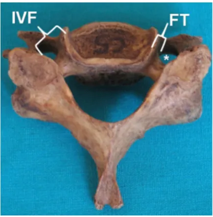
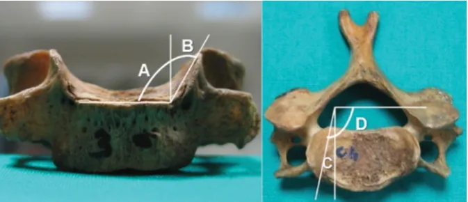
Furthermore, the distance of the apex of the UP to intervertebral foramen (IVF) and foramen trans-versarium (FT) was studied (Fig. 2). We also assessed inclination angle of UP relative to sagittal plane (B), angle between medial surface of UP and superior sur-face of vertebra (A+B), angle between long axis of UP and sagittal plane (C), angle between long axis of the UP and frontal plane (C+D) (Fig. 3). All measurements were made using an electronic digital calliper accurate to 0.1 mm. Angles were measured with a goniometer.
Statistical analysis
Statistical analysis was done with SPSS (Chi., IL, USA) software. Descriptive statistics were presented as mean ± standard deviation. Pearson correlation analy-sis was performed to determine the linear association between morphometric parameters in C3–T1 verte-brae; p < 0.05 was accepted as statistically significant.
RESULTS
Foramina transversaria of vertebrae C3, C4, C5
and C6 had bifid spinous process while FT of vertebra C7 had smaller, non-bifid and obvious spinous pro -cesses. Vertebra T1 had no FT, and joint surfaces for ribs on their bodies were notable. The width, height and length of UPs on vertebrae C3–T1, as well as the distances of their apex to the IVF and the FT are presented in Table 1.
Figure 1. The width (W), height (H) and length (L) of uncinate processes.
Figure 2. The uncinate process is found on the lateral margin of the superior endplate in close proximity to the intervertebral foramen (IVF) and foramen transversarium (FT); *foramen transversarium.
emphasis on their relevance to the regional anatomy,
and their clinical significance.
MATERIALS AND METHODS
A total of 75 vertebrae were used in this study. Of them, 20 were C3 vertebrae, 20 were C4, 9 were C5, 6 were C6, 8 were C7, and 12 were T1. Adult vertebrae present in the anatomy laboratory, of ages between
65 and 75 years were used. No gender classification was
levels [13, 29]. In our cases, highest length was found in UPs of vertebrae C4–C6.
In addition, height of UPs was reported in 10 stud-ies, indicating an increasing pattern from C3 to lower cervicals. This pattern was not observed in the studies of Civelek et al. [6] and Pait et al. [18], and UPs of C5 were found to be shorter compared to those of the adjacent vertebra C4 and C6. Moreover, 4 of 10 studies revealed shorter UPs of C7 compared to those of the adjacent C6 [6, 13, 15, 23]. The length of UPs revealed a range of minimum 2 mm at C7 to maximum 10.5 mm at C6 [22, 29]. In our cases, highest UP was observed at vertebra C4 (7.54 ± 1.39 mm on the right side and 7.35 ± 1.17 mm on the left side) followed by the vertebra C6 (7.42 ± ± 1.34 mm on the right side and 7.04 ± 1.25 mm on the left side). Indeed, a declining pattern of height after the vertebra C6 is not surprising, because no UP is observed after vertebra C7 and T1. In vertebra T1, UPs height was found to be the shortest compared to all cervical verte- bra (5.53 ± 0.85 mm on the right side and 4.88 ± ± 0.79 mm on the left side). Lower values at T1 confirm our
hypothesis above. It should further be added that, such a variation in the height is probably due to different types of samples ranging from fresh cadavers to dry vertebrae underlying the morphometric data reported in this study.
Kotani et al. [11] and Snyder et al. [26] showed that the posterior segment of UPs provide more sta-bility, compared to the anterior segment. Authors
believed this finding was due to the wider and longer
morphology of the posterior segment of UP compared to its anterior segment. The uncovertebral articula-tion was found to contribute in excess of 60% of the stability of the spinal motion segment in extension at C3–C4 [11].
Figure 3. Inclination angle of uncinate process relative to sagittal plane (B), angle between medial surface of uncinate process and superior surface of vertebra (A+B), angle between long axis of uncinate process and sagittal plane (C), angle between long axis of the uncinate pro-cess and frontal plane (C+D).
Morphometric measurements of vertebrae C3–T1 reveal an average (r = 0.536) positive correlation in the width of UPs; however, a negative correlation in height (r = –244) and length (r = –432). While an increasing pattern is observed from C3 to T1, this is exactly opposite in height and length except for a few exceptions. An assessment of the height and length trend reveals that such pattern is reversed particularly in vertebra C4 and C5, and occasionally in vertebra C6.
The inclination angle of UP relative to sagittal plane (B) was relatively constant, with T1 having the largest value. The average angle B was 41.39°, and the range was 17° to 85°. The angle between the long axis of the
UP and the sagittal plane (C) was increasing significantly
from C5 to T1. The average angle C was 20.74°, and the range was 6° to 65°. An overall evaluation of these an-gular measurements reveals that angle C has the highest correlation within the range of vertebrae from C3 to T1 (10.00 ± 1.79°; r = 0.794, p < 0.001, on the right, and 9.56 ± 1.52°; r = 0.750, p < 0.001, on the left). Similarly, the angle C+D displayed a high correlation within the same range at an angle of 100.00 ± 1.79° (r = 0.795, p < 0.001) on the right side, and 99.56 ± 1.55° (r = 0.759, p < 0.001) on the left side.
DISCUSSION
The UP is a phylogenetic residue of the costover-tebral joint in reptiles and birds [1]. Tubbs et al. [29] found that the height of the UP was 5–6 mm at the C4–6 levels, making the anterolateral window for de-compression of the neural foramen determinable by the height and width of the UP, and this is comparable
In the literature, it is reported that the width of UPs shows an increasing pattern from C3 to C7, similar to the height trend. Average widths are reported to be in the range of 4.6 mm at C3 to 7.4 mm at C7 [6, 13]. Next, two of six studies report a lower average width of UPs at C5 compared to C4, and one study reports a wider UP at C5 compared to C6 [6, 13, 18]. Yilmazlar et al. [32] attributed the increased width of the UP at C5 to spondylosis secondary to the increased cervical segmental motion at this level. In our cases, widest UPs were found at vertebrae C7 (5.38 ± 0.82 mm on the right side and 5.38 ± 0.54 mm on the left side) and at vertebrae T1 (6.33 ± 1.26 mm on the right side and 6.23 ± 0.88 mm on the left side). In our study, average width of UPs at vertebrae C4 (4.92 ± 1.21 mm on the right side, and 4.96 ± 1.22 mm on the left side) was found to be exceed-ing that of the adjacent vertebrae C3 and C5.
In the literature, also the anterior-posterior lengths of UPs are assessed, revealing an overall pattern of increase towards C7 from C3. Average lengths are reported to be in the range of 6.0 mm at C3 to 13.0 mm at C7 [4]. However, 2 of 5 studies report longer UPs of C7 compared to those of C6 [4, 19]. In our cases, measured lengths of C7 and T1 UPs were lower compared to that of vertebra C6. Indeed, length and height of UPs display a gradually decreasing pattern towards lower cervicals, namely after C6. This is quite expectable, as these protuberances are not observed after T1. Therefore, they are very obvious throughout the C3–C6 range. After C6, we see a de-cline in the morphometric values of UPs due to relative downsizing.
Tubbs et al. [29] classified the UP with regard to the
degree of impingement of the ipsilateral IVF. No typology was performed in our study. However, the proximity of uncinate process’ apex to IVF and to FT was studied. In our study, smallest distance between uncinate process’ upper margin and IVF was observed at C5. On the other hand, distance of uncinate process’ upper margin to FT was observed to be lowest at C5, and highest in C3.
Intervertebral foramen at the cervical spine forms an angle of 45° with rather the coronal plane, and extends forward from the vertebral channel [9, 20]. Anteromedial margin of IVF is constructed with the posterolateral segment of the uncovertebral joint [5, 11, 14, 21, 24, 27]. Posterior segment of the uncovertebral joint is associated with the front branches of nerve roots, and the lateral segment of the spinal cord [9]. As the nerve roots pass through the IVF they are found in the lower third of the space with the apex of the UP being above each root [9, 20]. The more anterior portion of
Table 1.
Morphometric measurements of uncinate processes (n = 63)
n Vertebra Width [mm] Height [mm] Length [mm] IVF [mm] FT [mm] Right Left Right Left Right Left Right Left Right Left 16 C3
4.25 ± 1.10
4.28 ± 1.08
6.50 ± 2.16
6.11 ± 1.85
9.00 ± 1.33
9.31 ± 1.66
6.56 ± 1.14
6.31 ± 0.85
3.16 ± 0.93
3.00 ± 0.93
13
C4
4.92 ± 1.26
4.96 ± 1.27
7.54 ± 1.45
7.35 ± 1.21
11.46 ± 1.56
11.19 ± 1.49
5.85 ± 1.81
5.77 ± 1.42
2.22 ± 1.63
2.14 ± 1.51
8
C5
4.63 ± 0.52
4.75 ± 0.38
6.56 ± 1.18
6.44 ± 1.18
9.00 ± 1.51
8.75 ± 1.65
5.56 ± 0.56
5.75 ± 0.85
2.38 ± 1.48
2.03 ± 1.37
6
C6
4.92 ± 0.20
5.50 ± 0.89
7.42 ± 1.46
7.04 ± 1.36
10.00 ± 1.26
8.67 ± 1.25
6.08 ± 1.83
6.17 ± 1.21
1.64 ± 0.91
1.38 ± 0.44
8
C7
5.38 ± 0.88
5.38 ± 0.58
6.38 ± 1.09
6.13 ± 1.53
8.88 ± 2.63
8.50 ± 2.33
5.88 ± 1.30
6.22 ± 1.33
2.53 ± 1.98
2.05 ± 1.94
12
T1
6.33 ± 1.32
6.23 ± 0.92
5.53 ± 0.89
4.88 ± 0.82
6.88 ± 1.09
7.48 ± 0.81
6.28 ± 1.10
6.77 ± 1.00
–
–
the uncovertebral joint has its lateral surface being in proximity to the vertebral artery and its accompanying venous plexus [33]. The distance between the medial margin of the FT — in which the vertebral artery and veins travel — and the UP increases from C3 to C7 [9, 10, 17, 31]. The segment of the vertebral artery that is related to the UP is the portion located between the transverse processes of adjacent vertebrae. Nour-bakhsh et al. [16] demonstrated that these segments demonstrate tortuosity in 13.4% of all intertransverse
segments. Tortuosity was greatest at the third to fifth
intertransverse spaces and was more likely to be present in segments demonstrating degeneration.
Panjabi et al. [19] measured the slope angle of UPs
with the sagittal plane and found a relatively fixed angle
peaking at C7. Average angle found was 40.3° [19]. In our study, this angle was found to be highest between cervical vertebrae, with 46.13 ± 15.6° on the right side and 47.25 ± 15.64° on the left side. However, an increase is noteworthy at C4 on the right side (48.08 ± 11.69°). Among all vertebrae with UP, maximum angle was 52.17 ± 8.02° on the right side and 47.67 ± 5.44° on the left side. Also in the literature, the angle between the long axis of UPs and the frontal plane was analysed, revealing
a significant increase from C5 to C7, averaging 91.02°.
In our study, such angle measured was highest between cervical vertebrae, with 114.63 ± 5.45° on the right side and 111.00 ± 3.81° on the left side in average. Again in our study, highest value was found in T1 as 138.75 ± ± 7.84° on the right side and 135.33 ± 10.53° on the left side. Saringer et al. [23] measured the angle between the long axis of UPs and the sagittal plane, and similarly reported an increasing pattern towards lower cervicals. Average angle measured was reported as 5.06°. And also in our study, there is an increase from C3 to T1.
Ugur et al. [31] and Bozbuga et al. [2] measured the angle between the medial surface of UPs and the upper surface of vertebra, and reported a greatly varying value in the 90–162° range. In our study, this angle was calculated as 123.66 ± 8.11° on the right side and 123.38 ± 7.30° on the left side in C3, and 135.5 ± 14.05° on the right side and 137.25 ± 15.64°
in the left side in C7. A positive, statistically significant
correlation was found between the vertebra sequence (from C3 to T1) and angle C (the angle between long axis of UP and sagittal plane) (r = 0.794, p < 0.001).
CONCLUSIONS
As a conclusion; UPs are defined as critical structures
in head and neck movements. Their interaction with the
inferior side of vertebrae makes up the uncovertebral joints. These uncovertebral joints are in a lifetime develop-ment from a rudidevelop-mentary joint up to a mature joint, and eventually degenerate. In some cases, these degraded joints become clinically visible due to the compressive effect of uncinate osteophytes. These compressive ef-fects culminate in nerve root compression in IVF besides vertebral artery compression as osteophytes hand down onto the lateral. Due to this condition, pain, paraesthesia,
reflex decay, muscle weakness and even symptomatic
vertebrobasilar failures may occur. Besides being an injury site in patients with severe head and neck trauma, UPs and uncovertebral joints were mistaken for torticollis in acute cases in young people. Despite being often ne-glected due to their relatively small size; uncovertebral joints have major contributions to the stability of the cervical spine. In our study, also the UP of vertebra T1 was studied in addition to the literature.
While a positive correlation was observed in the width of these protuberances downwardly towards lower cervi-cals, a declining pattern, as suggested by some authors, was found in height and length after C6. We hope that, supporting morphometric studies with radiologic efforts to exhaustively study UPs in a wider sample population would serve as a guide for neurosurgeons conducting uncinectomy and uncoforaminotomy.
REFERENCES
1. Bland JH, Boushey DR. Anatomy and physiology of the cervi-cal spine. Semin Arthritis Rheum. 1990; 20(1): 1–20, indexed
in Pubmed: 2218549.
2. Bozbuğa M, Oztürk A, Ari Z, et al. Surgical anatomic evalua-tion of cervical uncinate process for ventral and ventrolateral subaxial decompression. Okajimas Folia Anat Jpn. 1999;
76(4): 193–196, indexed in Pubmed: 10565202.
3. Brismée JM, Sizer PS, Dedrick GS, et al. Immunohistochemi-cal and histologiImmunohistochemi-cal study of human uncovertebral joints: a preliminary investigation. Spine. 2009; 34(12): 1257–1263,
doi: 10.1097/BRS.0b013e31819b2b5d, indexed in
Pub-med: 19455000.
4. Browne KM. The anatomy, spatial relationships, and role of uncovertebral articulations as the source of posterolateral cervical cartilage sequestrations. J Neurosurg Spine. 2010;
12(3): 270–274, doi: 10.3171/2009.10.SPINE09367, indexed
in Pubmed: 20192626.
5. Cave AJ, Griffiths JD, Whiteley MM. Osteo-arthritis deformans
of the Luschka jounts. Lancet. 1955; 268(6856): 176–179, indexed in Pubmed:13234316.
6. Civelek E, Kiris T, Hepgul K, et al. Anterolateral approach to the cervical spine: major anatomical structures and landmarks. Technical note. J Neurosurg Spine. 2007; 7(6):
669–678, doi: 10.3171/SPI-07/12/669, indexed in
Pub-med: 18074695.
7. Clausen JD, Goel VK, Traynelis VC, et al. Uncinate processes
-Surg. 1984; 103(3): 201–211, indexed in Pubmed:6497609. 21. Raynor RB. Anterior or posterior approach to the cervical
spine: An anatomical and radiographic evaluation and comparison. Neurosurgery. 1983; 12: 7–13.
22. Russo VM, Graziano F, Peris-Celda M, et al. The V(2) seg-ment of the vertebral artery: anatomical considerations and surgical implications. J Neurosurg Spine. 2011; 15(6):
610–619, doi: 10.3171/2011.7.SPINE1132, indexed in
Pub-med: 21905775.
23. Saringer WF, Reddy B, Nöbauer-Huhmann I, et al. Endoscopic anterior cervical foraminotomy for unilateral radiculopathy: anatomical morphometric analysis and preliminary clini-cal experience. J Neurosurg. 2003; 98(2 Suppl): 171–180,
indexed in Pubmed: 12650402.
24. Shen FH, Samartzis D, Khanna N, et al. Comparison of clinical and radiographic outcome in instrumented ante-rior cervical discectomy and fusion with or without direct uncovertebral joint decompression. Spine J. 2004; 4(6):
629–635, doi: 10.1016/j.spinee.2004.04.009, indexed in
Pubmed: 15541694.
25. Silberstein CE. The evolution of degenerative changes in the cervical spine and an investigation into the “joints of luschka”. Clin Orthop Relat Res. 1965; 40: 184–204, indexed
in Pubmed: 14304709.
26. Snyder JT, Tzermiadianos MN, Ghanayem AJ, et al. Effect of uncovertebral joint excision on the motion response of the cervical spine after total disc replacement. Spine. 2007;
32(26): 2965–2969, doi: 10.1097/BRS.0b013e31815cd482,
indexed in Pubmed: 18091488.
27. Tanaka N, Fujimoto Y, An HS, et al. The anatomic relation among the nerve roots, intervertebral foramina, and interver-tebral discs of the cervical spine. Spine (Phila Pa 1976). 2000;
25(3): 286–291, indexed in Pubmed: 10703098.
28. Taylor J, Twomey L, Levander B. Contrasts between cervical and lumbar motion segments. Crit Rev Phys Rehabil Med. 2000; 12: 345–371.
29. Tubbs RS, Rompala OJ, Verma K, et al. Analysis of the uncinate processes of the cervical spine: an anatomical study. J
Neu-rosurg Spine. 2012; 16(4): 402–407, doi: 10.3171/2011.12. SPINE11541, indexed in Pubmed: 22264177.
30. Tulsi RS, Perrett LV. The anatomy and radiology of the cervi-cal vertebrae and the tortuous vertebral artery. Aust Radiol.
1975; 19(3): 258–264, indexed in Pubmed: 1212130. 31. Uğur HC, Uz A, Attar A, et al. Anatomical projection of the
cervical uncinate process in ventral, ventrolateral, and poste-rior decompressive surgery. J Neurosurg. 2000; 93(2 Suppl):
248–251, indexed in Pubmed: 11012055.
32. Yilmazlar S, Ikiz I, Kocaeli H, et al. Details of fibroligamentous
structures in the cervical unco-vertebral region: an obscure
corner. Surg Radiol Anat. 2003; 25(1): 50–53, doi: 10.1007/ s00276-002-0087-5, indexed in Pubmed: 12819950. 33. Yilmazlar S, Kocaeli H, Uz A, et al. Clinical importance of
ligamentous and osseous structures in the cervical uncov-ertebral foraminal region. Clin Anat. 2003; 16(5): 404–410,
doi: 10.1002/ca.10158, indexed in Pubmed: 12903062.
cal spine: quantification using a finite element model of
the C5-C6 segment. J Orthop Res. 1997; 15(3): 342–347,
doi: 10.1002/jor.1100150305, indexed in Pubmed: 9246079. 8. Del Sasso L, Mondini A, Brambilla S, et al. Operative treat-ment of cervicobrachialgia and vertigo due to uncovertebral joint arthritis. Ital J Orthop Traumatol. 1991; 17(4): 498–504,
indexed in Pubmed: 1816155.
9. Ebraheim NA, Lu J, Biyani A, et al. Anatomic considerations for uncovertebral involvement in cervical spondylosis. Clin Orthop Relat Res. 1997(334): 200–206, indexed in
Pub-med: 9005914.
10. Ebraheim NA, Lu J, Brown JA, et al. Vulnerability of vertebral artery in anterolateral decompression for cervical spondylo-sis. Clin Orthop Relat Res. 1996(322): 146–151, indexed in
Pubmed: 8542690.
11. Kotani Y, McNulty PS, Abumi K, et al. The role of anteromedial foraminotomy and the uncovertebral joints in the stability of the cervical spine. A biomechanical study. Spine. 1998;
23(14): 1559–1565, indexed in Pubmed: 9682312. 12. Lee JY, Löhr M, Impekoven P, et al. Small keyhole transuncal
foraminotomy for unilateral cervical radiculopathy. Acta
Neurochir. 2006; 148(9): 951–958, doi: 10.1007/s00701-006-0812-7, indexed in Pubmed: 16804642.
13. Lu J, Ebraheim NA, Haman SP, et al. Cervical uncinate process: an anatomic study for anterior decompression of the cervical spine. Surg Radiol Anat. 1998; 20(4): 249–252, indexed in
Pubmed: 9787390.
14. Lyon E. Uncovertebral osteophytes and osteochondrosis of the cervical spine. J Bone Joint Surg Am. 1945; 27: 248–253.
15. Milne N. The role of zygapophysial joint orientation and uncinate processes in controlling motion in the cervical spine.
J Anat. 1991; 178: 189–201, indexed in Pubmed: 1810926. 16. Nourbakhsh A, Yang J, Gallagher S, et al. A safe approach
to explore/identify the V(2) segment of the vertebral artery during anterior approaches to cervical spine and/or arterial repairs: anatomical study. J Neurosurg Spine. 2010; 12(1):
25–32, doi: 10.3171/2009.7.SPINE08504, indexed in Pub-med:20043760.
17. Oh SH, Perin NI, Cooper PR. Quantitative three-dimen-sional anatomy of the subaxial cervical spine: implication for anterior spinal surgery. Neurosurgery. 1996; 38(6):
1139–1144, indexed in Pubmed: 8727144.
18. Pait TG, Killefer JA, Arnautovic KI. Surgical anatomy of the anterior cervical spine: the disc space, vertebral artery, and associated bony structures. Neurosurgery. 1996; 39(4):
769–776, indexed in Pubmed: 8880772.
19. Panjabi MM, Duranceau J, Goel V, et al. Cervical human vertebrae. Quantitative three-dimensional anatomy of the middle and lower regions. Spine. 1991; 16(8): 861–869,
indexed in Pubmed: 1948369.