REVIEW
Breast cancer brain metastases: the last
frontier
José Pablo Leone
1*and Bernardo Amadeo Leone
2Abstract
Breast cancer is a common cause of brain metastases, with metastases occurring in at least 10–16 % of patients.
Longer survival of patients with metastatic breast cancer and the use of better imaging techniques are associated
with an increased incidence of brain metastases. Unfortunately, patients who develop brain metastases tend to have
poor prognosis with short overall survival. In addition, brain metastases are a major cause of morbidity, associated
with progressive neurologic deficits that result in a reduced quality of life. Tumor subtypes play a key role in prognosis
and treatment selection. Current therapies include surgery, whole-brain radiation therapy, stereotactic radiosurgery,
chemotherapy and targeted therapies. However, the timing and appropriate use of these therapies is controversial
and careful patient selection by using available prognostic tools is extremely important. This review will focus on
cur-rent treatment options, novel therapies, future approaches and ongoing clinical trials for patients with breast cancer
brain metastases.
Keywords: Breast cancer, Brain metastasis, Metastatic breast cancer
© 2015 Leone and Leone. This article is distributed under the terms of the Creative Commons Attribution 4.0 International License (http://creativecommons.org/licenses/by/4.0/), which permits unrestricted use, distribution, and reproduction in any medium, provided you give appropriate credit to the original author(s) and the source, provide a link to the Creative Commons license, and indicate if changes were made. The Creative Commons Public Domain Dedication waiver (http://creativecommons. org/publicdomain/zero/1.0/) applies to the data made available in this article, unless otherwise stated.
Background
Breast cancer represents the second most frequent
cause of brain metastases after lung cancer, with
metas-tases occurring in 10–16 % of patients [
1
]. In addition,
autopsy studies have demonstrated another 10 % which
were asymptomatic [
2
]. The incidence of brain
metasta-ses seem to have increased in recent years, this is likely
due to prolonged survival of patients receiving more
efficient treatments and the availability of better
imag-ing techniques that lead to increased detection of brain
metastases.
The development of brain metastases is a complex
pro-cess, requiring invasion of the primary breast cancer cells
into surrounding tissue and vessels, traffic through the
circulatory system and colonization and growth in the
brain parenchyma [
3
,
4
]. In breast cancer, this process
takes a median of 32 months from the initial cancer
diag-nosis [
5
]; which shows that the breast cancer tumor cells,
unlike other cancer cells, need more time to develop
the ability to penetrate through the blood–brain
bar-rier (BBB) and colonize the brain. There is also a
selec-tive pressure that can make the brain a preferential site of
metastasis, as many of our currently available therapies
are unable to cross the BBB, even if this barrier is
dis-rupted by tumor invasion.
Previous studies have identified the subgroups of
patients with triple-negative and human epidermal
growth factor receptor 2 (HER2)-positive breast
can-cer as having an increased risk for the development of
brain metastases [
6
–
9
], with up to half of patients with
HER2-positive metastatic breast cancer experiencing
brain metastases over time [
10
]. Tumor subtypes are also
an important factor for the median time interval from
primary diagnosis to development of brain metastases;
a recent large study showed shorter intervals for
triple-negative and HER2-positive patients, and longer intervals
for estrogen receptor (ER) positive tumors [
11
].
Brain metastases in breast cancer patients represent a
catastrophic event that portends a poor prognosis, with
a median survival that ranges from 2 to 25.3 months
despite treatment [
5
,
12
–
14
]. In addition, brain
metas-tases are a major cause of morbidity, associated with
progressive neurologic deficits that result in a reduced
Open Access
*Correspondence: jose-leone@uiowa.edu
1 University of Iowa Holden Comprehensive Cancer Center, University of Iowa Hospitals and Clinics, C32 GH, 200 Hawkins Drive, Iowa City, IA 52242, USA
quality of life [
15
]. With the advent of better systemic
therapies, brain metastases constitute an increasing
clini-cal problem. This is particularly important in
HER2-pos-itive patients, in whom brain metastases can occur in the
setting of controlled extracranial disease [
16
]. In contrast,
it is common for patients with triple-negative breast
can-cer to develop brain metastases with concurrent
extrac-ranial disease progression [
17
]. Treatment options for
patients with breast cancer brain metastases are limited
and include surgical resection, whole-brain radiation
therapy (WBRT), stereotactic radiosurgery (SRS),
chem-otherapy and targeted therapy [
12
,
18
,
19
]. This review
will focus on the key issues of current treatment options,
comment on novel therapies and ongoing clinical trials
for patients with breast cancer brain metastases.
Prognostic factors
The prognosis of patients with breast cancer who develop
brain metastases is affected by several factors. Tumor
subtypes have been identified as a prognostic factor
for overall survival in brain metastases [
20
,
21
].
Triple-negative breast cancer patients have the shortest
sur-vival ranging from 3 to 4 months [
9
,
16
,
22
]. In contrast,
patients with HER2-positive tumors have longer survival
than those with triple-negative or luminal subtypes,
although their rates of brain metastases are higher [
9
,
16
,
23
].
Another important prognostic factor is the
perfor-mance status of the patient at the time of diagnosis of
brain metastases. Most studies have established the
util-ity of the Karnofsky Performance Status (KPS) as a tool to
assess prognosis and identified that patients with longer
survival have KPS scores
≥
70 [
13
,
14
,
24
]. In addition to
the KPS, patient’s age can also affect prognosis. Older
age at the time of initial breast cancer diagnosis has been
associated with shorter overall survival and shorter
sur-vival from the time of first tumor relapse [
5
,
25
]. Finally,
the burden of disease represented by the number of
brain metastases, as well as the presence of uncontrolled
extracranial disease have both been related with worse
prognosis [
23
,
24
,
26
].
One of the most frequently used tools for the
assess-ment of prognosis in brain metastases is the graded
prognostic assessment (GPA) [
27
]. This prognostic index
includes age, KPS score, number of brain metastases and
extracranial metastases. After its original validation [
28
],
the index was modified to create a breast cancer-specific
GPA that included tumor subtypes among its prognostic
factors [
11
,
14
]. However, number of brain metastases
was not incorporated into the final model. A recent study
validated the breast cancer-specific GPA and refined it
with the addition of number of brain metastases [
29
].
This represents a very useful tool for patient risk
assess-ment and selection for clinical trials.
Local therapy modalities
Surgical resection
Surgical resection of the brain metastasis is an
impor-tant treatment option in patients with single or few (
≤
3)
lesions. Particularly when the systemic disease is well
controlled and when the brain metastases are
sympto-matic. Although the anatomic location of the metastatic
lesion can be a limitation, surgical resection has
addi-tional advantages including the potential for immediate
improvement of focal deficits, relief of intracranial
hyper-tension and establishment of histological diagnosis in
patients with no other site of metastasis.
One of the first studies to evaluate the role of
sur-gical resection in brain metastasis was conducted by
Patchell et al. [
30
]. In this study, 48 patients with single
brain metastasis from any primary were randomized
to either surgical resection of the brain metastasis
fol-lowed by WBRT or needle biopsy folfol-lowed by WBRT.
Brain recurrence was less frequent in the surgery group
compared with the radiation group (20 vs. 52 %,
respec-tively;
P
< 0.02). Median overall survival was longer in the
surgery group (40 weeks) compared with the radiation
group (15 weeks) (
P
< 0.01). Neurological outcomes were
also improved with surgery where patients remained
functionally independent longer (median, 38 vs. 8 weeks
in the radiation group;
P
< 0.005).
A subsequent study randomized 63 patients with
sys-temic cancer and a single brain metastasis to surgical
resection plus WBRT vs. WBRT alone. The combined
modality led to longer overall survival and longer
func-tionally independent survival compared with WBRT
alone, particularly in patients with stable extracranial
disease (median overall survival 12 vs. 7 months,
respec-tively; median functionally independent survival 9 vs.
4 months, respectively) [
31
]. Patients with progressive
extracranial disease had similar outcomes irrespective
of treatment, a finding that was also observed in another
randomized trial [
32
]. Three non-randomized
stud-ies have also confirmed improvements in survival, brain
recurrence and neurological outcomes with surgical
resection in addition to WBRT [
33
–
35
].
Stereotactic radiosurgery
has an advantage over WBRT in that it avoids the feared
toxicity of neurocognitive decline that is associated with
the latter intervention [
36
–
38
].
The efficacy of SRS for local control of brain
metas-tases has been demonstrated in a study conducted by
Kondziolka et al. [
36
], were median time to local failure
in patients with two to four brain metastases was
sig-nificantly improved from 6 months with WBRT alone to
36 months with the addition of SRS (
P
=
0.0005). In spite
of this improvement in local control, overall survival was
unchanged and was related to the extent of extracranial
disease. The Radiation Therapy Oncology Group (RTOG)
confirmed the efficacy of SRS in addition to WBRT in
patients with one to three brain metastases and
docu-mented an overall survival improvement of 1.6 months
in the subgroup of patients with single unresectable brain
metastasis who received the combined therapy [
39
]. In
patients with solitary metastases who are treated with
surgical resection plus WBRT, the addition of SRS to the
tumor bed can improve local control [
40
].
Subsequent randomized studies that included brain
metastases from different cancers demonstrated that
patients with one to four lesions who were treated with
SRS alone had similar survival and improved
neurocog-nition compared with patients who received both SRS
and WBRT, however local control was inferior with SRS
alone [
41
,
42
]. These results were confirmed in a
meta-analysis [
43
]. A recent non-randomized non-inferiority
trial showed the efficacy of SRS without WBRT for
over-all survival in patients with five to ten brain metastases to
be not inferior to the same treatment in patients with two
to four lesions [
44
]. Despite the findings of this study and
others showing similar clinical outcomes [
45
–
47
],
cur-rently there is no randomized data to support the use of
SRS without WBRT in the treatment of brain metastases
for patients with >4 lesions.
Whole‑brain radiation therapy
One of the most important treatments available for brain
metastases is WBRT, particularly in the setting of
multi-ple brain lesions. This approach has two main goals—the
control of macroscopic metastases, and the eradication of
microscopic seeding of the brain. The majority of patients
are given conventional WBRT, a total dose of 30 Gy in 10
fractions with daily fractions of 3–4 Gy [
48
].
The benefit of WBRT after surgical resection has been
demonstrated in a prospective trial that randomized 95
patients who had single brain metastases to WBRT or
observation [
49
]. The study showed that patients in the
WBRT group had fewer recurrences both at the operative
site (10 vs. 46 %,
P
< 0.001) and at other sites in the brain
(14 vs. 37 %,
P
< 0.01), however overall survival was not
increased.
Substantial controversy exists about the role of WBRT
in patients with few (
≤
4) brain metastases. In this
set-ting, treatment with WBRT after surgical resection or
SRS resulted in fewer intracranial recurrences, but there
was no difference in overall survival [
41
,
50
]. Associated
toxicities with WBRT included worse neurocognitive
outcomes and quality of life [
42
,
51
]. However,
withhold-ing WBRT can lead to progressive disease in the brain,
which in turn could also negatively impact cognition [
52
,
53
]. This issue was addressed in a study conducted by
the North Central Cancer Treatment Group (NCCTG)
N0574 where patients with one to three brain metastases
were randomized to SRS or SRS plus WBRT, the study
showed more frequent decline in cognitive function with
the addition of WBRT despite better brain control [
54
].
Therefore, delaying or avoiding the administration of
WBRT in metastatic breast cancer after surgical
resec-tion or SRS through the careful use of effective systemic
therapies, could provide substantial benefits in terms
of quality of life, particularly in patients with high GPA
scores in whom survival is expected to be longer [
14
,
29
].
This approach results even more appealing when one
considers that none of the above mentioned randomized
trials have shown overall survival gain with the addition
of WBRT.
Systemic therapies
The mainstay of systemic treatment for breast cancer
brain metastases is cytotoxic chemotherapy; however,
there are currently additional options for targeted
ther-apy. Therefore, it is crucial to consider the tumor subtype
not only for prognosis but also to understand the
differ-ent options for systemic therapies.
Hormone receptor‑positive
Patients with ER-positive brain metastases derive
sub-stantial benefit from systemic chemotherapy. Niwinska
et al. reported improvements in median survival from
3 to 14 months with the addition of systemic therapy in
patients with luminal breast cancer [
20
]. A similar result
was seen in another study where the median overall
sur-vival of patients with luminal breast cancer was improved
from 7.1 to 14.3 months with chemotherapy [
55
].
support the hypothesis that in patients with metastatic
ER-positive breast cancer who have asymptomatic
sys-temic disease and locally treated brain metastases
treat-ment with endocrine therapy could be considered prior
to systemic chemotherapy. Clinical trials evaluating this
approach should be conducted.
HER2‑positive
Patients with HER2-positive metastatic breast cancer
have experienced a dramatic improvement in overall
survival with optimal the utilization of HER2 targeted
therapy [
63
]. Unfortunately, the advances in systemic
treatments for these patients came in hand with an
increase in the rate of brain metastases, which now
poses a significant threat [
10
]. The efficacy of anti-HER2
therapy to control systemic disease for longer periods of
time has exposed the ability of the HER2-positive breast
cancer cells to seed the brain parenchyma and develop
brain metastases. Most chemotherapy agents and HER2
targeted therapies do not cross the intact BBB or are
pumped out of the central nervous system (CNS) by
P-glycoproteins present in the BBB, therefore they may
not reach sufficient therapeutic levels to eradicate
met-astatic cells [
1
]. For example, in patients without brain
metastases, the ratio of trastuzumab in plasma to
tras-tuzumab in cerebrospinal fluid is >300:1 [
64
,
65
]. The
brain then, can serve as a sanctuary where those cells that
have the ability to seed can escape the cytotoxic efficacy
of systemic therapy. However, tumor growth in the brain
as well as cranial surgery and brain radiotherapy can
dis-rupt the BBB and allow access of systemic drugs to the
tumor. This concept has been proven by a number of
labeled-trastuzumab imaging studies [
66
,
67
]. Also,
sev-eral clinical studies have shown that the combination of
chemotherapy with trastuzumab improved survival, even
after the development of brain metastases [
68
–
70
]. This
benefit is presumed to be mainly due to improved control
of systemic disease [
71
].
Lapatinib, a small molecule with potential ability to
cross the BBB, has been extensively tested in the
treat-ment of HER2-positive brain metastases. As a single
agent, lapatinib has shown response rates in the brain
ranging from 2.6 to 6 % in heavily pre-treated patients
[
72
,
73
]. However, when added to capecitabine, response
rates increase to 20 to 33 % [
73
–
77
]. The highest
effi-cacy is observed in previously untreated patients, where
the combination of lapatinib and capecitabine produces
an objective response rate of 65.9 %, with a median
time to progression of 5.5 months and a 1-year survival
rate >70 % [
78
]. This drug combination has also shown
to reduce the rate of brain metastases as the first site
of progression from 6 % with capecitabine alone to 2 %
with capecitabine and trastuzumab (
P
=
0.045) [
79
]. The
efficacy of lapatinib to prevent brain metastases was
fur-ther tested in the CEREBEL trial, where patients with
HER2-positive metastatic breast cancer without CNS
metastases were randomized to lapatinib or trastuzumab
in combination with capecitabine. The primary end point
of the study was incidence of CNS metastases as first site
of relapse. The study was terminated early and showed
no difference between arms for the incidence of CNS
metastases (3 % for lapatinib vs. 5 % for trastuzumab,
P
=
0.36), however progression-free survival and overall
survival were longer with trastuzumab and capecitabine
[
80
]. Despite the low incidence of CNS metastases seen
during the study, it is important to notice that 4.7 % of
all screened patients were excluded due to detection of
asymptomatic brain metastases.
In the EMILIA trial, Trastuzumab emtansine (T-DM1),
a novel antibody–drug conjugate, improved overall
survival compared with lapatinib plus capecitabine in
patients with previously treated HER2-positive
meta-static breast cancer [
81
]. A recent retrospective,
explor-atory analysis of this trial focusing on patients with
baseline CNS metastases, showed that the rate of CNS
progression was similar for both arms, however median
overall survival in patients with CNS metastases at
base-line was significantly improved with T-DM1 (26.8 vs.
12.9 months,
P
=
0.008) [
82
]. Similar results were seen
in the CLEOPATRA trial, where patients with
HER2-positive first line metastatic breast cancer experienced
significant improvements in progression-free and
over-all survival with pertuzumab, trastuzumab and docetaxel
compared with placebo, trastuzumab and docetaxel [
63
].
In this trial, an exploratory analysis of the incidence
and time to development of CNS metastases as first site
of disease progression, also showed that the incidence
was similar between the two arms, however the time to
development of CNS metastases was significantly
pro-longed in the pertuzumab arm from 11.9 to 15 months
(
P
=
0.0049) [
83
]. Taken together, the data from EMILIA
and CLEOPATRA underscore the importance of
sys-temic disease control for improving overall survival in
patients with brain metastases.
whose systemic disease is progressive at the time of brain
metastasis diagnosis, treatment should include
HER2-targeted therapy according to the algorithms for
treat-ment of HER2-positive metastatic breast cancer [
85
].
Triple‑negative
Patients with brain metastases from triple-negative
breast cancer unfortunately lack targeted therapies and
chemotherapy is currently their only systemic option.
Some of the initial studies of patients with brain
metas-tases have shown objective response rates of around 50 %
with traditional chemotherapy combinations [
86
,
87
]. A
study evaluating cisplatin with etoposide showed 38 %
response rate in the brain [
88
]. Topotecan and
temozola-mide have failed to show responses as single agent [
89
,
90
]. However, when temozolamide was combined with
cisplatin had 40 % response rate [
91
], and showed 18 %
response rate when combined with capecitabine [
92
].
The experience with single agent capecitabine is limited
to mostly retrospective studies [
93
].
It is important to keep in mind that while different
chemotherapies will defer in their ability to penetrate
the BBB, most brain metastases will significantly disrupt
this barrier. Therefore the ability to deliver systemic
chemotherapy to the brain metastasis is not much
dif-ferent from the ability to deliver chemotherapy to that
tumor anywhere else in the body. Hence, treatment
efficacy is more closely related to tumor
chemosen-sitivity than to the drug ability to cross an intact BBB
and this hypothesis has been proven in several of the
above mentioned studies [
86
–
88
]. Brain metastases
tend to be chemotherapy-resistant because they tend
to occur late in the natural history of breast cancer and
by that point many times the breast cancer is already
chemotherapy-resistant.
Suggested treatment approach
Based on the evidence reviewed above, we suggest the
following management approach:
a. For patients with a single brain metastasis, surgical
resection can improve overall survival, particularly in
symptomatic patients when systemic disease is well
controlled. The addition of SRS to the tumor bed or
WBRT improve local control.
b. For patients with one to four brain metastases, SRS
with or without WBRT should be considered to
improve local control. If WBRT is added, we
recom-mend to delay its administration as much as possible
to prevent neurocognitive decline, which is
particu-larly important in the absence of overall survival
ben-efit. Surgery can be considered for large or
sympto-matic lesions.
c. For patients with more than four brain metastases,
WBRT can be the treatment of choice to palliate
symptoms and improve local control. There are no
randomized trials to support the use of SRS in this
setting.
d. For patients with progressive systemic disease at the
time of development of brain metastases, a change in
systemic therapy should be considered based on the
tumor subtype.
e. For patients with non-progressive systemic disease
at the time of development of brain metastases,
sys-temic therapy should not be changed.
f. For each patient, the choice of systemic therapy
should be considered based on the tumor subtype.
g. For patients with poor prognosis, options include
WBRT and/or best supportive care.
Novel approaches and future directions
Given the paucity of effective treatment options for
patients with breast cancer brain metastases, this
cur-rently represents an area of great potential for future
research. The use of bevacizumab has shown good results
in patients with glioblastoma and has made the
strat-egy of blocking the vascular endothelial growth factor
(VEGF) pathway an interesting alternative to treat brain
metastases. Two studies evaluating this approach have
been reported. One showed CNS response rate of 63 %
for bevacizumab and carboplatin; and the other one
showed a response rate of 60 % for bevacizumab,
etopo-side and cisplatin [
94
,
95
]. It is important to consider that
VEGF blockade rises a number of controversies. One of
the most concerning being that meta-analyses have failed
to show an overall survival benefit with the use of
bevaci-zumab in metastatic breast cancer, which resulted in the
US Food and Drug Administration (FDA) withdrawal of
the conditional approval of the drug.
The high affinity folate receptor (HFR) is a novel
tar-get present in 33 % of breast cancers for which there
are available drugs being evaluated. Despite of the
ini-tial excitement, a recent study showed very low levels of
expression in brain metastases [
96
]. Another approach
that is currently under study in patients with breast
can-cer brain metastases is targeting the
phosphatidylino-sitol 3-kinase (PI3K)—mammalian target of rapamycin
(mTOR) pathway. This is one of the most commonly
altered pathways not only in metastatic breast cancer
but also in brain metastases [
97
]. Everolimus is being
evaluated in combination with capecitabine and lapatinib
(NCT01783756) and in combination with vinorelbine
and trastuzumab (NCT01305941) in patients with
HER2-positive brain metastases.
cancer, there have been efforts to develop strategies for
prevention of brain metastases in the high-risk subgroups.
Much of these have evolved around the concept of using
prophylactic cranial irradiation (PCI) in patients with these
breast cancer subtypes, similar to what is currently done
for patients with small cell lung cancer. However, as
men-tioned earlier in this article, the timing of development of
brain metastases in patients with HER2-positive and
triple-negative breast cancer is not the same, nor is the same their
survival after development of metastatic disease. In
addi-tion, given the cognitive effects of brain radiotherapy, there
is significant controversy around the optimal timing of PCI.
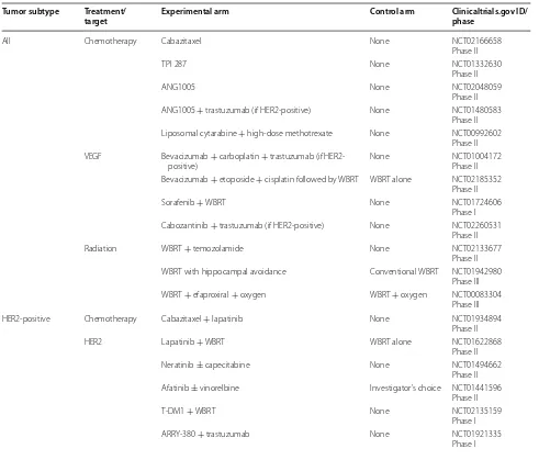
Table
1
shows a summary of currently ongoing studies
evaluating different treatment strategies in breast cancer
brain metastases.
Conclusions
Brain metastases are an increasing problem in breast
cancer. They represent an unmet need for which more
efficacious therapies are urgently required. A better
understanding of the molecular underpinnings of CNS
progression is needed and ongoing studies analyzing
matched tissue from primary and brain metastases
will hopefully shed light on this. Traditionally, patients
with brain metastases were excluded from clinical
tri-als evaluating systemic therapies and we are left with
the unanswered question of how efficacious those
therapies would be for patients with brain metastases.
To this end, the large number of ongoing breast
can-cer-specific brain metastases trials is a step in the right
direction.
Table 1 Ongoing clinical trials in breast cancer brain metastases
Tumor subtype Treatment/target Experimental arm Control arm Clinicaltrials.gov ID/phase
All Chemotherapy Cabazitaxel None NCT02166658
Phase II
TPI 287 None NCT01332630
Phase II
ANG1005 None NCT02048059
Phase II
ANG1005 + trastuzumab (if HER2-positive) None NCT01480583
Phase II Liposomal cytarabine + high-dose methotrexate None NCT00992602
Phase II VEGF Bevacizumab + carboplatin + trastuzumab (if
HER2-positive) None NCT01004172Phase II
Bevacizumab + etoposide + cisplatin followed by WBRT WBRT alone NCT02185352 Phase II
Sorafenib + WBRT None NCT01724606
Phase I Cabozantinib + trastuzumab (if HER2-positive) None NCT02260531
Phase II
Radiation WBRT + temozolamide None NCT02133677
Phase II
WBRT with hippocampal avoidance Conventional WBRT NCT01942980
Phase III
WBRT + efaproxiral + oxygen WBRT + oxygen NCT00083304
Phase III
HER2-positive Chemotherapy Cabazitaxel + lapatinib None NCT01934894
Phase II
HER2 Lapatinib + WBRT WBRT alone NCT01622868
Phase II
Neratinib ± capecitabine None NCT01494662
Phase II
Afatinib ± vinorelbine Investigator’s choice NCT01441596
Phase II
T-DM1 + WBRT None NCT02135159
Phase I
ARRY-380 + trastuzumab None NCT01921335
Authors’ contributions
JPL designed the manuscript, did the literature search and prepared the manuscript. BAL revised the manuscript. Both authors wrote the manuscript. Both authors read and approved the final manuscript.
Author details
1 University of Iowa Holden Comprehensive Cancer Center, University of Iowa Hospitals and Clinics, C32 GH, 200 Hawkins Drive, Iowa City, IA 52242, USA. 2 Grupo Oncológico Cooperativo del Sur (GOCS), Rivadavia 360, 8300 Neu-quén, Argentina.
Acknowledgements
We want to thank the University of Iowa Libraries for the support for this publication.
Competing interests
The authors declare that they have no competing interests.
Received: 16 September 2015 Accepted: 9 November 2015
References
1. Lin NU, Bellon JR, Winer EP. CNS metastases in breast cancer. J Clin Oncol Off J Am Soc Clin Oncol. 2004;22(17):3608–17. doi:10.1200/JCO.2004.01.175. 2. Arslan C, Dizdar O, Altundag K. Systemic treatment in
breast-cancer patients with brain metastasis. Expert Opin Pharmacother. 2010;11(7):1089–100. doi:10.1517/14656561003702412.
3. Weil RJ, Palmieri DC, Bronder JL, Stark AM, Steeg PS. Breast cancer metas-tasis to the central nervous system. Am J Pathol. 2005;167(4):913–20. doi:10.1016/S0002-9440(10)61180-7.
4. Nguyen DX, Bos PD, Massague J. Metastasis: from dissemination to organ-specific colonization. Nat Rev Cancer. 2009;9(4):274–84. doi:10.1038/ nrc2622.
5. Leone JP, Lee AV, Brufsky AM. Prognostic factors and survival of patients with brain metastasis from breast cancer who underwent craniotomy. Cancer Med. 2015;4(7):989–94. doi:10.1002/cam4.439.
6. Gabos Z, Sinha R, Hanson J, Chauhan N, Hugh J, Mackey JR, et al. Prognostic significance of human epidermal growth factor receptor positivity for the development of brain metastasis after newly diagnosed breast cancer. J Clin Oncol Off J Am Soc Clin Oncol. 2006;24(36):5658–63. doi:10.1200/JCO.2006.07.0250.
7. Tham YL, Sexton K, Kramer R, Hilsenbeck S, Elledge R. Primary breast can-cer phenotypes associated with propensity for central nervous system metastases. Cancer. 2006;107(4):696–704. doi:10.1002/cncr.22041. 8. Pestalozzi BC, Zahrieh D, Price KN, Holmberg SB, Lindtner J, Collins J,
et al. Identifying breast cancer patients at risk for central nervous system (CNS) metastases in trials of the International Breast Cancer Study Group (IBCSG). Ann Oncol Off J Eur Soc Med Oncol/ESMO. 2006;17(6):935–44. doi:10.1093/annonc/mdl064.
9. Nam BH, Kim SY, Han HS, Kwon Y, Lee KS, Kim TH, et al. Breast cancer subtypes and survival in patients with brain metastases. Breast Cancer Res BCR. 2008;10(1):R20. doi:10.1186/bcr1870.
10. Aversa C, Rossi V, Geuna E, Martinello R, Milani A, Redana S, et al. Meta-static breast cancer subtypes and central nervous system metastases. Breast. 2014;23(5):623–8. doi:10.1016/j.breast.2014.06.009.
11. Sperduto PW, Kased N, Roberge D, Chao ST, Shanley R, Luo X, et al. The effect of tumor subtype on the time from primary diagnosis to development of brain metastases and survival in patients with breast cancer. J Neuro-Oncol. 2013;112(3):467–72. doi:10.1007/ s11060-013-1083-9.
12. Lee SS, Ahn JH, Kim MK, Sym SJ, Gong G, Ahn SD, et al. Brain metastases in breast cancer: prognostic factors and management. Breast Cancer Res Treat. 2008;111(3):523–30. doi:10.1007/s10549-007-9806-2.
13. Ogawa K, Yoshii Y, Nishimaki T, Tamaki N, Miyaguni T, Tsuchida Y, et al. Treatment and prognosis of brain metastases from breast cancer. J Neuro-Oncol. 2008;86(2):231–8. doi:10.1007/s11060-007-9469-1. 14. Sperduto PW, Kased N, Roberge D, Xu Z, Shanley R, Luo X, et al. Effect of
tumor subtype on survival and the graded prognostic assessment for
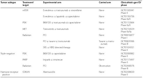
Table 1 continued
Tumor subtype Treatment/
target Experimental arm Control arm Clinicaltrials.gov ID/phase
mTOR Everolimus + trastuzumab + vinorelbine None NCT01305941
Phase II
Everolimus + lapatinib + capecitabine None NCT01783756
Phase Ib/II
PI3K BKM120 + trastuzumab ± capecitabine None NCT01132664
Phase Ib/II
MET Tesevatinib + trastuzumab None NCT02154529
Phase Ib/IIa
Radiation PCI None NCT00916877
Phase I PCI + taxane + trastuzumab Taxane +
trastu-zumab NCT00639366Phase III
SRS + HER2 directed therapy None NCT01924351
Phase II
Triple-negative PI3K BKM120 + capecitabine None NCT02000882
Phase II
PARP Iniparib + irinotecan None NCT01173497
Phase II
Radiation PCI Observation NCT02448576
Phase III Hormone
receptor-positive CDK4/6 Abemaciclib None NCT02308020Phase II
CDK cyclin-dependent kinase, HER2 human epidermal growth factor receptor 2, mTOR mammalian target of rapamycin, PARP poly ADP ribose polymerase, PCI
patients with breast cancer and brain metastases. Int J Radiat Oncol Biol Phys. 2012;82(5):2111–7. doi:10.1016/j.ijrobp.2011.02.027.
15. Klos KJ, O’Neill BP. Brain metastases. Neurol. 2004;10(1):31–46. doi:10.1097/01.nrl.0000106922.83090.71.
16. Dawood S, Broglio K, Esteva FJ, Ibrahim NK, Kau SW, Islam R, et al. Defining prognosis for women with breast cancer and CNS metastases by HER2 status. Ann Oncol Off J Eur Soc Med Oncol/ESMO. 2008;19(7):1242–8. doi:10.1093/annonc/mdn036.
17. Lin NU, Claus E, Sohl J, Razzak AR, Arnaout A, Winer EP. Sites of distant recurrence and clinical outcomes in patients with metastatic triple-nega-tive breast cancer: high incidence of central nervous system metastases. Cancer. 2008;113(10):2638–45. doi:10.1002/cncr.23930.
18. Gil-Gil MJ, Martinez-Garcia M, Sierra A, Conesa G, Del Barco S, Gonzalez-Jimenez S, et al. Breast cancer brain metastases: a review of the literature and a current multidisciplinary management guideline. Clin Transl Oncol Off Publ Fed Span Oncol Soc Natl Cancer Inst Mexico. 2014;16(5):436–46. doi:10.1007/s12094-013-1110-5.
19. Niwinska A, Pogoda K, Murawska M, Niwinski P. Factors influencing survival in patients with breast cancer and single or solitary brain metastasis. Eur J Surg Oncol J Eur Soc Surg Oncol Br Assoc Surg Oncol. 2011;37(7):635–42. doi:10.1016/j.ejso.2011.05.002.
20. Niwinska A, Murawska M, Pogoda K. Breast cancer brain metastases: differences in survival depending on biological subtype, RPA RTOG prognostic class and systemic treatment after whole-brain radiotherapy (WBRT). Ann Oncol Off J Eur Soc Med Oncol/ESMO. 2010;21(5):942–8. doi:10.1093/annonc/mdp407.
21. Niikura N, Hayashi N, Masuda N, Takashima S, Nakamura R, Watanabe K, et al. Treatment outcomes and prognostic factors for patients with brain metastases from breast cancer of each subtype: a multicenter retrospec-tive analysis. Breast Cancer Res Treat. 2014;147(1):103–12. doi:10.1007/ s10549-014-3090-8.
22. Anders CK, Deal AM, Miller CR, Khorram C, Meng H, Burrows E, et al. The prognostic contribution of clinical breast cancer subtype, age, and race among patients with breast cancer brain metastases. Cancer. 2011;117(8):1602–11. doi:10.1002/cncr.25746.
23. Melisko ME, Moore DH, Sneed PK, De Franco J, Rugo HS. Brain metastases in breast cancer: clinical and pathologic characteristics associated with improvements in survival. J Neuro-oncol. 2008;88(3):359–65. doi:10.1007/ s11060-008-9578-5.
24. Lentzsch S, Reichardt P, Weber F, Budach V, Dorken B. Brain metastases in breast cancer: prognostic factors and management. Eur J Cancer. 1999;35(4):580–5.
25. Hung MH, Liu CY, Shiau CY, Hsu CY, Tsai YF, Wang YL, et al. Effect of age and biological subtype on the risk and timing of brain metastasis in breast cancer patients. PloS One. 2014;9(2):e89389. doi:10.1371/journal. pone.0089389.
26. Nieder C, Marienhagen K, Astner ST, Molls M. Prognostic scores in brain metastases from breast cancer. BMC Cancer. 2009;9:105. doi:10.1186/1471-2407-9-105.
27. Sperduto PW, Berkey B, Gaspar LE, Mehta M, Curran W. A new prognostic index and comparison to three other indices for patients with brain metastases: an analysis of 1,960 patients in the RTOG database. Int J Radiat Oncol Biol Phys. 2008;70(2):510–4. doi:10.1016/j.ijrobp.2007.06.074. 28. Sperduto CM, Watanabe Y, Mullan J, Hood T, Dyste G, Watts C, et al. A vali-dation study of a new prognostic index for patients with brain metasta-ses: the Graded Prognostic Assessment. J Neurosurg. 2008;109(Suppl):87– 9. doi:10.3171/JNS/2008/109/12/S14.
29. Subbiah IM, Lei X, Weinberg JS, Sulman EP, Chavez-MacGregor M, Tripathy D, et al. Validation and development of a modified breast graded prog-nostic assessment as a tool for survival in patients with breast cancer and brain metastases. J Clin Oncol Off J Am Soc Clin Oncol. 2015;33(20):2239– 45. doi:10.1200/JCO.2014.58.8517.
30. Patchell RA, Tibbs PA, Walsh JW, Dempsey RJ, Maruyama Y, Kryscio RJ, et al. A randomized trial of surgery in the treatment of single metas-tases to the brain. N Engl J Med. 1990;322(8):494–500. doi:10.1056/ NEJM199002223220802.
31. Vecht CJ, Haaxma-Reiche H, Noordijk EM, Padberg GW, Voormolen JH, Hoekstra FH, et al. Treatment of single brain metastasis: radiotherapy alone or combined with neurosurgery? Ann Neurol. 1993;33(6):583–90. doi:10.1002/ana.410330605.
32. Mintz AH, Kestle J, Rathbone MP, Gaspar L, Hugenholtz H, Fisher B, et al. A randomized trial to assess the efficacy of surgery in addition to radiotherapy in patients with a single cerebral metastasis. Cancer. 1996;78(7):1470–6.
33. Sause WT, Crowley JJ, Morantz R, Rotman M, Mowry PA, Bouzaglou A, et al. Solitary brain metastasis: results of an RTOG/SWOG protocol evalua-tion surgery +RT versus RT alone. Am J Clin Oncol. 1990;13(5):427–32. 34. Ampil FL, Nanda A, Willis BK, Nandy I, Meehan R. Metastatic disease in the cerebellum. The LSU experience in 1981–1993. Am J Clin Oncol. 1996;19(5):509–11.
35. Rades D, Kieckebusch S, Haatanen T, Lohynska R, Dunst J, Schild SE. Surgi-cal resection followed by whole brain radiotherapy versus whole brain radiotherapy alone for single brain metastasis. Int J Radiat Oncol Biol Phys. 2008;70(5):1319–24. doi:10.1016/j.ijrobp.2007.08.009.
36. Kondziolka D, Patel A, Lunsford LD, Kassam A, Flickinger JC. Stereotactic radiosurgery plus whole brain radiotherapy versus radiotherapy alone for patients with multiple brain metastases. Int J Radiat Oncol Biol Phys. 1999;45(2):427–34.
37. Sneed PK, Suh JH, Goetsch SJ, Sanghavi SN, Chappell R, Buatti JM, et al. A multi-institutional review of radiosurgery alone vs. radiosurgery with whole brain radiotherapy as the initial management of brain metastases. Int J Radiat Oncol Biol Phys. 2002;53(3):519–26.
38. Linskey ME, Andrews DW, Asher AL, Burri SH, Kondziolka D, Robinson PD, et al. The role of stereotactic radiosurgery in the management of patients with newly diagnosed brain metastases: a systematic review and evidence-based clinical practice guideline. J Neuro-oncol. 2010;96(1):45– 68. doi:10.1007/s11060-009-0073-4.
39. Andrews DW, Scott CB, Sperduto PW, Flanders AE, Gaspar LE, Schell MC, et al. Whole brain radiation therapy with or without stereotactic radio-surgery boost for patients with one to three brain metastases: phase III results of the RTOG 9508 randomised trial. Lancet. 2004;363(9422):1665– 72. doi:10.1016/S0140-6736(04)16250-8.
40. Roberge D, Petrecca K, El Refae M, Souhami L. Whole-brain radiotherapy and tumor bed radiosurgery following resection of solitary brain metasta-ses. J Neuro-oncol. 2009;95(1):95–9. doi:10.1007/s11060-009-9899-z. 41. Aoyama H, Shirato H, Tago M, Nakagawa K, Toyoda T, Hatano K, et al.
Ste-reotactic radiosurgery plus whole-brain radiation therapy vs steSte-reotactic radiosurgery alone for treatment of brain metastases: a randomized con-trolled trial. Jama. 2006;295(21):2483–91. doi:10.1001/jama.295.21.2483. 42. Chang EL, Wefel JS, Hess KR, Allen PK, Lang FF, Kornguth DG, et al.
Neuro-cognition in patients with brain metastases treated with radiosurgery or radiosurgery plus whole-brain irradiation: a randomised controlled trial. Lancet Oncol. 2009;10(11):1037–44. doi:10.1016/S1470-2045(09)70263-3. 43. Tsao M, Xu W, Sahgal A. A meta-analysis evaluating stereotactic
radio-surgery, whole-brain radiotherapy, or both for patients presenting with a limited number of brain metastases. Cancer. 2012;118(9):2486–93. doi:10.1002/cncr.26515.
44. Yamamoto M, Serizawa T, Shuto T, Akabane A, Higuchi Y, Kawagishi J, et al. Stereotactic radiosurgery for patients with multiple brain metastases (JLGK0901): a multi-institutional prospective observational study. Lancet Oncol. 2014;15(4):387–95. doi:10.1016/S1470-2045(14)70061-0. 45. Chang WS, Kim HY, Chang JW, Park YG, Chang JH. Analysis of
radiosurgi-cal results in patients with brain metastases according to the number of brain lesions: is stereotactic radiosurgery effective for multiple brain metastases? J Neurosurg. 2010;113(Suppl):73–8.
46. Yamamoto M, Kawabe T, Sato Y, Higuchi Y, Nariai T, Barfod BE, et al. A case-matched study of stereotactic radiosurgery for patients with multiple brain metastases: comparing treatment results for 1–4 vs ≥5 tumors: clinical article. J Neurosurg. 2013;118(6):1258–68. doi:10.3171/2013.3.JNS121900. 47. Ojerholm E, Lee JY, Kolker J, Lustig R, Dorsey JF, Alonso-Basanta M.
Gamma Knife radiosurgery to four or more brain metastases in patients without prior intracranial radiation or surgery. Cancer Med. 2014;3(3):565– 71. doi:10.1002/cam4.206.
48. Kaal EC, Niel CG, Vecht CJ. Therapeutic management of brain metastasis. Lancet Neurol. 2005;4(5):289–98. doi:10.1016/S1474-4422(05)70072-7. 49. Patchell RA, Tibbs PA, Regine WF, Dempsey RJ, Mohiuddin M, Kryscio RJ,
et al. Postoperative radiotherapy in the treatment of single metastases to the brain: a randomized trial. Jama. 1998;280(17):1485–9.
or surgical resection of one to three cerebral metastases: results of the EORTC 22952-26001 study. J Clin Oncol Off J Am Soc Clin Oncol. 2011;29(2):134–41. doi:10.1200/JCO.2010.30.1655.
51. Soffietti R, Kocher M, Abacioglu UM, Villa S, Fauchon F, Baumert BG, et al. A European Organisation for research and treatment of cancer phase III trial of adjuvant whole-brain radiotherapy versus observation in patients with one to three brain metastases from solid tumors after surgical resec-tion or radiosurgery: quality-of-life results. J Clin Oncol Off J Am Soc Clin Oncol. 2013;31(1):65–72. doi:10.1200/JCO.2011.41.0639.
52. Li J, Bentzen SM, Renschler M, Mehta MP. Regression after whole-brain radiation therapy for brain metastases correlates with survival and improved neurocognitive function. J Clin Oncol Off J Am Soc Clin Oncol. 2007;25(10):1260–6. doi:10.1200/JCO.2006.09.2536.
53. Aoyama H, Tago M, Kato N, Toyoda T, Kenjyo M, Hirota S, et al. Neuro-cognitive function of patients with brain metastasis who received either whole brain radiotherapy plus stereotactic radiosurgery or radiosurgery alone. Int J Radiat Oncol Biol Phys. 2007;68(5):1388–95. doi:10.1016/j. ijrobp.2007.03.048.
54. Brown PD, Asher AL, Ballman KV, Farace E, Cerhan JH, Anderson SK, et al. NCCTG N0574 (Alliance): a phase III randomized trial of whole brain radia-tion therapy (WBRT) in addiradia-tion to radiosurgery (SRS) in patients with 1–3 brain metastases. J Clin Oncol Off J Am Soc Clin Oncol. 2015;33(suppl; abstr LBA4).
55. Kaplan MA, Isikdogan A, Koca D, Kucukoner M, Gumusay O, Yildiz R, et al. Biological subtypes and survival outcomes in breast cancer patients with brain metastases (study of the Anatolian Society of Medical Oncology). Oncology. 2012;83(3):141–50. doi:10.1159/000338782.
56. Colomer R, Cosos D, Del Campo JM, Boada M, Rubio D, Salvador L. Brain metastases from breast cancer may respond to endocrine therapy. Breast Cancer Res Treat. 1988;12(1):83–6.
57. Pors H, von Eyben FE, Sorensen OS, Larsen M. Longterm remission of mul-tiple brain metastases with tamoxifen. J Neuro-oncol. 1991;10(2):173–7. 58. Stewart DJ, Dahrouge S. Response of brain metastases from breast cancer
to megestrol acetate: a case report. J Neuro-oncol. 1995;24(3):299–301. 59. Madhup R, Kirti S, Bhatt ML, Srivastava PK, Srivastava M, Kumar S. Letro-zole for brain and scalp metastases from breast cancer–a case report. Breast. 2006;15(3):440–2. doi:10.1016/j.breast.2005.07.006.
60. Goyal S, Puri T, Julka PK, Rath GK. Excellent response to letrozole in brain metastases from breast cancer. Acta Neurochir. 2008;150(6):613–4 (dis-cussion 4-5). doi:10.1007/s00701-008-1576-z.
61. Ito K, Ito T, Okada T, Watanabe T, Gomi K, Kanai T, et al. A case of brain metastases from breast cancer that responded to anastrozole mono-therapy. Breast J. 2009;15(4):435–7. doi:10.1111/j.1524-4741.2009.00756.x. 62. Lien EA, Wester K, Lonning PE, Solheim E, Ueland PM. Distribution of
tamoxifen and metabolites into brain tissue and brain metastases in breast cancer patients. Br J Cancer. 1991;63(4):641–5.
63. Swain SM, Baselga J, Kim SB, Ro J, Semiglazov V, Campone M, et al. Pertu-zumab, trastuPertu-zumab, and docetaxel in HER2-positive metastatic breast cancer. N Engl J Med. 2015;372(8):724–34. doi:10.1056/NEJMoa1413513. 64. Pestalozzi BC, Brignoli S. Trastuzumab in CSF. J Clin Oncol Off J Am Soc
Clin Oncol. 2000;18(11):2349–51.
65. Stemmler HJ, Heinemann V. Central nervous system metastases in HER-2-overexpressing metastatic breast cancer: a treatment challenge. Oncol. 2008;13(7):739–50. doi:10.1634/theoncologist.2008-0052.
66. Dijkers EC, Oude Munnink TH, Kosterink JG, Brouwers AH, Jager PL, de Jong JR, et al. Biodistribution of 89Zr-trastuzumab and PET imaging of HER2-positive lesions in patients with metastatic breast cancer. Clin Pharmacol Ther. 2010;87(5):586–92. doi:10.1038/clpt.2010.12. 67. Tamura K, Kurihara H, Yonemori K, Tsuda H, Suzuki J, Kono Y, et al.
64Cu-DOTA-trastuzumab PET imaging in patients with HER2-positive breast cancer. J Nucl Med Off Publ Soc Nucl Med. 2013;54(11):1869–75. doi:10.2967/jnumed.112.118612.
68. Bartsch R, Rottenfusser A, Wenzel C, Dieckmann K, Pluschnig U, Altorjai G, et al. Trastuzumab prolongs overall survival in patients with brain metas-tases from Her2 positive breast cancer. J Neuro-oncol. 2007;85(3):311–7. doi:10.1007/s11060-007-9420-5.
69. Brufsky AM, Mayer M, Rugo HS, Kaufman PA, Tan-Chiu E, Tripathy D, et al. Central nervous system metastases in patients with HER2-positive meta-static breast cancer: incidence, treatment, and survival in patients from registHER. Clin Cancer Res Off J Am Assoc Cancer Res. 2011;17(14):4834– 43. doi:10.1158/1078-0432.CCR-10-2962.
70. Yap YS, Cornelio GH, Devi BC, Khorprasert C, Kim SB, Kim TY, et al. Brain metastases in Asian HER2-positive breast cancer patients: anti-HER2 treatments and their impact on survival. Br J Cancer. 2012;107(7):1075–82. doi:10.1038/bjc.2012.346.
71. Park YH, Park MJ, Ji SH, Yi SY, Lim DH, Nam DH, et al. Trastuzumab treat-ment improves brain metastasis outcomes through control and durable prolongation of systemic extracranial disease in HER2-overexpressing breast cancer patients. Br J Cancer. 2009;100(6):894–900. doi:10.1038/ sj.bjc.6604941.
72. Lin NU, Carey LA, Liu MC, Younger J, Come SE, Ewend M, et al. Phase II trial of lapatinib for brain metastases in patients with human epidermal growth factor receptor 2-positive breast cancer. J Clin Oncol Off J Am Soc Clin Oncol. 2008;26(12):1993–9. doi:10.1200/JCO.2007.12.3588.
73. Lin NU, Dieras V, Paul D, Lossignol D, Christodoulou C, Stemmler HJ, et al. Multicenter phase II study of lapatinib in patients with brain metastases from HER2-positive breast cancer. Clin Cancer Res Off J Am Assoc Cancer Res. 2009;15(4):1452–9. doi:10.1158/1078-0432.CCR-08-1080.
74. Sutherland S, Ashley S, Miles D, Chan S, Wardley A, Davidson N, et al. Treatment of HER2-positive metastatic breast cancer with lapatinib and capecitabine in the lapatinib expanded access programme, including efficacy in brain metastases–the UK experience. Br J Cancer. 2010;102(6):995–1002. doi:10.1038/sj.bjc.6605586.
75. Metro G, Foglietta J, Russillo M, Stocchi L, Vidiri A, Giannarelli D, et al. Clini-cal outcome of patients with brain metastases from HER2-positive breast cancer treated with lapatinib and capecitabine. Ann Oncol Off J Eur Soc Med Oncol/ESMO. 2011;22(3):625–30. doi:10.1093/annonc/mdq434. 76. Iwata H, Narabayashi M, Ito Y, Saji S, Fujiwara Y, Usami S, et al. A phase II
study of lapatinib for brain metastases in patients with HER2-overexpress-ing breast cancer followHER2-overexpress-ing trastuzumab based systemic therapy and cranial radiotherapy: subset analysis of Japanese patients. Int J Clin Oncol. 2013;18(4):621–8. doi:10.1007/s10147-012-0444-2.
77. Ro J, Park S, Kim S, Kim TY, Im YH, Rha SY, et al. Clinical outcomes of HER2-positive metastatic breast cancer patients with brain metastasis treated with lapatinib and capecitabine: an open-label expanded access study in Korea. BMC Cancer. 2012;12:322. doi:10.1186/1471-2407-12-322. 78. Bachelot T, Romieu G, Campone M, Dieras V, Cropet C, Dalenc F, et al.
Lapatinib plus capecitabine in patients with previously untreated brain metastases from HER2-positive metastatic breast cancer (LANDSCAPE): a single-group phase 2 study. Lancet Oncol. 2013;14(1):64–71. doi:10.1016/ S1470-2045(12)70432-1.
79. Cameron D, Casey M, Press M, Lindquist D, Pienkowski T, Romieu CG, et al. A phase III randomized comparison of lapatinib plus capecitabine versus capecitabine alone in women with advanced breast cancer that has pro-gressed on trastuzumab: updated efficacy and biomarker analyses. Breast Cancer Res Treat. 2008;112(3):533–43. doi:10.1007/s10549-007-9885-0. 80. Pivot X, Manikhas A, Zurawski B, Chmielowska E, Karaszewska B, Allerton
R, et al. CEREBEL (EGF111438): a phase III, randomized, open-label study of lapatinib plus capecitabine versus trastuzumab plus capecitabine in patients with human epidermal growth factor receptor 2-posi-tive metastatic breast cancer. J Clin Oncol Off J Am Soc Clin Oncol. 2015;33(14):1564–73. doi:10.1200/JCO.2014.57.1794.
81. Verma S, Miles D, Gianni L, Krop IE, Welslau M, Baselga J, et al. Trastu-zumab emtansine for HER2-positive advanced breast cancer. N Engl J Med. 2012;367(19):1783–91. doi:10.1056/NEJMoa1209124.
82. Krop IE, Lin NU, Blackwell K, Guardino E, Huober J, Lu M, et al. Trastuzumab emtansine (T-DM1) versus lapatinib plus capecitabine in patients with HER2-positive metastatic breast cancer and central nervous system metas-tases: a retrospective, exploratory analysis in EMILIA. Ann Oncol Off J Eur Soc Med Oncol/ESMO. 2015;26(1):113–9. doi:10.1093/annonc/mdu486. 83. Swain SM, Baselga J, Miles D, Im YH, Quah C, Lee LF, et al. Incidence
of central nervous system metastases in patients with HER2-positive metastatic breast cancer treated with pertuzumab, trastuzumab, and docetaxel: results from the randomized phase III study CLEOPATRA. Ann Oncol Off J Eur Soc Med Oncol/ESMO. 2014;25(6):1116–21. doi:10.1093/ annonc/mdu133.
85. Giordano SH, Temin S, Kirshner JJ, Chandarlapaty S, Crews JR, Davidson NE, et al. Systemic therapy for patients with advanced human epidermal growth factor receptor 2-positive breast cancer: American Society of Clinical Oncology clinical practice guideline. J Clin Oncol Off J Am Soc Clin Oncol. 2014;32(19):2078–99. doi:10.1200/JCO.2013.54.0948. 86. Rosner D, Nemoto T, Lane WW. Chemotherapy induces regression of
brain metastases in breast carcinoma. Cancer. 1986;58(4):832–9. 87. Boogerd W, Dalesio O, Bais EM, van der Sande JJ. Response of brain
metastases from breast cancer to systemic chemotherapy. Cancer. 1992;69(4):972–80.
88. Franciosi V, Cocconi G, Michiara M, Di Costanzo F, Fosser V, Tonato M, et al. Front-line chemotherapy with cisplatin and etoposide for patients with brain metastases from breast carcinoma, nonsmall cell lung carcinoma, or malignant melanoma: a prospective study. Cancer. 1999;85(7):1599–605. 89. Lorusso V, Galetta D, Giotta F, Rinaldi A, Romito S, Brunetti C, et al.
Topotecan in the treatment of brain metastases. A phase II study of GOIM (Gruppo Oncologico dell’Italia Meridionale). Anticancer Res. 2006;26(3B):2259–63.
90. Trudeau ME, Crump M, Charpentier D, Yelle L, Bordeleau L, Matthews S, et al. Temozolomide in metastatic breast cancer (MBC): a phase II trial of the National Cancer Institute of Canada—Clinical Trials Group (NCIC-CTG). Ann Oncol Off J Eur Soc Med Oncol/ESMO. 2006;17(6):952–6. doi:10.1093/annonc/mdl056.
91. Christodoulou C, Bafaloukos D, Linardou H, Aravantinos G, Bamias A, Carina M, et al. Temozolomide (TMZ) combined with cisplatin (CDDP) in
patients with brain metastases from solid tumors: a Hellenic Cooperative Oncology Group (HeCOG) Phase II study. J Neuro-oncol. 2005;71(1):61–5. doi:10.1007/s11060-004-9176-0.
92. Rivera E, Meyers C, Groves M, Valero V, Francis D, Arun B, et al. Phase I study of capecitabine in combination with temozolomide in the treat-ment of patients with brain metastases from breast carcinoma. Cancer. 2006;107(6):1348–54. doi:10.1002/cncr.22127.
93. Ekenel M, Hormigo AM, Peak S, Deangelis LM, Abrey LE. Capecitabine therapy of central nervous system metastases from breast cancer. J Neuro-oncol. 2007;85(2):223–7. doi:10.1007/s11060-007-9409-0. 94. Lin NU, Gelman RS, Younger WJ, Sohl J, Freedman RA, Sorensen AG, et al.
Phase II trial of carboplatin (C) and bevacizumab (BEV) in patients (pts) with breast cancer brain metastases (BCBM). J Clin Oncol Off J Am Soc Clin Oncol. 2013;31(suppl; abstr 513).
95. Lu YS, Chen WW, Lin CH, Tseng LM, Yeh DC, Wu PF, et al. Bevacizumab, etoposide, and cisplatin (BEEP) in brain metastases of breast cancer progressing from radiotherapy: Results of the first stage of a multicenter phase II study. J Clin Oncol Off J Am Soc Clin Oncol. 2012;30(suppl; abstr 1079).
96. Leone JP, Bhargava R, Theisen BK, Hamilton RL, Lee AV, Brufsky AM. Expression of high affinity folate receptor in breast cancer brain metasta-sis. Oncotarget. 2015;6(30):30327–33. doi:10.18632/oncotarget.4639. 97. Adamo B, Deal AM, Burrows E, Geradts J, Hamilton E, Blackwell KL, et al.
Phosphatidylinositol 3-kinase pathway activation in breast cancer brain metastases. Breast Cancer Res BCR. 2011;13(6):R125. doi:10.1186/bcr3071.
Submit your next manuscript to BioMed Central
and take full advantage of:
• Convenient online submission
• Thorough peer review
• No space constraints or color figure charges
• Immediate publication on acceptance
• Inclusion in PubMed, CAS, Scopus and Google Scholar
• Research which is freely available for redistribution