Abstract
Objective: Current criteria for performing relaparoto-my for suspected peritonitis are non explicit and based on non-quantitative, subjective arguments or hospital practice. The aim of this study was to determine the value of routinely used clinical and diagnostic parame-ters in early detection of postoperative, diffuse peri-tonitis (PP). Furthermore, the prognosis and outcome after early indication for relaparotomy in patients with PP compared to community-aquired peritonitis (CAP) was evaluated.
Methods: Between 1999 and 2008, a total of 251 pa-tients with diffuse secondary peritonitis either postop-erative (PP) or community acquired (CAP) were ana-lyzed retrospectively. PP (n = 114) and CAP (n = 137) were compared regarding physical examination, MPI-Score, APACHE II-MPI-Score, evidence of organ failure, laboratory parameters, diagnostic instruments and clinical course. The treatment regimen comprised sur-gical source control (with/without programmed lavage), abdominal closure and relaparotomy on de-mand, broad spectrum antibiotic therapy and intensive care support.
Results: The APACHE II-Score (20 CAP vs. 22 PP, p = 0.012), MPI-Score (27 CAP vs. 30 PP, p = 0.001) and the number of lavages differed significantly. Posi-tive phyiscal testing and signs of sepsis [abdominal pain (81.6% PP vs. CAP 97.1%, p = 0.03), rebound tenderness (21.9% vs. 35.8%, p = 0.02), fever (35.1% vs. 51.8%, p = 0.03)] occurred significantly less often in the PP patients than in the CAP group. Conven-tional radiography (66.2%) and ultrasonography (44.3%) had a lower diagnostic sensitivity than did ab-dominal CT-scan (97.2%). Mortality was higher in the PP group but did not differ significantly between the two groups (47.4% PP vs. 35.8% CAP, p = 0.06).
Conclusion: The value of physical tests and laboratory parameters in diagnosing abdominal sepsis is limited. CT-scanning revealed the highest diagnostic accuracy. A treatment regimen of early relaprotomy appears to be the most reasonable strategy for as early discovery of postoperative peritonitis as possible.
Key words: peritonitis - abdominal sepsis - relaparoto-my – diagnosis – treatment
I
NTRODUCTIONSecondary peritonitis accounts for approximately 90% of all peritonitis cases in western countries [1]. Within this group diffuse postoperative peritonitis (PP) and abdominal sepsis are common concerns following sur-gical interventions. The current literature indicates a rate of between 30 and 42% for diffuse postoperative peritonitis within the subgroup of secondary peritoni-tis [2-4]. Despite the development of antibiotics and significant improvement in intensive care support, morbidity is high and mortality rates remain between 30-66% [5-10].
The surgical treatment of PP is primarily aimed at defining source control, followed by debridement of fibrin bedding and abdominal lavage of contaminants and infectious fluids. Nevertheless, the prognosis and outcome of patients with PP is directly related to early diagnosis and stringent treatment interventions. Re-cently, encouraging data have been published favoring a relaparotomy-on-demand strategy [11]. Current cri-teria for performing relaparotomy are non-explicit and are based on non-quantitative, subjective arguments or hospital doctrines. Furthermore, it is known that fail-ure of initial antibiotic therapy in patients with com-plicated intraabdominal infections is associated with higher mortality rates [12]. Multiple scoring systems predicting the development of severe, life-threatening abdominal sepsis have been established but frequently fail to prognosticate the early onset of peritonitis and therefore miss the ideal time point for intervention. Reliable clinical parameters as well as precise diagnos-tic predictors which allow for precise detection of PP would thus be of paramount importance.
The aim of this retrospective study was to clarify the value of routinely used clinical and diagnostic pa-rameters in early detection of PP compared to com-munity acquired peritonitis (CAP). Furthermore, the prognosis and outcome after early indication for rela-parotomy in patients with PP was evaluated.
M
ATERIAL ANDM
ETHODSA total of 251 patients with diffuse secondary peri-tonitis treated between May 1999 and April 2008 at
Eur J Med Res (2009) 14: 491-496 © I. Holzapfel Publishers 2009
D
IFFUSE
P
OSTOPERATIVE
P
ERITONITIS
– V
ALUE OF
D
IAGNOSTIC
P
ARAMETERS AND
I
MPACT OF
E
ARLY
I
NDICATION FOR
R
ELAPAROTOMY
F. G. Bader1, 2, M. Schröder1, P. Kujath1, E. Muhl1, 3, H.-P. Bruch1, C. Eckmann1 1Department of Surgery, University of Schleswig-Holstein, Campus Lübeck, Lübeck, Germany
2Karolinska Institutet, Karolinska Biomics Center (KBC), Stockholm, Sweden
the Department of Surgery, University of Schleswig-Holstein Campus Lübeck, were evaluated retrospec-tively. All consecutive cases within this time-frame were included. This group of patients comprised 114 individuals with PP and 137 patients with CAP, the lat-ter serving as a control group.
All data regarding short and long term medical his-tory, physical examination, MPI-score, APACHE II-score, laboratory values, imaging procedures (ultra-sound, CT-scan, contrast-media imaging, mesenteric angiography), intraoperative findings, operative proce-dures and postoperative course were collected in our ‘peritonitis-database’ annually.
Diffuse secondary peritonitis was defined as intra-operative evidence of inflammation of the peritoneal surface and/or contaminants/infectious peritoneal fluid in all quadrants due to an intestinal perforation. CAP and PP were defined accordingly. In addition, PP was defined as a direct (e.g. anastomotic leakage) or indirect (e.g. perforated gastric ulcer after hemi-colectomy) complication of a previously performed abdominal surgery. All cases of localized perito-nitis were excluded. Furthermore, patients with sus-pected peritonitis but negative findings at the time of laparotomy were not included in the peritonitis data-base.
Diagnostic procedures in the case of suspected sec-ondary diffuse peritonitis included abdominal ultra-sound, CT-scan and radiological imaging using con-trast-media as well as mesenteric angiography (when appropriate). Indication for relaparotomy after posi-tive findings in CT-scan were based on the following citeria: Evidence of leakage, intraabdomnal air after more than five days postoperatively, and/or massive collection of intraabdominal fluid. The diagnostic sig-nificance was defined as ‘correct’ when correlating with intraoperative findings.
The APACHE II-score, the values for C-reactive protein (CRP), white blood cells (WBC), lactate and antithrombine III (AT III) were all documented daily for the first 7 days and twice a week thereafter until the patients were discharged.
The surgical treatment was primarily aimed at defin-itive source control, followed by gentle debridement of fibrin bedding and abdominal lavage with lactated Ringer`s-solution and Polyhexanide. Relaparotomy was performed immediately following positive radio-logical examination and/or indicative clinical/ labora-tory signs. In PP, negative radiological findings and persistent symptoms of sepsis for longer than 24 hours were also indications for relaparotomy.
After the intraoperative diagnosis of secondary peritonitis, relaparotomy was performed every 24-48 hours on a scheduled basis (at least once), until intra-operative findings allowed for an abdominal closure. In case of dehiscent abdominal fascia, a Dexon®
-mesh was used.
STATISTICALANALYSIS
To determine differences between the two groups (PP versus CAP), the χ2-test and Mann-Whitney U-test
were used when appropriate. P < 0.05 was considered as being statistically significant. Statistical analyses were performed using Statistical Package for Social Science (SPSS®, version 12.0) for Windows (SPSS®,
Chicago, Illinois, USA).
R
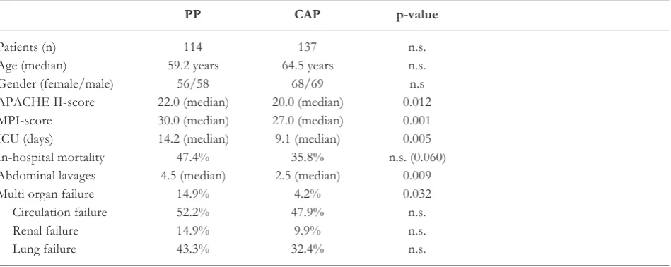
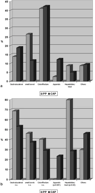
ESULTSOf 251 patients treated for diffuse secondary peritoni-tis 114 (45.4%) were defined as PP and 137 (54.6%) as CAP. The median daily evaluated APACHE II-score for all patients was 21.0. Within the PP group the APACHE II-score was significantly higher (median 22.0) compared to in the CAP group (median 20.0) (p = 0.012). The mortality rate was also higher within the PP group, but was not significantly different compared to that of the CAP patients (p = 0.06). The character-istics for both study groups are presented in Table 1. The underlying cause for secondary diffuse peritonitis regarding the anatomic origin is depicted in Figure 1a.
Statistical analyses revealed no significant differ-ences regarding mortality and anatomic origin of
sec-Table 1.Characteristics of the study population: Biographical data and characteristics for PP and CAP groups.
PP CAP p-value
Patients (n) 114 137 n.s.
Age (median) 59.2 years 64.5 years n.s.
Gender (female/male) 56/58 68/69 n.s
APACHE II-score 22.0 (median) 20.0 (median) 0.012 MPI-score 30.0 (median) 27.0 (median) 0.001 ICU (days) 14.2 (median) 9.1 (median) 0.005 In-hospital mortality 47.4% 35.8% n.s. (0.060) Abdominal lavages 4.5 (median) 2.5 (median) 0.009
Multi organ failure 14.9% 4.2% 0.032
Circulation failure 52.2% 47.9% n.s.
Renal failure 14.9% 9.9% n.s.
Lung failure 43.3% 32.4% n.s.
ondary peritonitis between the two groups except for the appendix [4/17(CAP) vs. 0/3(PP), p = 0.01] and the hepatobiliary tract [2/7(CAP) vs. 8/10(PP), p = 0.03] (Fig. 1b).
A total of 93 patients (81.6%) within the PP group and 133 patients (97.1%) in the CAP group presented with abdominal pain at the time of physical examina-tion (p = 0.03). Abdominal rebound tenderness
oc-Fig. 1.a Anatomical origin of secondary peritonitis. No statistically significant dif-ferences between PP and CAP (p>0.05).
b.Anatomical origin of secondary perito-nitis and mortality.
a
curred in 25 (PP, 21.9%) and 49 (CAP, 35.8%) tients, respectively (p = 0.02). Furthermore, 40 pa-tients (35.1%) within the PP group and 71 papa-tients (51.8%) within the CAP group, respectively, presented with fever exceeding 38.5 °C (p = 0.029) (Fig. 2).
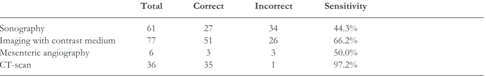
Over 95% of all patients presented with elevated infection parameters (WBC, CRP) but with no statisti-cally significant difference between the two groups. Similarly, lactate and AT III values revealed no signifi-cant differences between the CAP and PP groups, re-spectively. Differences in clinical and paraclinical para-meters for both groups are given in Table 2. PP was predicted correctly using diagnostic imaging in 97.2% (CT-scan), 66.2% (Radiographs with contrast-medium) and 44.3% (Sonography), respectively (Table 3).
The causes for peritonitis in the PP group were as follows: Anastomotic leakage 50.9% (n = 58), perfora-tions 22.8% (n = 26), mesenteric ischemia 11.4% (n = 13) and others 14.9% (n = 17).
The number of abdominal lavages was significantly higher in the PP group (4.5 median) compared to the CAP group (2.5 median) (p = 0.009). In the CAP group one or two lavages were significantly more fre-quent than within the PP group (62.0% vs. 40.7%; p = 0.001). More than four lavages were performed more frequently in the PP group (27.4% vs. 10.2%; p< 0.001).
D
ISCUSSIONEarly detection and diagnosis in secondary peritonitis, either postoperative or community-acquired, is critical for defining the most effective treatment intervention. Clinical monitoring should be aimed at early identifica-tion of the source of the complicaidentifica-tion before sec-ondary organ failure aggravates the clinical situation. The complication itself does not frequently represent the major problem, but is more likely a consequence of late diagnosis and therefore of insufficient therapy. Thus the identification of early predictive indicators of peritonitis and abdominal sepsis is of utmost im-portance [10, 11, 13-16].
In our study we demonstrate that routinely used clinical and paraclinical parameters are of limited pre-dictive value in the diagnosis of PP when compared to the group of patients with CAP. Slightly more than 50% of patients within the PP group presented with abdominal pain or tenderness to palpation accompa-nied by fever at the time of physical examination. Fur-thermore, clinical signs of peritonitis such as rigidity or
Fig. 2. Graphs depict the differences of clinical parameters. Clinical signs differ significantly between the two groups and are therefore of limited diagnotic value.
Table 2.Clinical and paraclinical parameters: Differences in clinical and paraclinical parameters of CAP and PP patients.
PP CAP p-value
Abdominal pain 93 (81.6%) 133 (97.1%) 0.03
Abdominal rebound tenderness 25 (21.9%) 49 (35.8%) 0.02
Paralytic Ileus 40 (35.0%) 33 (24.1%) n.s.
Fever(> 38.5 °C) 40 (35.1%) 71 (51.8%) 0.029
WBC [/nl] 17.3 (median) 18.0 (median) n.s.
CRP [mg/l] 221.2 (median) 205.1 (median) n.s.
Lactate [mmol/l] 3.0 (median) 3.4 (median) n.s.
AT III (%) 54.1 (median) 59.6% (median) n.s.
Albumin (g/L) 22.3 (median) 25.7 (median) 0.03
Immunosuppression 18 (15.8%) 8 (5.8%) 0.019
MOF 17(14.9%) 6 (4.2%) 0.043
Table 3.Diagnostic significance in PP.
Total Correct Incorrect Sensitivity
Sonography 61 27 34 44.3%
Imaging with contrast medium 77 51 26 66.2%
Mesenteric angiography 6 3 3 50.0%
CT-scan 36 35 1 97.2%
rebound tenderness were only evident in 21.9% of these patients. This could be explained by the predomi-nant aggression catabolism as well as by the post-operative administration of analgetic drugs [17]. These patients are therefore frequently diagnosed after signs of sepsis have already occurred [5, 7, 10, 15, 17-19].
The low accuracy of physical tests in diagnosing postoperative peritonitis reflects the questionable val-ue of a so-called ‘experienced surgeon’. Of course, clinical judgment remains important but seems to fre-quently fail in times of effective postoperative analge-sia and sedation of intubated patients [15, 19]. This is underlined by the fact that physical tests are not in-cluded in the ‘top-ten ranked’ variables for inpatient mortality of relaparotomy outcome in peritonitis pa-tients [13]. The current situation is thus that physical tests do not yield meaningful variables with respect to prediction of ongoing abdominal sepsis.
Analysis of laboratory infection parameters such as leukocytosis and C-reactive protein revealed no signifi-cant difference between the two groups. Furthermore, levels of AT III and lactate as parameters for mesente-ric ischemia and coagulopathy within the course of sep-sis were also not significantly different. Other diagnos-tic parameters such as interleukin 6 and tumor necrosis factorα(TNFα) levels are implicated as being impor-tant in the early onset of sepsis but do not correlate with outcome [20-22]. Moreover, their short half-lives make their diagnostic applicability questionable [23].
There has been great interest in the diagnostic and prognostic potential of procalcitonin in abdominal sepsis. Even though existing data support the sensitivi-ty and specificisensitivi-ty as an early marker for sepsis, its value is discussed controversial [8, 24, 25]. Recently, deter-mination of procalcitonin levels in peritonitis was as-sociated with a low sensitivity and specificity [26]. In severe abdominal inflammation such as peritonitis, other sources such as catheter infections, pulmonary or urinary tract infections have to be taken into ac-count in critically ill patients.
Once secondary peritonitis is suspected clinically, further investigations such as abdominal ultrasound and radiograph-based imaging techniques are necessary to confirm the presence and source of peritonitis. In our study the use of abdominal ultrasonography only revealed a sensitivity of 46%, possibly due to com-monly occurring paralysis and/or meteorism of the in-testines. Undoubtedly, mesentericography and radi-ograph imaging using contrast dyes have their value. Within this study, CT-scan revealed a sensitivity of 97.2% in detecting formation and source of peritonitis and was therefore the most precise and valuable imag-ing technique. Other studies support the utilization of CT-scanning in the diagnosis of secondary PP [27, 28]. An optimal scoring system should fulfill the follow-ing criteria: Objective, readily measurable under rou-tine conditions, simple, easy available but also reliable, specific for the function of the organ considered and independent on the type of patient and therapeutic in-tervention. With the exception of the abdominal CT-scan, the investigated clinical and laboratory parame-ters unfortunately did not meet these criteria [13]. Due to the retrospective character of the study only sensi-tivity and not specificity can be presented.
The mortality rate for patients within the PP group was 47.4% and therefore slightly higher than that for patients with CAP (35.8%, p = 0.060). The current lit-erature reports mortality rates between 25 and 68% for secondary peritonitis, whereas the rates for the subgroup of PP unexceptionally exceed those for CAP [3-5, 11, 29]. Even though there is a trend to-wards higher mortality in the PP group, rates did not differ significantly from the CAP group, which is a novel finding. Thus our results do not support an ob-servation strategy in uncertain cases of peritonits without pathological findings on CT-scan as it has been advocated by others [14, 30]
The lack of statistical difference in mortality rates between the two groups are probably caused by the most early intervention, even in doubtful cases. The significantly higher number of lavages necessary in the PP group indicates that these patients are either diag-nosed too late (despite most early indication for rela-parotomy) or are more susceptible to bacteraemia due to predominant immunosuppression. Nevertheless, the re-laparotomy strategy was not an outcome mea-sure in this study. Thus, no clear recommendations can be given from our data.
There is no ideal control group for patients with diffuse PP to assess the value of diagnostic parame-ters. A comparison with uncomplicated recovering pa-tients having undergone elective abdominal surgery would give apparently clear results but would not re-flect the group of importance – postoperative pa-tients in the ICU in critical condition with the ques-tion of whether to perform relaparotomy or not. We therefore consider the group of patients with diffuse CAP to be the most appropriate due to the compara-ble extent of peritonitis and the severity of sepsis signs. The separation between diffuse CAP and PP is undoubtfully artificial. It might be more appropriate to compare patients with surgically proven peritonitis and patients suspicious for peritonitis but without surgical findings at the time of laparotomy. Due to the retrospective character of this study it is almost impossible to reevaluate the latter group. Evaluating -on the other hand – this more “ideal” study collective in a randomized controlled setting might increase the clinical awareness and therefore leading to an evalua-tion bias. Nevertheless, earlier diagnosis and consecu-tive treatment in these patients is frequently per-formed because of the more frequently apparent clin-ical symptoms. This has to be taken into account criti-cally for the assessment of the results revealed by this study.
C
ONCLUSIONAcknowledgments: Prof. Robert Harris, Karolinska Institutet, Stockholm is greatefully acknowledged for critical reviewing the manuscript and his helpful comments.
R
EFERENCES1. Kujath P, Rodloff A: Peritonitis. Second edition, page 36-41. ed Second edition Bremen, London, Boston, UNI-MED, 2005.
2. Buchler MW, Baer HU, Brugger LE, Feodorovici MA, Uhl W, Seiler C: [Surgical therapy of diffuse peritonitis: debridement and intraoperative extensive lavage]. Chirurg 1997;68:811-815.
3. Pacelli F, Doglietto GB, Alfieri S, Piccioni E, Sgadari A, Gui D, Crucitti F: Prognosis in intra-abdominal infec-tions. Multivariate analysis on 604 patients. Arch Surg 1996;131:641-645.
4. Pusajo JF, Bumaschny E, Doglio GR, Cherjovsky MR, Lipinszki AI, Hernandez MS, Egurrola MA: Postopera-tive intra-abdominal sepsis requiring reoperation. Value of a predictive index. Arch Surg 1993;128:218-222; dis-cussion 223.
5. Anderson ID, Fearon KC, Grant IS: Laparotomy for ab-dominal sepsis in the critically ill. Br J Surg 1996;83:535-539.
6. Kirschner: Die Behandlung der akuten eitrigen freien Bauchfellentzündung. Arch klin Chir 1926;142:253-311. 7. McLauchlan GJ, Anderson ID, Grant IS, Fearon KC:
Outcome of patients with abdominal sepsis treated in an intensive care unit. Br J Surg 1995;82:524-529.
8. Rau BM, Frigerio I, Buchler MW, Wegscheider K, Bassi C, Puolakkainen PA, Beger HG, Schilling MK: Evalua-tion of procalcitonin for predicting septic multiorgan fail-ure and overall prognosis in secondary peritonitis: a prospective, international multicenter study. Arch Surg 2007;142:134-142.
9. Unalp HR, Kamer E, Kar H, Bal A, Peskersoy M, Ali Onal M: Urgent abdominal re-explorations. World J Emerg Surg 2006;1:10.
10. van Ruler O, Lamme B, Gouma DJ, Reitsma JB, Boer-meester MA: Variables associated with positive findings at relaparotomy in patients with secondary peritonitis. Crit Care Med 2007;35:468-476.
11. Lamme B, Boermeester MA, Belt EJ, van Till JW, Gouma DJ, Obertop H: Mortality and morbidity of planned rela-parotomy versus relarela-parotomy on demand for secondary peritonitis. Br J Surg 2004;91:1046-1054.
12. Edelsberg J, Berger A, Schell S, Mallick R, Kuznik A, Os-ter G: Economic consequences of failure of initial antibi-otic therapy in hospitalized adults with complicated intra-abdominal infections. Surg Infect (Larchmt) 2008;9:335-347.
13. Lamme B, Mahler CW, van Ruler O, Gouma DJ, Reitsma JB, Boermeester MA: Clinical predictors of ongoing in-fection in secondary peritonitis: systematic review. World J Surg 2006;30:2170-2181.
14. Marshall JC, Innes M: Intensive care unit management of intra-abdominal infection. Crit Care Med 2003;31:2228-2237.
15. Mulier S, Penninckx F, Verwaest C, Filez L, Aerts R, Fieuws S, Lauwers P: Factors affecting mortality in gener-alized postoperative peritonitis: multivariate analysis in 96 patients. World J Surg 2003;27:379-384.
16. van Ruler O, Mahler CW, Boer KR, Reuland EA, Gooszen HG, Opmeer BC, de Graaf PW, Lamme B, Gerhards MF, Steller EP, van Till JW, de Borgie CJ, Gouma DJ, Reitsma JB, Boermeester MA: Comparison of on-demand vs planned relaparotomy strategy in pa-tients with severe peritonitis: a randomized trial. Jama 2007;298:865-872.
17. Makela J, Kairaluoma MI: Relaparotomy for postopera-tive intra-abdominal sepsis in jaundiced patients. Br J Surg 1988;75:1157-1159.
18. Hinsdale JG, Jaffe BM: Re-operation for intra-abdominal sepsis. Indications and results in modern critical care set-ting. Ann Surg 1984;199:31-36.
19. Holzheimer RG, Gathof B: Re-operation for complicated secondary peritonitis - how to identify patients at risk for persistent sepsis. Eur J Med Res 2003;8:125-134.
20. Fang XM, Schroder S, Hoeft A, Stuber F: Comparison of two polymorphisms of the interleuk1 gene family: in-terleukin-1 receptor antagonist polymorphism contributes to susceptibility to severe sepsis. Crit Care Med 1999;27:1330-1334.
21. Harbarth S, Holeckova K, Froidevaux C, Pittet D, Ricou B, Grau GE, Vadas L, Pugin J: Diagnostic value of pro-calcitonin, interleukin-6, and interleukin-8 in critically ill patients admitted with suspected sepsis. Am J Respir Crit Care Med 2001;164:396-402.
22. Majetschak M, Flohe S, Obertacke U, Schroder J, Staubach K, Nast-Kolb D, Schade FU, Stuber F: Relation of a TNF gene polymorphism to severe sepsis in trauma patients. Ann Surg 1999;230:207-214.
23. Reith HB, Mittelkotter U, Debus ES, Kussner C, Thiede A: Procalcitonin in early detection of postoperative com-plications. Dig Surg 1998;15:260-265.
24. Reith H, Mittelkötter U, Wagner R, Thiede A: Procalci-tonin (PCT) in patients with abdominal sepsis. Intensive Care Med 2000;26(suppl2):159-164.
25. Schroder J, Staubach KH, Zabel P, Stuber F, Kremer B: Procalcitonin as a marker of severity in septic shock. Lan-genbecks Arch Surg 1999;384:33-38.
26. Lam MF, Leung JC, Lam CW, Tse KC, Lo WK, Lui SL, Chan TM, Tam S, Lai KN: Procalcitonin fails to differen-tiate inflammatory status or predict long-term outcomes in peritoneal dialysis-associated peritonitis. Perit Dial Int 2008;28:377-384.
27. Evans HL, Raymond DP, Pelletier SJ, Crabtree TD, Pruett TL, Sawyer RG: Diagnosis of intra-abdominal in-fection in the critically ill patient. Curr Opin Crit Care 2001;7:117-121.
28. Rotstein OD, Meakins JL: Diagnostic and therapeutic challenges of intraabdominal infections. World J Surg 1990;14:159-166.
29. Nathens AB, Rotstein OD, Marshall JC: Tertiary peritoni-tis: clinical features of a complex nosocomial infection. World J Surg 1998;22:158-163.
30. Marshall JC, Vincent JL, Fink MP, Cook DJ, Rubenfeld G, Foster D, Fisher CJ, Jr., Faist E, Reinhart K: Mea-sures, markers, and mediators: toward a staging system for clinical sepsis. A report of the Fifth Toronto Sepsis Roundtable, Toronto, Ontario, Canada, October 25-26, 2000. Crit Care Med 2003;31:1560-1567.
Received: May 2, 2009 / Accepted: June 9, 2009
Address for correspondence:
Dr. med. Franz G. Bader, MD, PhD Department of Surgery
University of Schleswig-Holstein Campus Lübeck
Ratzeburger Allee 160 23538 Lübeck Germany