Abstract
The ulnar nerve is a branch of the C8 and T1 nerve roots and arises from the medial cord of the brachial plexus. It supplies the intrinsic muscles of the hand and assists the median nerve in functioning of the flexors. Also known as the musician’s nerve, it is the second most common nerve involved in compressive neuropathy following the median nerve. Common sites of entrapment include cubital tunnel at the elbow, the ulnar groove in the humerus and the Guyon’s canal at the wrist. Patients present with altered sensation in the ulnar fourth and the fifth digit and the medial side of arm with loss of function of intrinsic muscles of the hand, the flexor carpi ulnaris and ulnar fibres of flexor digitorum superficialis in more severe cases. Diagnosis relies on clinical examination, electrodiagnostic studies and imaging findings. Plain radiographs are used to identify fracture sites, callus, or tumours as cause of compression. Technological advances in ultrasonography have allowed direct visualisation of the involved nerve with assessment of exact site, extent and type of injury. It yields unmatched information about anatomical details of the nerve. MR imaging adds to soft tissue details and helps in characterising the lesion. This pictorial review aims to illustrate a wide spectrum of causes of ulnar neuropathies as seen on ultrasound and MRI and emphasises upon the importance of imaging modalities in the diagnosis of neuropathies.
Keywords:Imaging in the ulnar mononeuropathy, High-resolution ultrasound of ulnar neuropathy, MR ulnar neuropathy
Key points
Over the last decade, neuromuscular ultrasound and MR neurography have emerged as useful tools for the diagnosis of peripheral nerve disorders.
The ulnar nerve is the second most common nerve involved in compressive neuropathies of the upper limb. This pictorial review aims to demonstrate various ulnar nerve pathologies as diagnosed using imaging.
Imaging helps in identifying the exact anatomical details about the involved nerve which provides an edge over the information provided by the electrodiagnostic tests.
Awareness about the pathology and extent of involvement helps in proper clinical decision making and timely management.
Introduction
Following the carpal tunnel syndrome, compressive neur-opathy of the ulnar nerve at the cubital tunnel is the most common cause of compressive neuropathy [1,2]. Conven-tionally, neuropathies have been diagnosed by clinical examination, Tinel’s sign and electrodiagnostic (nerve conduction velocity and electromyography) findings which provide information about the nerve involved and the pos-sible site of injury but do not provide any accurate ana-tomical information. Introduction of high-frequency ultrasound probes have made direct visualisation of per-ipheral nerves possible, thus providing anatomical details about the nerve. Ultrasonographic findings of peripheral nerves were first reviewed by Fornage in 1988 [3]. Since then, technological advances like increased frequency and variable sizes of footprints of linear transducers have escalated the use of ultrasound in peripheral nerve pathologies. Exact site, extent and type of involvement, local cause of neuropathy, continuity of nerve and architectural distortion can be identified for accurate diagnosis and planning the management. Ultrasound is quick, cost effective, has no contraindications and * Correspondence:[email protected]
Aakanksha Agarwal and Abhishek Chandra are joint first authors.
†Aakanksha Agarwal and Abhishek Chandra contributed equally to this work.
1Department of Radiodiagnosis, SMS Medical College, Jaipur, Rajasthan, India 3A 235, Shivanand Marg, Malviya Nagar, Jaipur, India
Full list of author information is available at the end of the article
provides detailed imaging of the entire length of the nerve. MR imaging adds to soft tissue details, change in muscles supplied by the affected nerve and helps in characterising the nerve lesion. In-spite of these advan-tages, imaging remains underutilised in cases of periph-eral neuropathy. The aim of this pictorial essay is to demonstrate the use of imaging in diagnosing the ulnar nerve neuropathies due to various aetiologies. Various previous publications have mentioned methods to iden-tify the nerve in specific anatomical locations and vari-ous peripheral nerve pathologies, centred around the Guyon’s canal and Cubital tunnel [3–7]. With this re-view, we provide a neoteric review which combines ultrasound and MR imaging of commonly encountered causes of ulnar neuropathies.
Path of the ulnar nerve
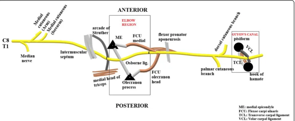
The nerve arises from the medial cord of the brachial plexus carrying fibres from the C8-T1 nerve roots. It lies posteromedial to the brachial artery in the anterior com-partment of the upper arm and descends down to enter the posterior compartment by piercing the medial intermuscu-lar septum (Fig.1). This is a potential site of compression of the nerve under the‘Arcade of Struther’[8] which is de-scribed as a fibrous canal on the medial aspect of the mid-dle- and lower-third of the arm, consisting of the medial head of the triceps brachii muscle and its aponeurotic ex-pansion, which extends into the intermuscular septum and internal brachial ligament and covers part of the ulnar nerve. Thereafter, the ulnar nerve runs behind the medial epicondyle with the superior ulnar collateral vessels Fig. 1Diagrammatic representation of the pathway of the ulnar nerve from origin up to terminal bifurcation at wrist. The elbow and wrist are the most common sites of ulnar nerve entrapment
under the ‘Cubital tunnel’ [9–11]. The roof of the cu-bital tunnel is formed by the Osborne’s ligament or the Cubital retinaculum which is a ligament spanning from the medial epicondyle to the olecranon process, con-tinuous with the fascia connecting the humeral and ulnar heads of the flexor carpi ulnaris (FCU). Alternatively, the
roof may be formed by the anconeus epitrochearlis muscle [12]. The floor is formed by the medial collateral ligament (MCL) and elbow joint capsule, while the medial epicon-dyle and olecranon form the walls on either side [13]. Flexion of the elbow decreases the height, area and sagittal curvature of the tunnel, with maximum nerve Fig. 3aProbe position for localisation of the ulnar nerve at the wrist.bThe nerve (arrowhead) is localised in the short axis in the Guyon’s canal between the pisiform and hook of hamate, lying adjacent to the ulnar artery (colour Doppler signal).cMR image showing the normal ulnar nerve (yellow arrow) in the Guyon’s canal: pisiform (P) and the hook of hamate (H) form the radial and ulnar boundaries. The roof is formed by the volar carpal ligament (red arrow) while the hypothenar muscles and transverse carpal ligament (red arrowhead) form the floor
compression occurring at 135° of flexion according to a study by James et al. [14]. At this level, the ulnar nerve gives off an articular branch to the elbow joint.
Thereafter, the ulnar nerve descends into the forearm, passing between the two heads of the flexor carpi ulnaris muscle (FCU) [15]. The nerve provides motor supply to the FCU and medial half of the flexor digitorum profundus. It continues distally along the ulna, lying deep to the FCU. About 5 cm distal to the medial epicondyle, the ulnar nerve pierces the flexor-pronator aponeurosis which is the fibrous common origin of the flexor and pronator muscles. Another aponeurosis extends between the flexor digitorum superfi-cialis of the ring finger and the humeral head of the flexor carpi ulnaris, known as the ligament of Spinner, which at-taches to the medial epicondyle and can cause kinking of the nerve following an anterior transposition. Near the wrist, the ulnar nerve moves lateral to the FCU, courses medial to the ulnar artery to enter the palm through the Guyon’s canal. The volar carpal ligament forms the roof of the canal while the transverse carpal ligament and the hypothenar muscles form the floor. The medial and lateral walls are formed by pisiform, pisohamate ligament, abductor digiti minimi muscle belly on the ulnar side and the hook of ham-ate on the radial side [16]. Near the wrist, the motor innerv-ation of the ulnar nerve includes the thenar muscles: the adductor policis, deep head of flexor pollicis brevis (FPB); dorsal and palmar interossei and 3rd and 4th lumbricals, hypothenar muscles: abductor digiti minimi, opponens digiti minimi and flexor digiti minimi. The ulnar nerve gives dor-sal and palmar cutaneous branches and a few superficial ter-minal branches to provide sensation to the ulnar fourth and entire fifth finger and the medial aspect of the forearm.
Technical details of imaging
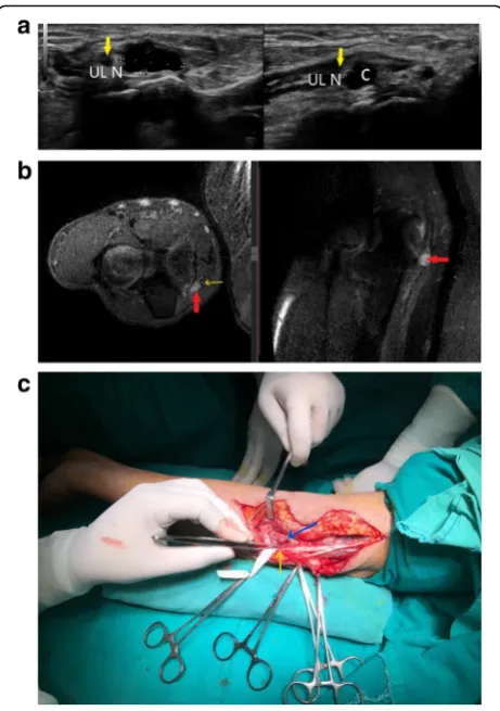
The normal peripheral nerve has a honeycomb appearance which is readily identified on imaging. The central spots Fig. 4aShort axis and long axis view of ulnar nerve (yellow arrow)
which appears hypoechoic. There is a cystic lesion (between callipers) causing direct compression over the ulnar nerve.bPD FS axial and sagittal image showing a hyperintense ulnar nerve (yellow arrow) getting compressed by a hyperintense cyst (red arrow).cPer operative picture showing the ulnar nerve (yellow arrow) and the cyst (blue arrow). Histopathology revealed it to be a ganglion cyst
are the nerve fascicles which are formed by bundles of nerve fibres surrounded by the perineurium. Each nerve fibre is also surrounded by an endoneurium which is not visible on imaging with the currently available resolution. The fascicles are in turn embedded and surrounded by epineurium which forms the sheath of the peripheral nerve [17]. Normal maximum cross-sectional area of the ulnar nerve at the level of cubital tunnel in a study by Wiesler et al. proved to be 0.065 cm2[18].
High-resolution ultrasound
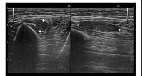
A high-frequency linear probe is required for scanning peripheral nerves. We used a probe with frequency set-tings to 14 Hz. The nerve should be identified in cross section as it has a characteristic honeycomb appearance which helps in demarcating it from the surrounding soft tissue. The nerve is more echogenic to surrounding muscle and less echogenic to surrounding tendon. After identifying the nerve in its short axis at predetermined anatomical locations, it can be traced proximally and dis-tally throughout its length. The ulnar nerve is easily
located at the elbow, passing under the cubital tunnel (Fig. 2) and at the wrist where it lies under the Guyon’s canal (Fig.3). Pathological segment of the nerve will show loss of the normal honeycomb structure, decreased echo-genicity, discontinuity, focal enlargement or neuroma for-mation. Surrounding compressive lesions, muscle and soft tissue oedema, joint effusions can also be identified.
MR imaging
We used a 3T MRI scanner with a dedicated protocol for visualisation of the ulnar nerve. T1, T2 fat saturated, PD fat saturated sequences were acquired with slice thickness of 3 mm and interslice gap of 0.3 mm. Gadolinium-based contrast was used for post contrast images if indicated. Axial sections are of utmost import-ance as the characteristic honeycomb appearimport-ance is eas-ily identified. We acquired axial sections of T1, T2 fat-saturated and PD fat-saturated sequences in addition to sagittal T1, PD fat-saturated and coronal T2 slices. Knowledge of the anatomical details of the course of the nerve is indispensable to identify the nerve in its short Fig. 6aAnechoic cystic lesion (red arrowheads) adjacent to the ulnar nerve (yellow arrow) at the elbow with underlying cortical erosions (red arrow). The cystic lesion appears to arise from the nerve, suggestive of an intraneural ganglion cyst.bT2-weighted axial section shows hyperintense cystic lesion (red arrowheads) which post operatively was diagnosed as ganglion cyst arising from the nerve sheath of the ulnar nerve (yellow arrow). The nerve is hyperintense due to compressive neuropathy. Also noted are osteophytes (red arrow) and joint effusion (blue arrowhead) suggestive of arthritic changes.cT2-weighted coronal image shows the cystic lesion (red arrowheads) arising from the adjacent ulnar nerve (yellow arrow) lying along the course of the nerve [54]. Also noted are osteophytes (red arrow)
axis. T1-weighted images help in identification of the nerve in its short axis as the peripheral rim of fat in the nerve sheath appears hyperintense while the fascicles appear as hypointense dots within. Epineural or perineural fat re-mains bright on T2WI which can mask pathological changes in the nerves. Fat suppression is thus needed for identification of pathological segment of the nerve on T2 and PD sequences [19,20]. Focal enlargement, hyperinten-sity and altered fascicular patterns are signs of neuropathy. The denervated muscles begin to appear hyperintense on T2-weighted images 48 h after nerve injury. These changes are seen in axonotmetic and neurotmetic injuries. MRI may not clearly differentiate between disruption injuries and contusion of nerve [21]. Muscle atrophy with persistent hyperintensity is seen in failure of regeneration [20]. Fatty infiltration is seen in chronic denervation. Orthopaedic implants may limit the use of MRI due to susceptibility artefacts.
Clinical presentation of ulnar neuropathy
Ulnar neuropathy can result in sensory symptoms only or can cause progressively worsening motor deficit. It usually starts as altered sensation in the ulnar nerve sensory distri-bution, including the ulnar fourth and entire fifth finger
which may progress to involve the motor fibres leading to muscular weakening and muscular atrophy over time [12, 22]. Elbow flexion can result in exaggerated symp-toms as the cubital tunnel height and area decrease on flexion with resultant nerve compression [14]. Elbow flexion during sleep can result in night symptoms severe enough to cause awakening. The fibres of the in-trinsic muscles of hand are located more peripherally in the nerve while the FCU and FDP fibres are more cen-trally placed. Chronic severe injury progressively leads to weakening of the FCU and FDP resulting in the typ-ical ‘Ulnar claw hand’ [12]. When the ulnar nerve is divided at the wrist, only the opponens pollicis, superfi-cial head of the flexor pollicis brevis and lateral 2 lumb-ricals are functioning, all of which are supplied by the median nerve.
Aetiology of ulnar nerve pathology
Ulnar neuropathy can occur due to entrapment of the nerve at anatomical sites, chronic irritation of the nerve due to local causes or transection of the nerve following penetrating injuries.
The most common site of ulnar nerve entrapment is at the elbow, within the cubital tunnel or the epicondylar Fig. 8aThickened hypoechoic ulnar nerve (yellow arrow) getting compressed by orthopaedic implant—K wire (red arrow).bRing down artefact on colour Doppler shows the orthopaedic implant (red arrowhead) compressing the hypoechoic ulnar nerve which also shows increased vascularity suggestive of neuritis (yellow arrow).cPlain radiograph of the same patient shows the implant following intervention for an olecranon fracture.dColour Doppler image of the same patient showing hypervascularity of the ulnar nerve (yellow arrow) which co-relates with higher grade of neuritis due to compression or inflammation [41]
groove [23] while the second most common site is at the wrist, commonly in the Guyon’s canal [24–27].
Another common cause of ulnar neuropathy is the
‘tardy ulnar nerve palsy’ as described by Panas et al. [28–30] in which chronic nerve irritation occurs due to prior trauma or osteoarthritis.
Common sites of nerve compression include a site in the upper arm where the ulnar nerve pierces the intermuscular septum, beneath the arcade of Struther, alongside the med-ial epicondyle particularly in patients with cubitus valgus deformity, in the ulnar groove, within the cubital tunnel and between the two heads of FCU. More distally, it can get compressed where it exits the FCU and perforates a fascial layer between the flexor digitorum superficialis and the flexor digitorum profundus [31,32].
Guyon’s canal is the commonest site of direct nerve trauma following penetrating injuries as the nerve is superficial in this region [27]. Three zones have been identified in relation to the ulnar nerve pathology near the Guyon’s canal—zone 1: encompassing the area prox-imal to the bifurcation of the ulnar nerve; zone 2:
encompassing the motor branch of the nerve after it has bifurcated; and zone 3: encompassing the superficial or sensory branch of the bifurcated nerve.
Other aetiologies include compression during general anaesthesia, blunt trauma, deformities, transient occlu-sion of brachial artery, cigarette smoking, entrapment within scar tissue, local hematomas, subdermal contra-ceptive implants and following venepuncture [33–37]. Thermal and burn injuries are another cause of ulnar nerve palsy which may also include other nerves in the region of the burn. Tumour and tumour like lesions involving the ulnar nerve include peripheral nerve sheath tumours (PNSTs), fibrolipomatous hypertrophy, intraneural lipomas, intraneural ganglion, true neuromas and pseudo neuromas.
Diagnosis of ulnar nerve pathologies
Clinical examination and electrodiagnostic studies (nerve conduction velocity (NCV) and electromyography (EMG)) are the conventional diagnostic tools in neurop-athies as they provide essential information about the Fig. 10aGrey scale ultrasound image shows a hypoechoic, significantly thickened ulnar nerve (ULN) in the cubital tunnel with underlying joint effusion (E) and cortical irregularity (CC). The medial epicondyle (ME) and olecranon process (O) forming boundaries of the cubital tunnel are also seen.bT2-weighted MR image of the patient shows hyperintensity of the ulnar nerve (yellow arrow) with surrounding arthritic
changes—subchondral cyst (red arrow), marrow oedema with cortical irregularity (blue arrow) and joint effusion (red arrowheads).cIntra operative picture of the patient showing the thickened ulnar nerve (black arrow)
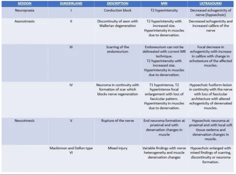
type of dysfunction and help in clinical monitoring. Tinel’s sign helps in assessing the possible site of injury and to assess response to treatment. Nerve injuries have been classified by Seddon in 1943 and expanded by Sunderland in 1951. Mackinnon and Dellon in 1992 added grade VI injury to Sunderland grading scheme which was defined as a mixed type of injury which de-notes various types of injuries across the cross section of the nerve [38–40]. Table 1 illustrates this classification along with imaging findings in different types of nerve injuries. Imaging helps by providing unmatched anatom-ical details about the nerve involved. Plain radiographs show fracture sites, soft tissue swellings and orthopaedic
implants. It may also demonstrate a fat stripe of lipoma or a bony lesion in proximity to the course of the nerve. High-resolution ultrasound and MRI help in direct visu-alisation of the nerve with anatomical delineation of the pathological segment. Hypo-echogenicity of the nerve on ultrasound and hyperintensity on MRI are markers of neuropathy. Normal nerves do not show any intra-neural vascularity. Appreciation of intra-neural vascularity is a marker of neural compression or inflam-mation and co-relates with severity of affection of nerve [41]. These imaging modalities also provide essential in-formation about the surrounding soft tissue and the muscles innervated by the injured nerve.
Fig. 12aThickened, hypoechoic ulnar nerve with increased intra neural vascularity on colour Doppler, signifying ulnar neuritis.bUlnar nerve (white arrow head) is hypointense and located anteromedial to the normal location anterior to the medial epicondyle (ME) in a case with anterior intramuscular transposition of ulnar nerve. There is also associated joint effusion (red arrow) which appears hypointense on this T1-weighted image.cUlnar nerve (white arrow head) appears hyperintense and thickened on T2 coronal image lying above the medial epicondyle, passing lateral to it instead of its normal location in the cubital tunnel. Also noted are osteophytes (red arrow)
Fig. 13aA patient with lepromatous leprosy and paraesthesia in ulnar distribution with a hypoechoic ulnar nerve (between callipers).bAnother patient with more advanced nerve damage, as evident by loss of fascicular architecture and irregularly, thickened ulnar nerve (between callipers).
Imaging findings in a wide spectrum of ulnar nerve pathologies
Ulnar nerve compression at the elbow Due to ganglion cyst
Ganglion cysts are pseudocysts with no epithelial lin-ing of their own. These are non-neoplastic lesions filled with gelatinous material and originate from ten-don sheath, ligament, bursa, joint capsule or subchon-dral bone. Rarely, they may present in an intramuscular location, away from the joint with no synovial communication [42, 43]. Ganglion cysts of the nerve sheath are uncommon. There are reports in the literature of ulnar nerve compression by extra-neural ganglion cysts at the elbow and less commonly in the Guyon’s canal [44].
Figure 4 shows a simple cystic lesion on ultrasound causing ulnar nerve compression at the elbow. The nerve was hypoechoic on HRUS and hyperintense on MRI signifying neuropathic changes. Per operatively, neural compression by a ganglion cyst lying within the cubital tunnel under the fascial sheath of the FCU muscle was confirmed.
Due to compression by fracture segment
Fracture of the medial epicondyle or the olecranon process of ulna can impinge upon the ulnar nerve in the cubital tunnel. This cortical breach can be well visualised on plain radiographs while compression of the nerve can be seen on HRUS. Figure7shows a case with ulnar com-pression by fracture fragment at the elbow.
Due to orthopaedic implants
Metallic K wires are frequently used in banding of frac-tures of the olecranon process. These may impinge upon the nerve causing chronic nerve irritation. Figure 8 shows a case with ulnar nerve impingement by a K wire used for tension band wiring of the olecranon process.
Tardy ulnar nerve palsy
Tardy ulnar nerve palsy is a clinical condition charac-terised by delayed onset ulnar neuropathy due to nerve compression. It has a variety of etiological factors in-cluding callus of old healed fractures around the elbow, arthritis, effusions and synovial thickening of the elbow joint, congenital anomalies, adhesion, recurrent disloca-tion of the elbow, etc. Tardy nerve palsy can also result from cubitus varus deformity following old supracondy-lar fracture of humerus [45,46].
Figure 9 shows a case with ulnar neuropathy due to underlying joint effusion and synovial thickening in a pa-tient with juvenile rheumatoid arthritis. The ulnar nerve Fig. 14Hypoechoic ulnar nerve (between yellow arrowheads) is
noted at the wrist in a patient with penetrating injury to the forearm. The nerve is discontinuous (red arrow) with fluid filled (F) tract extending from skin to the nerve
was displaced out of the cubital tunnel due to underlying soft tissue.
Age-related arthritic changes, as seen in Fig.10are an-other probable cause of ulnar neuropathy in patients with tardy ulnar nerve palsy. Also seen is increased neural vascularity.
Age-related joint effusion, subchondral cysts, and syn-ovial thickening can cause displacement of the ulnar nerve out of the cubital tunnel, as seen in Fig.11.
Figure12shows another case with history of tardy ulnar nerve palsy. The nerve was transposed anteriorly to relieve compression. The patient developed neuritis post opera-tively which was subsequently managed with steroids and analgesics.
Ulnar neuropathy in leprosy
One of the principal causes of ulnar neuropathy in the Indian subcontinent is infection by lepra bacilli. Nerve involvement in leprosy may vary from involvement of an intradermal nerve in the cutaneous patch to a major
lesion in the peripheral or the cranial nerve trunk [47]. Nerve damage in leprosy may present itself as silent neuropathy without overt signs and symptoms or as clinically manifest disease which may present as weak-ness, atrophy or contracture.
Imaging features may range from a hypoechoic nerve with loss of fascicular architecture to frank abscess formation and intra neural hypervascularity. Figure13shows cases of ulnar nerve involvement in different stages of leprosy.
Traumatic ulnar nerve palsy at wrist
The ulnae nerve has a superficial course near the wrist where it passes under the Guyon’s canal. It can be com-monly injured in penetrating injuries to the wrist. Figure14 shows a case with discontinuity in ulnar nerve following a penetrating injury to forearm. Another case with glass cut injury shows a neuroma in continuity in the ulnar nerve in Fig.15along with soft tissue oedema.
Peripheral nerve sheath tumours involving the ulnar nerve [48,49]
Peripheral nerve sheath tumours (PNSTs) include neuro-fibroma and schwannoma which may be benign or malignant. Deep-seated neurofibromas may have neuro-logical symptoms while superficial neurofibromas are usu-ally small painless masses. Plain radiography is usuusu-ally normal in PNST while high-resolution ultrasound shows a hypoechoic fusiform mass in continuity with the parent nerve. MRI is the imaging modality used to differentiate between PNST and traumatic neuromas. PNST are char-acterised by T1 iso-hypointense, T2 hyperintense fusiform mass in continuity with the nerve which may show low signal intensity in the centre on T2-weighted images cor-responding to the higher fibro-collagen content in the centre which co-relates with the organised histological architecture. Another sign is the split fat sign best seen on Fig. 16aGrey scale image showing characteristic fusiform swelling in continuity of the ulnar nerve, suggestive of peripheral nerve sheath tumour.bHypointense lesion in continuity with ulnar nerve with fascicular pattern (yellow arrowheads) is seen with surrounding thin rim of fat (white arrow)—the split fat sign.cPD FS image shows a fusiform swelling which is hyperintense (red arrow) with internal hypointense areas, suggestive of collagenous stroma. Also noted is the ulnar nerve (yellow arrow) forming a tail.dPost contrast T1 FS image shows vivid enhancement in the PNST, differentiating it from a neuroma
showed enhancement in 88% of traumatic neuromas, which might be attributed to a broken blood-nerve barrier [50]. Figure16shows a PNST in continuity with the ulnar nerve in the arm in a patient with soft tissue swelling and no neural symptoms. Neurofibroma in ulnar nerve is seen in Fig.17in a patient with neurofibromatosis.
Management
Conservative treatment of ulnar neuropathy focuses on pain relief, inflammation reduction, and rehabilitation. Pa-tients are advised to avoid aggravating activities, use night splints and use NSAIDs and steroids to decrease inflam-mation [12, 15]. Surgery is indicated when conservative management fails or for decompression and in cases of primary repair of the nerve.
Further advances in MRI
Diffusion tensor imaging and tractography are now being used for peripheral nerve pathologies. Jengojen et al. [51] used diffusion tensor imaging and tractography to assess acute changes in radial and median nerve following com-pression and demonstrated changes in radial nerve metrics. Razek et al. [52] carried out a prospective study in 39 pa-tients with mild to moderate carpal tunnel syndrome and found positive correlation between findings of diffusion ten-sor imaging, electrodiagnostic tests and clinical assessment.
Neuropathic segments tend to have a lower fractional anisotropy value as compared to the normal segment [53].
Advantages and limitations of ultrasound
Use of high-resolution ultrasound in imaging of periph-eral neuropathy is a relatively new use of this age-old modality. Ultrasound is a rapid, cost effective, widely available modality with no contraindications. It allows quick assessment of the nerve throughout its length and provides unmatched data about anatomical details of the affected nerve. Technical issues like inability to image through bone, orthopaedic implants, poor resolution of deeper nerves with higher frequency transducers, sub-optimal scan in patients with higher fatty tissue due to increase in depth and similar echogenicity limit its use to a certain extent. Operator dependence and need for in-depth knowledge of the anatomical details is another limiting factor.
aetio-pathogenesis of the palsy, facilitating better clinical decision making and patient management. This pictorial essay describes the hallmark of imaging findings in a wide spectrum of ulnar nerve pathologies as diagnosed using imaging modalities.
Acknowledgements
We would like to thank Dr. Mamta Agarwal, Consultant Radiologist, Jaipur for her guidance and Mr. Shobey John and Mr. Johnson NJ, technicians, SONI CT MRI, SMS medical college for their contribution.
Authors’contributions
AA and AC were involved in conception of the design of the review, formulating the first draft and gathering necessary information and data. UJ and NS were involved in literature search and critical corrections of the manuscript. All authors read and approved the final manuscript.
Competing interests
The authors declare that they have no competing interests.
Publisher’s Note
Springer Nature remains neutral with regard to jurisdictional claims in published maps and institutional affiliations.
Author details
1Department of Radiodiagnosis, SMS Medical College, Jaipur, Rajasthan, India. 2
Department of Orthopaedics, SMS Medical College, Jaipur, Rajasthan, India.
3A 235, Shivanand Marg, Malviya Nagar, Jaipur, India.
Received: 16 November 2018 Accepted: 4 February 2019
References
1. Bozentka DJ (1998) Cubital tunnel syndrome pathophysiology. Clin Orthop Relat Res (351):90-4 (ISSN: 0009-921X)
2. Robertson C, Saratsiotis J (2005) A review of compressive ulnar neuropathy at the elbow. J Manipulative Physiol Ther 28(5):345
3. Fornage BD (1988) Peripheral nerves of the extremities: imaging with US. Radiology 167:179–182
4. Schafhalter-Zoppoth I, Gray AT (2005) The musculocutaneous nerve: ultrasound appearance for peripheral nerve block. Reg Anesth Pain Med 30: 385–390
5. Chien AJ, Jamadar DA, Jacobson JA, Hayes CW, Louis DS (2003) Sonography andMR imaging of posterior interosseous nerve syndrome with surgical correlation. AJR Am J Roentgenol 181(1):219–221
6. Mondelli M, Morena P, Ballerina M, Rossi S, Giannini F (2005)
Mononeuropathies of the radial nerve: clinical and neurotrophic findings in 91 consecutive cases. J Electromyogr Kinesiol 15(4):377–383
7. Miller TT, Reins WR (2010) Nerve entrapment syndromes of the elbow, forearm, and wrist. AJR Am J Roentgenol 195(3):585–594
8. Caetano EB, Sabongi Neto JJ, Vieira LA, Caetano MF (2017) The arcade of Struthers: an anatomical study and clinical implications. Rev Bras Ortop 52(3):331–336
10. Granger A, Sardi JP, Iwanaga J et al (2017) Osborne’s ligament: a review of its history, anatomy, and surgical importance. Cureus 9(3):e1080 11. Kazuteru Doi (2009) Chapter 8 - Nerves. In: Wei FC, Mardini S (Eds.) Flaps
and reconstructive surgery. W.B. Saunders, pp 71–79.https://doi.org/10. 1016/B978-0-7216-0519-7.00008-3
12. Folberg CR, Weiss AP, Akelman E (1994) Cubital tunnel syndrome. Part I: presentation and diagnosis. Orthop Rev 23(2):136–144
13. Huang JH, Samadani U, Zager EL (2004) Ulnar nerve entrapment neuropathy at the elbow: simple decompression. Neurosurgery 55(5): 1150–1153
14. James J, Sutton LG, Werner FW, Basu N, Allison MA, Palmer AK (2011) Morphology of the cubital tunnel: an anatomical and biomechanical study with implications for treatment of ulnar nerve compression. J Hand Surg Am 36(12):1988–1995
15. Posner MA (2000) Compressive neuropathies of the ulnar nerve at the elbow and wrist. Instr Course Lect 49:305–317
16. Shen L, Masih S, Patel DB, Matcuk GR Jr (2016) MR anatomy and pathology of the ulnar nerve involving the cubital tunnel and Guyon’s canal. Clin Imaging 40(2):263–274.https://doi.org/10.1016/j.clinimag.2015.11.008 17. Grant GA, Goodkin R, Kliot M (1999) Evaluation and surgical management of
peripheral nerve problems. Neurosurgery 44:825–839 discussion 839–840 18. Wiesler ER, Chloros GD, Cartwright MS, Shin HW, Walker FO (2006)
Ultrasound in the diagnosis of ulnar neuropathy at the cubital tunnel. J Hand Surg Am 31(7):1088–1093.https://doi.org/10.1016/j.jhsa.2006.06.007 19. Kuntz C 4th, Blake L, Britz G et al (1969) Magnetic resonance neurography
of peripheral nerve lesions in the lower extremity. Neurosurgery 39:750–756 discussion 756–757
20. Bendszus M, Wessig C, Solymosi L, Reiners K, Koltzenburg M (2004) MRI of peripheral nerve degeneration and regeneration: correlation with electrophysiology and histology. Exp Neurol 188:171–177
21. Umans H, Kessler J, de la Lama M, Magge K, Liebling R, Negron J (2010) Sonographic assessment of volar digital nerve injury in the context of penetrating trauma. AJR Am J Roentgenol 194:1310–1313 22. Bradshaw DY, Shefner JM (1999) Ulnar neuropathy at the elbow. Neurol Clin
17(3):447–461 v-vi
23. Campbell WW, Pridgeon RM, Riaz G, Astruc J, Sahni KS (1991) Variations in anatomy of the ulnar nerve at the cubital tunnel: pitfalls in the diagnosis of ulnar neuropathy at the elbow. Muscle Nerve 14(8):733–738
24. Szabo RM, Steinberg DR (1994) Nerve entrapment syndromes in the wrist. J Am Acad Orthop Surg 2(2):115–123
25. Elhassan B, Steinmann SP (2007) Entrapment neuropathy of the ulnar nerve. J Am Acad Orthop Surg 15(11):672–681
26. Pearce C, Feinberg J, Wolfe SW (2009) Ulnar neuropathy at the wrist. HSS J 5(2):180–183 quiz 184-5
27. Olney RK, Wilbourn AJ, Miller RG (1983) Ulnar neuropathy at or distal to the wrist (abstr). Neurology 33(Suppl 2):185
28. Panas J (1878) Sur une cause peu connue de paralysie du nerf cubital. Archives Generales de Medecine
29. Murphy JB (1914) Neuroma of the ulnar nerve, result of cicatricial compression following unrecognised fracture. Clin John B Murphy 3:369
30. Brickner WM (1924) Late ulnar nerve palsy following elbow fracture in the adult. J Bone Jt Surg 6:477–481
31. Halikis MN, Taleisnik J, Szabo RM (2001) Compression neuropathies of the upper extremity. In: Chapman MW (ed) Chapman’s Orthopaedic Surgery, 3rd edn. Lippincott Williams & Wilkins, Philadelphia, PA, pp 40–99
32. Campbell WW (2000) Ulnar neuropathy at the elbow. Muscle Nerve 23(4):450–452
33. Swenson JD, Hutchinson DT, Bromberg M, Pace NL (1998 Sep) Rapid onset of ulnar nerve dysfunction during transient occlusion of the brachial artery. Anesth Analg 87(3):677–680
34. Marin R, McMillian D (1998) Ulnar neuropathy associated with subdermal contraceptive implant. South Med J 91(9):875–878
35. Masoorli S, Angeles T, Barbone M (1998) Danger points. How to prevent nerve injuries from venipuncture. Nursing 28(9):34–39 quiz 40 36. Chang C, Shen M (1998) Mononeuropathy multiplex in hemophilia: an
electrophysiologic assessment. Eur Neurol 40(1):15–18 37. Richardson JK, Jamieson SC (2004) Cigarette smoking and ulnar
mononeuropathy at the elbow. Am J Phys Med Rehabil 83(9):730–734 38. Seddon HJ (1943) Three types of nerve injuries. Brain 66:237–288
39. Sunderland S (1951) A classification of peripheral nerve injuries producing loss of function. Brain 74:491–516
40. Mackinnon S, Dellon AL (1988) Diagnosis of nerve injury. Surgery of the peripheral nerve. Thieme, NY, pp 74–78
41. Ghasemi-Esfe AR, Khalilzadeh O, Vaziri-Bozorg SM et al (2011) Color and power Doppler US for diagnosing carpal tunnel syndrome and determining its severity: a quantitative image processing method. Radiology 261:499–506
42. Gül M, Özkaya U,Parmaksızoğlu AS,Sökücü S, Kabukçuoğlu Y (2008). Ganglion cysts and feet caused by low case. (Case report)Şişli Children's Hospital. Medical bulletin, 42:4
43. Rozbruch SR, Chang V, Bohne WH, Deland JT (1998) Ganglion cysts of the lower extremity: an analysis of 54 cases and review of the literature. Orthopedics 21(2):141–148
44. Brooks DM (1952) Nerve compression by simple ganglia. J Bone Joint Surg Br 34B:391–400
45. Ferlic DC, Ries MD (1990) Epineural ganglion of the ulnar nerve at the elbow. J Hand Surg Am 15(6):996–998. https://doi.org/10.1016/0363-5023(90)90031-l
46. Thiyam R, Lalchandani R (2015) Tardy ulnar nerve palsy after fracture non-union medial epicondyle of humerus—an unusual case. J Clin Orthop Trauma 6(2):137–139
47. Ooi WW, Srinivasan J (2004) Leprosy and the peripheral nervous system: basic and clinical aspects. Muscle Nerve 30:393–409
48. Chee DW, Peh WC, Shek TW (2011) Pictorial essay: imaging of peripheral nerve sheath tumours. Can Assoc Radiol J 62(3):176–182
49. Agarwal A, Chandra A, Jaipal U, Saini N (2018) A panorama of radial nerve pathologies—an imaging diagnosis: a step ahead. Insights Imaginghttps:// doi.org/10.1007/s13244-018-0662-x
50. Ahlawat S, Belzberg AJ, Montgomery EA, Fayad LM (2015) MRI features of peripheral traumatic neuromas. Eur Radiol 26(4):1204–1212.https://doi.org/ 10.1007/s00330-015-3907-9
51. Jengojan S, Kovar F, Breitenseher J, Weber M, Prayer D, Kasprian G (2015) Acute radial nerve entrapment at the spiral groove: detection by DTI-based neurography. Eur Radiol 25(6):1678–1683
52. Razek AAKA, Shabana AAE, El Saied TO, Alrefey N (2017) Diffusion tensor imaging of mild-moderate carpal tunnel syndrome: correlation with nerve conduction study and clinical tests. Clin Rheumatol 36:2319–2324 53. Bäumer P, Pham M, Ruetters M et al (2014) Peripheral neuropathy: detection
with diffusion-tensor imaging. Radiologyhttps://doi.org/10.1148/radiol. 14132837