PRIMARY RESEARCH
C-Phycocyanin exerts anti-cancer effects
via the MAPK signaling pathway in MDA-MB-231
cells
Liangqian Jiang, Yujuan Wang
†, Guoxiang Liu
†, Huihui Liu
†, Feng Zhu
†, Huanhuan Ji
†and Bing Li
*Abstract
Background: Triple-negative breast cancer is a biological subtype of breast cancer, which is unresponsive to con-ventional chemotherapies and has a poor prognosis. C-Phycocyanin (C-PC), a marine natural purified from Spirulina platensis, has been investigated that has anti-cancer function. The mitogen activated protein kinase (MAPK) pathway plays a crucial role in the development and progression of cancer. Therefore, we would like to study the anti-cancer effects of C-phycocyanin in the treatment of triple-negative breast cancer, and explore the role of MAPK pathway in the anti-tumor effects of C-phycocyanin.
Methods: Cell proliferation, cell cycle, cell apoptosis and cell migration were explored in breast cancer MDA-MB-231 cell lines. AKT, MAPK and membrane death receptor signaling were evaluated in MDA-MB-231 cell lines.
Results: Our study indicated that C-phycocyanin inhibited cell proliferation and reduced colony formation ability of MDA-MB-231 cells. Furthermore, C-phycocyanin induced cell cycle G0/G1 arrest by decreasing protein expression levels of Cyclin D1 and CDK-2 and increasing protein expression levels of p21 and p27. In addition, C-phycocyanin induced cell apoptotic by activating cell membrane surface death receptor pathway. Besides, C-phycocyanin down-regulated the protein expression levels of cyclooxygenase-2, and further inhibited MDA-MB-231 cells migration. We also found cell death induced by C-phycocyanin was carried through the MAPK signaling pathways. C-Phycocyanin was able to induce MDA-MB-231 cell apoptosis by activating p38 MAPK and JNK signaling pathways while inhibiting ERK pathway.
Conclusions: C-Phycocyanin exerted anti-cancer activity via the MAPK signaling pathway in MDA-MB-231 cells. Keywords: C-Phycocyanin, Cell cycle arrest, Apoptosis, COX-2, MAPK, MDA-MB-231 cells
© The Author(s) 2018. This article is distributed under the terms of the Creative Commons Attribution 4.0 International License (http://creativecommons.org/licenses/by/4.0/), which permits unrestricted use, distribution, and reproduction in any medium, provided you give appropriate credit to the original author(s) and the source, provide a link to the Creative Commons license, and indicate if changes were made. The Creative Commons Public Domain Dedication waiver (http://creativecommons.org/ publicdomain/zero/1.0/) applies to the data made available in this article, unless otherwise stated.
Introduction
The most common diagnosed cancer for women is breast cancer, meanwhile, it also has a high mortality rate in cancer worldwide for women. In the world, it was an esti-mated 1.7 million cases of breast cancer, and the num-ber of deaths was 521,900 cases in 2012. It is serious that breast cancer alone holds 25% of all female cancer cases and 15% of female cancer cases die from breast cancer in 2012 [1]. It is expected that breast cancer will account for
30% of all new diagnosed cancer and the new mortality rate of breast cancer will hold 14% of all diagnosed can-cer among females in 2017 [2]. The triple-negative breast cancer (TNBC) is a common biological subtype of breast cancer and the pathologic features are the progesterone receptor (PR)-negative, the estrogen receptor (ER)-nega-tive and the HER2-nega(ER)-nega-tive [3, 4]. The cancer progression of TNBC patients usually contains higher early recur-rence rates, poorer disease-specific survival, more aggressive process, and higher visceral and central nerv-ous system metastases [4]. Although clinical physicians currently lack treatment strategies for TNBC patients, the discovery of new drugs and the development of selec-tive therapy provide great hope for the future.
Open Access
*Correspondence: libing_516619@163.com
†Yujuan Wang, Guoxiang Liu, Huihui Liu, Feng Zhu and Huanhuan Ji
contributed equally to this work
Recently, more and more researches report that marine natural products have promising anticancer activity, meanwhile, with little or no side effects [5]. C-Phycocy-anin (C-PC), a marine protein purified from Spirulina platensis is, has been confirmed that has the function of inhibiting tumorigenesis. A wide range of pharmaco-logical studies have confirmed that C-phycocyanin has many functions, such as anti-cancer [6], antioxidant [7], anti-inflammatory activity [7] and light-induced cytotox-icity [8] and many more. More studies have shown that C-phycocyanin exerts anti-cancer effect in various cancer cell types (such as lung cancer [9], liver cancer [6], colon cancer [10], breast cancer [8] and Leukemia [11] and so on) in vitro and in vivo.
It is widely accepted that The mitogen activated protein kinase (MAPK) pathway plays a key role in the develop-ment and progression of cancer [12], which include cell proliferation, senescence, differentiation, migration, apoptosis and many more [13–15]. There are three major
MAPK cascades in humans: c-Jun N-terminal kinase
(JNK), extracellular signal-regulated kinase (ERK1/2) and p38 MAPK. JNK can function as a pro-apoptotic kinase in response to a variety of extracellular stimuli, including chemotherapeutic drugs, tumor necrosis factor (TNF), UV irradiation and cytokines. Some studies had proved that the JNK pathway activates caspases and regulates apoptosis-related proteins, including Bcl-2 and Bax [16]. The ERK activation is associated with the pathogenesis, progression, and oncogenic behavior of human breast cancer and colorectal cancer [17, 18]. The effect of p38 MAPK signaling is diverse, and p38 MAPK has been shown to promote cell death or enhance cell growth and survival [19, 20]. Thus, the MAPK pathway is one impor-tant signaling pathway associated with breast cancer pro-gression [21, 22].
In our study, we investigated the role of C-phycocyanin as an anti-breast cancer agent on triple-negative breast cancer MDA-MB-231 cells in vitro and uncovered the molecular mechanism of anti-cancer activity. We found that C-phycocyanin effectively inhibited MDA-MB-231 cell proliferation, induced cell apoptotic and triggered G0/G1 cell cycle arrest. Furthermore, the molecular mechanism of cell cycle arrest caused by C-phycocyanin might be attributed to down-regulate the expression of Cyclin D1 and CDK2, and at the same time up-regulate the protein expression levels of p21 and p27 in MDA-MB-231 cells. Moreover, we uncovered that C-phycocy-anin-mediated apoptosis was regulated by the inhibition of the ERK pathway and the activation of the JNK path-way and p38 MAPK pathpath-way.
Methods
Materials
C-Phycocyanin was extracted and purified in our lab, and dissolved in PBS as a stock solution and conserved at − 20 °C [23]. The cell cycle and apoptosis analysis kit and annexin V-FITC/PI apoptosis detection kit were pur-chased from Shanghai YEASEN Biotechnology Co., Ltd., Shanghai, China. The TUNEL detection kit was obtained from Beyotime Biotechnology, Shanghai, China. CCK8 and all other chemicals were of analytic grade and were also purchased from Beijing Solarbio Science & Technol-ogy, Beijing, China. Mouse anti-human COX-2, Cyclin D1, Cyclin E, CDK2, CDK4, p21, p27, Fas, cleaved-cas-pase 3, pro-cascleaved-cas-pase 3, ERK1/2, p-ERK1/2, JNK, p-JNK, p38 MAPK, p-p38 MAPK, AKT, and p-AKT monoclonal antibodies were obtained from Santa Cruz Biotechnol-ogy (Santa Cruz, CA, USA). Antibodies against β-actin and all the second antibodies were purchased from Sigma-Aldrich.
Cell culture
Human breast cancer cell line MDA-MB-231 was obtained from the Cell Bank of Chinese Academy of Sciences (Shanghai). MDA-MB-231 was cultured in high glucose DMEM supplemented with 10% (v/v) FBS, 100 mg/ml streptomycin and 100 units/ml penicillin in a humidified incubator with 5% CO2/95% air atmosphere at 37 °C.
Cell viability assay
The effect of C-phycocyanin on MDA-MB-231 cell was detected using CCK8 assays. MDA-MB-231 cells (1 × 10⁴ cells per well) were plated into 96-well cell culture plates for 24 h. Then the medium was replaced with fresh medium with various concentrations of C-phycocyanin (0, 50, 100, 150, 200, 250, 300 μg/ml) for 24 or 48 h. After treatment, CCK8 was added into the medium according to manufacturer’s instructions for 2 h. Finally, the absorb-ance value was measured at 490 nm and the absorbabsorb-ance value was positively correlated with cell viability.
Clonogenic assay
Analysis of cell cycle and apoptosis by flow cytometry
The synchronized cells treated with different concen-trations of C-phycocyanin (0, 100, 200 μg/ml) were col-lected using 0.25% trypsin, centrifuged (800 rpm), and washed with cold PBS twice. The synchronized cells were resuspended in pre-cooling 70% ethanol at 4 °C for 4 h. The synchronized cells were incubated with propidium iodide solution (20 μg/ml PI, 0.1% Triton X-100 stain-ing solution, 0.1 mg/ml RNase A) for 30 min. The DNA contents distribution was determined by the BD Bio-sciences FACSCanto II Analyzer. The number of cells per sample was at least 2 × 10⁴. The analysis of apoptosis was detected using Annexin V-FITC apoptosis detection kit according to the manufacturer’s recommendations, the MDA-MB-231 cells with or without C-phycocyanin treatment was collected using 0.25% Trypsin, centri-fuged (800 rpm), and washed with cold PBS twice. Then cells were resuspended in 1× binding buffer at a density of 1 × 10⁶ cells/ml. 5 μl Annexin V-FITC and 10 μl pro-pidium iodide (PI) were added and incubated for 30 min at room temperature in the dark. The cell apoptosis was determined by the BD Biosciences FACSCanto II Ana-lyzer. All experiments were repeated three or more times.
Terminal dUTP nick‑end labeling (TUNEL) assay
For the detection of C-phycocyanin-treated MDA-MB-231 DNA integrity, MDA-MDA-MB-231 cells were detected with the TUNEL detection kit. The collected cells were washed with PBS, and then fixed with 4% para-formaldehyde/PBS solution (pH7.4) at room temperature for 15 min. The cells were immersed into 0.3% Triton X-100 PBS solution at room temperature for 15 min, incubated with 50 μl labeling reaction mixture (5 μl TdT Enzyme and 45 μl TUNEL fluorescent labeling buffer) in a 37 °C humidified chamber for 60 min, then washed with PBS twice. After labeling, MDA-MB-231 cells were counterstained with DAPI and visualized under a fluores-cence microscopy.
cDNA synthesis and real‑time PCR
Total RNA was extracted using TRIzol reagent (Invit-rogen) according to the manufacturer’s instruction. The cDNA was synthesized with 1 μg total RNA and random primers by reverse transcription. The mRNA expression levels of genes were detected by real-time quantitative reverse transcription PCR in the ABI 7900HT real-time PCR system. Human glyceraldehyde 3-phosphate dehy-drogenase (GAPDH) was normalized to the endogenous reference gene. The levels of COX-2 and GAPDH mRNA were measured by the SYBR Green I assay. COX-2 was amplified by using the primers with the sequence
5′-GATACTCAGGCAGAGATGATCTACCC-3′
(for-ward) and 5′-AGACCAGGCACCAGACCAAAGA-3′
(reverse). The GAPDH primer was 5′
-ACCCAGAA-GACTGTGGATGG-3′ (forward) and 5′
-CAGT-GAGCTTCCCGTTCAG-3′ (reverse). The reaction was
carried out under the following conditions: 95 °C for 30 s, 45 cycles at 95 °C for 5 s, and 58 °C for 30 s. Values of each group mRNA level were calculated as 2−ΔΔCt levels and performed at least four times.
Western blot analysis
The concentrations of protein samples were detected using a BCA protein assay kit (Beyotime Biotechnology, Shanghai, China). Firstly, 40 μg protein was mixed with SDS loading buffer (5×) and then boiled for 10 min. Then the protein samples were separated by 10% SDS-PAGE, transferred to PVDF membrane (Millipore), blocked with 5% nonfat dry milk in TBS-Tween 20 (0.1%, v/v) for 1 h at room temperature and then incubated with specific pri-mary antibodies at 4 °C for more than 12 h. After washed for three times with TBS-Tween 20 (0.1%, v/v), the mem-brane was incubated with the appropriate horseradish peroxidase secondary antibody for 1 h. After washed with TBS-Tween 20 (0.1%, v/v) for three times, the blots were detected using an enhanced chemiluminescence (Millipore).
Immunofluorescence analysis
MDA-MB-231 cells were treated with 100 μg/ml C-phy-cocyanin for 24 h. Cells were fixed with 4% paraform-aldehyde in PBS for 15 min, then permeabilized with 0.5% Triton X-100 for 15 min, and blocked with 10% goat serum for 1 h. Incubation with primary rabbit anti-human β-actin (diluted 1:250) was done overnight at 4 °C. After washing, cells were exposed to FITC-conju-gated goat anti-rabbit second antibody (diluted 1:500). Washed with PBS, the nuclei were counterstained with DAPI for 7 min before imaging. Immunofluorescence analysis was detected by ImageXpress® Micro, Molecular
Devices, USA.
Scratch assay
MDA-MB-231 cells (5 × 10⁵ cells per well) were plated into 6-well cell culture plates for 24 h. Scratches were created using micropipette tips, then replaced the cul-ture medium with or without C-phycocyanin. Cells were incubated for 24 h. The scratch closure was scanned by phase-contrast microscopy and captured at the 0 and 24 h. The percentage of migration was calculated accord-ing to the formulae:
Transwell migration assay
MDA-MB-231 cells were collected in DMEM medium without FBS with a final concentration of 106 cells/
ml. Then 100 μl cells were added into the chambers. The chambers were transferred to wells of 24-well plate containing 650 μl DMEM with 10% FBS. The transwell chambers were incubated for 24 h. After washing twice with PBS, chambers were fixed by 4% paraformaldehyde solution, and stained with 0.5% crystal violet for 15 min. Then the top-side of the chambers was wiped twice with a Q-tip. Each chamber was captured at a random loca-tion. Cell number was counted for each field of images and averaged for each chamber.
Statistical analysis
For each measurement, three or four independent exper-iments were performed. Study results were represented as means ± the standard deviations. Statistical analyses were carried out by the Student’s t test or the one-way analysis of variance (ANOVA) using a statistical software package (SPSS, USA). P < 0.05 was considered as statisti-cally significant. Statistical significance was also taken as *P < 0.05, and **P < 0.01.
Results
C‑phycocyanin inhibited the proliferation of breast cancer MDA‑MB‑231 cells
The cytotoxic effect of C-phycocyanin on breast cancer MDA-MB-231 cells was firstly evaluated by the CCK8 cell viability assay. As shown in Fig. 1a, the cell viability was reduced in a dose-dependent manner in MDA-MB-231 cell line. The IC50 value of C-phycocyanin was 229.0 μg/ ml with C-phycocyanin treatment for 24 h, while the IC50 value was 189.4 μg/ml after 48 h treatment. When MDA-MB-231 cells were treated with C-phycocyanin, we observed a change in cell growth state that the cell mor-phology changed from long spindle-shaped to shorter spindle-shaped and flatter appearance. Furthermore, cell growth density was gradually thinning and cell growth status was significantly worse (Fig. 1b). In order to fur-ther study the inhibitory effect of C-phycocyanin on breast cancer MDA-MB-231 cells, we carried out clono-genic assay. The clonoclono-genic assay showed that the colony formation was significant reduced when MDA-MB-231 cells were treated with C-phycocyanin, and the colony formation was reduced in a dose-dependent manner in MDA-MB-231 cell line (Fig. 1c). In view of the above results, our data suggested that C-phycocyanin could potently prevent the proliferation of breast cancer MDA-MB-231 cells.
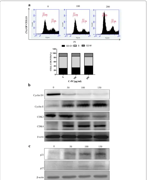
C‑Phycocyanin induced G0/G1 cell cycle arrest in MDA‑MB‑231 cells
Since C-phycocyanin inhibited MDA-MB-231 cells pro-liferation, we further explored the effects of C-phycocy-anin on cell cycle progress. The synchronized cells were
treated with C-phycocyanin for 24 h, and then followed by flow cytometry analysis to examine the cell cycle pro-gress. As shown in Fig. 2a, the G1 peaks increased from 29.6 to 32.6%, and finally changed to 40.9% after treat-ment with 100 and 200 μg/ml C-phycocyanin, respec-tively. In addition, the accumulation of cells in the G1 phase was accompanied by a decrease in the population of cells in the S phase. Indeed, the S peaks decreased from 55.5 to 53.3% and then 42.8% after treatment with 100 and 200 μg/ml C-phycocyanin, respectively. There-fore, these results indicated that C-phycocyanin induced G0/G1 phase cell cycle arrest in MDA-MB-231 cells.
C-Phycocyanin treatment in MDA-MB-231 cells could be observed to have a profound effect on cell-cycle pro-gression due to an increased population of cells in the G1 phase. To further investigate the proposed mechanism of C-phycocyanin inducing G1 phase arrest of MDA-MB-231 cells, we tested the levels of cell cycle regulatory proteins involved in G1 to S transition which included Cyclins (Cyclin D1 and Cyclin E), cyclin dependent kinases (CDK2 and CDK4) and CDKIs (CDK inhibitor p21 and p27). It has been well characterized that CDK4 is associated with d-type Cyclins while CDK2 is associ-ated with E-type Cyclins [24]. To investigate biological pathways of G1 arrest induced by C-phycocyanin via the regulatory protein expression, MDA-MB-231 cells were synchronized and treated with C-phycocyanin for 24 h. After treatment, the protein expression levels of Cyclin D1 and CDK2 decreased in a dose-dependent manner, as shown in Fig. 2b, while the expression levels of Cyclin E increased. Meanwhile, the protein expression levels of CDK4 first increased and then decreased in C-phycocya-nin-treated MDA-MB-231 cells.
Furthermore, CDKIs (CDK inhibitor p21 and p27) were often associated with the suppression of CDKs activity by forming CDK-CDKI complexes [25]. The effects of C-phycocyanin on the expression of p21 and p27 were detected by western blot, as shown in Fig. 2c. Obviously, the protein levels of p21 and p27 were upregulated in a dose-dependent manner (Fig. 2c). All these results sug-gested C-phycocyanin could inhibit the expressions of Cyclin D1 and CDK2, meanwhile increase p21 and p27 proteins expression in MDA-MB-231 cells.
C‑Phycocyanin induced cellular apoptosis of breast cancer MDA‑MB‑231 cells
0 50 100
150 200 250
0 50 100 150 200 250
0 200 400 600
** **
** **
NO. OF
COLONI
ES
Drug IC50 values( g/ml)
24h 48h
C-PC 229.0 189.4
150 200 250
0 50 100
24h
0 50 100 150 200 250 300 0.0
0.5 1.0 1.5
*
** ** **
** **
%
Ce
ll
Vi
ab
ilit
y
48h
0 50
100 150 200 250 300
0.0 0.5 1.0 1.5
** **
** ** ** **
%
Ce
ll
Vi
ab
ilit
y
a
b
c
PI
0 100 200
(%) OF CELLS
0
100 200 0
20 40 60 80 100 120
G0-G1 S G2-M
CELL COUNTS(%)
0 50 100 150
Cyclin D1
-actin
CDK4 CDK2 Cyclin E
0 50 100 150
-actin p21
p27
a
b
c
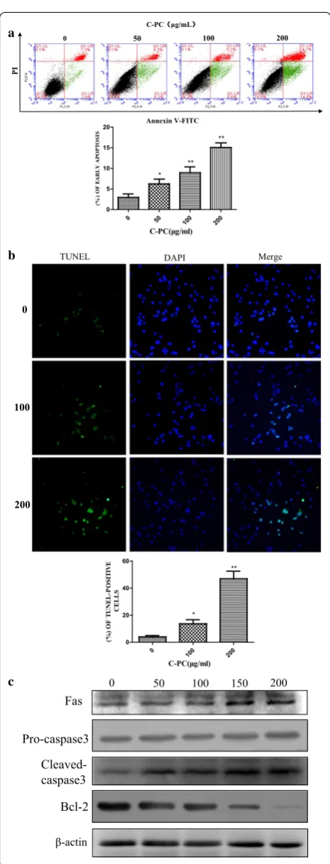
percentage of early apoptotic cells increased gradually from 2.9 to 6.2, 8.6 and 14.8% with the increase of C-phy-cocyanin concentrations. Consistent with this result, the percentage of late apoptotic cells increased gradually from 7.3% in untreated control group to 13.7% in treated cells. In order to further study the pro-apoptosis effect of C-phycocyanin on breast cancer MDA-MB-231 cells, we carried out TUNEL assay. The TUNEL assay showed that the TUNEL-positive cells were significant increased in a dose-dependent manner when MDA-MB-231 cells were treated with C-phycocyanin (Fig. 3b). There are many studies have revealed that membrane death receptor pathways played an important role in the progress of cell apoptosis. Moreover, to further gain a deeper insight into the mechanism of apoptosis induced by C-phycocyanin, we next tested the death receptor pathway. The results of Western blot showed that C-phycocyanin upregu-lated the protein levels of Fas and cleaved-caspase 3 while down-regulated the protein level of Bcl-2 in MDA-MB-231 cells (Fig. 3c). Altogether, these data indicated that the C-phycocyanin could induce the MDA-MB-231 cells apoptosis.
C‑Phycocyanin inhibited the migration of breast cancer MDA‑MB‑231 cells
Recently, some researches had found that C-phycocyanin could inhibit epithelial–mesenchymal transition (EMT) by down-regulating the expression of vimentin, type 1 collagen and fibronectin, and up-regulating the expres-sion of E-cadherin in TGF-β1-treated cells [26]. Thus, we wanted to determine the effects of C-phycocyanin on the migration of breast cancer MDA-MB-231 cells. The wound scratch assay results showed that the migra-tion activity of C-phycocyanin-treated MDA-MB-231 cells decreased in comparison to control cells. In wound scratch migration assays, the migration activity of C-phy-cocyanin-treated MDA-MB-231 cells was only 53% of the untreated control cells (Fig. 4a). Further, we verified the effect of C-phycocyanin on the metastatic activ-ity by the transwell migration assay. Similarly, the tran-swell migration assays were able to show that there is an decreased the migration ability in C-phycocyanin-treated
100 200
0 50
C-PC µg/mL
Annexin V-FITC
PI
0
100
200
TUNEL DAPI Merge
0 50 100 200
0 5 10 15 20
* **
**
Cleaved- caspase3 Pro-caspase3
Bcl-2 Fas
-actin
0 50 100 150 200
0 100 200
0 20 40 60
* **
a
b
c
Fig. 3 Effect of C-phycocyanin on cell apoptosis in MDA-MB-231 cells. a Analysis of MDA-MB-231 cells apoptosis by flow cytometry using Annexin V-FITC and PI. Quantitative representation of early apoptotic cells after C-phycocyanin treatment for 24 h. b Analysis of MDA-MB-231 cells apoptosis by TUNEL assay. Quantitative represen-tation of TUNEL-positive cells after C-phycocyanin treatment for 24 h. c The expressions of Fas, pro-caspase 3, cleaved-caspase 3 and Bcl-2 were determined by western blot
MDA-MB-231 cells (Fig. 4b). In order to further investi-gate whether C-phycocyanin was involved in the migra-tion of MDA-MB-231 cells, the MDA-MB-231 cells with high ability of metastasis and mesenchymal-like phenotype were treated with C-phycocyanin. We found mesenchymal cells features were reduced while epithe-lial morphological features were increased in C-phyco-cyanin-treated MDA-MB-231 cells. For example, the morphological features of C-phycocyanin-treated cells changed from the long spindle-shaped appearance to the shorter spindle-shaped and flatter appearance by the immunofluorescence assay (Fig. 4c). Collectively these results suggested that C-phycocyanin could inhibit the migration of breast cancer MDA-MB-231 cells.
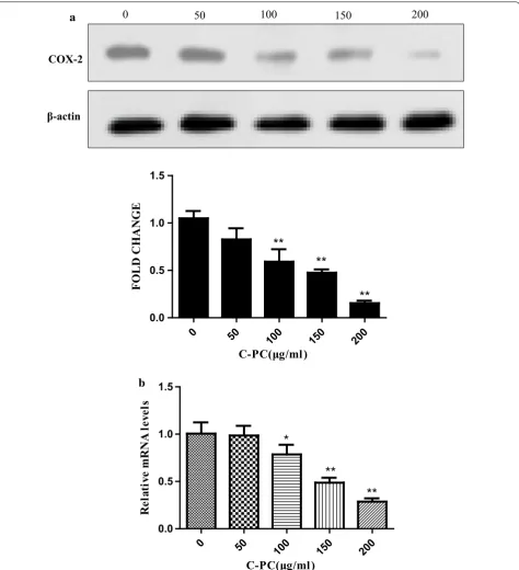
C‑Phycocyanin inhibited the COX‑2 expression of breast cancer MDA‑MB‑231 cells
It has been reported that COX-2 is high expression in tri-ple-negative breast cancer and correlates with poor sur-vival outcomes [27]. Recently, some studies have found that COX-2 is closely associated with tumor forma-tion and progression, as well as tumor angiogenesis and metastasis [28]. It has been reported that C-phycocyanin as COX-2 inhibitor can dock with VEGF1 and inhibit colon cancer through the angiogenic pathway [29]. Thus, we determined the levels of COX-2 protein and mRNA by Western blot and qPCR, respectively. C-Phycocyanin treatment showed an effective decrease in COX-2 protein and mRNA levels in a dose-dependent manner (Fig. 5a, b).
MAPK pathway was involved in the anti‑cancer effects of C‑phycocyanin on breast cancer MDA‑MB‑231 cells
The AKT and MAPK signal pathway have been reported to play crucial roles in proliferation and apoptosis pro-gress. In particular, down-regulation expression of the ERK could prevent cancer cell proliferation, while up-regulation expression of the JNK could promote cancer cell apoptosis. In addition, it was also reported that p38 MAPK could block cell proliferation, or promote cell apoptosis. To ascertain whether the AKT and MAPK signal pathways participated in the anti-tumor effects of C-phycocyanin, so we examined the phosphoryla-tion of MAPKs and AKT in C-phycocyanin-treated MDA-MB-231 cells. As shown in Fig. 6a, b, C-phycocy-anin regulated the phosphorylation of MAPKs in a dose-dependent manner, while did not significantly alter the phosphorylation of AKT. In addition, C-phycocyanin did not significantly alter the expression levels of total AKT and MAPKs. C-Phycocyanin increased the levels of p-JNK and p-p38 and decreased the level of p-ERK in a dose-dependent manner in the MDA-MB-231 cells (Fig. 6a).
In order to further verify the role of MAPK in the anti-tumor effect of C-phycocyanin, we treated the C-phyco-cyanin-treated MDA-MB-231 cells with 50 μM SB203580 (p38 MAPK inhibitor) or 50 μM SP600125 (JNK inhibi-tor). And through CCK-8 experiment, we found the inhibition of p38 MAPK and JNK suppress the C-phyco-cyanin-induced cell death when MDA-MB-231 cells were treated with SB203580 and SP600125 in Fig. 6c. Then we treated MDA-MB-231 cells with 10, 25, 50 μM PD98059 (ERK1/2 inhibitor), we found the inhibition of ERK could suppress the COX-2 expression, enhances the p21 and p27 expression, and further suppress CDK4 and Cyc-lin E expression when MDA-MB-231 cells were treated with PD98059 in Fig. 6d. Therefore, MAPK pathway was involved in the anti-cancer effects of C-phycocyanin on breast cancer MDA-MB-231 cells.
Discussion
Triple-negative breast cancer is a biological subtype of breast cancer, whose hallmarks is lack the expression of the breast cancer prognostic markers ER, PR and HER2 [3, 4]. Triple-negative breast cancer represents significant clinical challenge due to the abilities of highly aggressive and resistant to conventional endocrine therapy. While we currently lack targeted therapeutic strategies for triple-negative breast cancer, the discovery of novel bio-markers and selective therapies targeting these biomark-ers offer potential promise for the future. So far, more researchers have found that C-phycocyanin has the anti-cancer function, which can inhibit tumor cell prolifera-tion, induce tumor cell cycle arrest and promote tumor cell apoptosis and autophagy [6, 28]. Thus, C-phycocya-nin can function as a potential anti-cancer drug. But the current study on triple-negative breast cancer has barely been reported in detail. In our study, we have confirmed that C-phycocyanin could effectively inhibit the prolifer-ation, induce tumor G0/G1 cell cycle arrest and promote the apoptosis of the triple-negative breast cancer MDA-MB-231 cells in a dose-dependent manner. These results proved that C-phycocyanin has the anti-cancer effects on MDA-MB-231 cells.
0h
24h
CONTROL 50 100
CON 50 100
0 50 100 150
*
**
CONTROL 50 100
24h
24h
CONTRO
L 50 100
0.0 0.2 0.4 0.6
**
ACTIN C-PC DAPI MERGE
0
100 a
b
c
checkpoints. Our results showed that C-phycocyanin induced the G0/G1 cell cycle arrest. After C-phycocy-anin treatment, the protein expression levels of Cyclin D1 and CDK2 decreased in a dose-dependent manner
in C-phycocyanin-treated MDA-MB-231 cells. Further-more, the protein levels of CDKIs (CDK inhibitor p21 and p27) were upregulated by C-phycocyanin treatment in a dose-dependent manner. All these results suggested
COX-2
-actin
0 50 100 150 200
0 50
100 150 200
0.0 0.5 1.0 1.5
**
**
**
FOLD CHANG
E
0 50
100 150 200
0.0 0.5 1.0 1.5
*
**
**
a
b
C-phycocyanin could inhibit the expressions of Cyclin D1, CDK2 and CDK4 and meanwhile increase p21 and p27 proteins expression in MDA-MB-231 cells.
Apoptosis is a complementary mechanism of cell pro-liferation. There are two major apoptotic pathways: one is the mitochondrial/cytochrome C (endogenous) pathway, in which the endogenous signal ultimately activates Cas-pase-9 and Caspase-3; the other pathway is the cell mem-brane surface death receptor (exogenous) pathway, which finally activates Caspase-8 and Caspase-3 [31]. Caspase-3 is activated in most apoptotic signaling pathways [32], and Caspase-3 will eventually induce apoptosis [33]. Thus, the key of tumor therapy is how to induce tumor cell apoptosis. We found that C-phycocyanin could up-regulate the protein levels of Fas and cleaved-caspase 3 while down-regulated the protein level of Bcl-2 in a dose dependent manner in C-phycocyanin-treated MDA-MB-231 cells. It indicated that the antitumor effect of C-phycocyanin on the MDA-MB-231 cells was mediated by induction of apoptosis.
C-Phycocyanin can function as an inhibitor of COX-2 which plays a crucial role in tumor progression and chemical resistance [34, 35]. It has been studied that inhibitors of COX-2 up-regulated E-cadherin expres-sion in colon cancer cell lines [36]. In non-small cell lung cancer (NSCLC), the expression of COX-2 was positively correlated with tumor metastasis and invasion. The biological function of COX-2 is converting arachi-donic acid to prostaglandins. Further studies found that exogenous prostaglandin E2 (PGE2) could significantly reduce E-cadherin expression in the NSCLC cells. Over-expression of COX-2 or PGE2 treatment can up-regulate ZEB1 and Snail (transcriptional inhibitors of E-cadherin) in NSCLC cells. In addition, PGE2 enhanced the binding of ZEB1 and Snail to E-cadherin at the chromatin level. Therefore, COX-2 and PGE2 are closely interrelated with tumor metastasis and invasion [37]. C-Phycocyanin has a therapeutic effect on the metastasis and invasion of tumor cells. In TGF-β1-treated MCF-7 and A549 cells, C-phycocyanin reduced the expression of fibronectin,
t-AKT
0 50 100 150 200
p-AKT
-actin
0 50 100 150 200
p-ERK1/2 t-ERK1/2
p-p38 MAPK
t-JNK t-p38 MAPK
-actin p-JNK
C-PC( g/ml) SB203580( M) SP600125( M)
- 200 - - 200 200 - - 50 - 50 - - - - 50 - 50 0.0
0.5 1.0 1.5
Ce
ll
viab
ility(
%)
**
# #
** ** **
*
p21
0 10 25 50
Cyclin D1
p27 CDK4 CDK2 COX-2
Cyclin E
-actin
PD98095( M)
a
b
c
d
Fig. 6 Involvement of the MAPK pathway in C-phycocyanin-induced cell death. a Effect of C-phycocyanin on the PI3K/AKT signaling in MDA-MB-231 cells. After treatments, the expression levels of p-AKT and total AKT were analyzed by Western blotting. b Effect of C-phycocyanin on the MAPK signaling in MDA-MB-231 cells. After treatments, the levels of ERK1/2, JNK and p38 MAPK and their phos-phorylated forms were analyzed by Western blotting. c The effects of inhibitors (SB203580 or SP600125) on cell viability were evaluated by CCK-8 assay. Significant difference compared with control group are indicated as *P < 0.05, **P < 0.005, and # P > 0.05. d The effects of PD98059 on the levels of COX-2, CDK2, CDK4, Cyclin D1, Cyclin E, p21 and p27 were evaluated by Western blotting
vimentin and type 1 collagen, which increased the expression of E-cadherin. Thus, C-phycocyanin inhib-ited TGF-β1-induced EMT in MCF-7 and A549 cells [26]. Our results demonstrated that C-phycocyanin treat-ment effectively decreased in COX-2 protein and mRNA expression in a dose-dependent manner, furthermore inhibit the migration of breast cancer MDA-MB-231 cells.
As we all know the mitogen activated protein kinase (MAPK) pathway plays an important role in the devel-opment and progression of cancer [21]. When the cells encountered a variety of stimuli, such as UV irradiation, cytokines, tumor necrosis factor (TNF) and chemo-therapeutic drugs and so on, JNK can be activated and function as a pro-apoptotic kinase. Some studies had documented that the JNK pathway activated caspases and regulated apoptosis-related proteins, such as Bax, Bcl-2 and p53. Further researcher had shown JNK acted on the mitochondrial death pathway. There are growing evidence indicated that the ERK pathway is in connection with the pathogenesis and progression of human breast cancer [17]. The role of p38 MAPK signaling is diverse, mainly depending on the stimulus and cell type. Some-times, p38 MAPK had been shown to promote cell death,
sometimes, while p38 MAPK enhanced cell growth and survival [19, 20]. According to our study, C-phyco-cyanin regulated the phosphorylation of MAPKs in a dose-dependent manner, while C-phycocyanin did not significantly alter total MAPKs. After treatment, C-phy-cocyanin increased the levels of p-JNK and p-p38, and decreased the level of p-ERK in a dose-dependent man-ner in the MDA-MB-231 cells. In a word, MAPK pathway was involved in the anti-cancer effects of C-phycocyanin on breast cancer MDA-MB-231 cells.
Conclusions
Our study indicated that C-phycocyanin in triple-negative breast cancer MDA-MB-231 cells (i) inhibits the proliferation of tumor cell (ii) induces tumor cell cycle G0/G1 arrest (iii) promotes tumor cell apopto-sis through the cell membrane surface death receptor (exogenous) pathway (iv) inhibits the COX-2 expression and tumor cell metastasis (v) down-regulates ERK sign-aling pathways and up-regulates JNK and p38 MAPK signaling pathways to induce tumor cell death (Fig. 7). These results proved that C-phycocyanin could serve as a promising anti-cancer therapeutic agent on triple-neg-ative breast cancer.
Abbreviations
C-PC: C-phycocyanin; TNBC: triple-negative breast cancer; PR: progesterone receptor; ER: estrogen receptor; MAPK: mitogen activated protein kinase; JNK: c-Jun N-terminal kinase; ERK: extracellular signal-regulated kinase; CDK: cyclin dependent kinase; CDKI: cyclin dependent kinase inhibitor; COX-2: cyclooxy-genase-2; PGE2: prostaglandin E2.
Authors’ contributions
BL and LJ designed the experiments. LJ performed the experiments and wrote the articles. YW, GL and HL helped to perform the experiments and collected the data, FZ and HJ participated in the statistical analysis. All authors read and approved the final manuscript.
Acknowledgements
Not applicable.
Competing interests
The authors declare that they have no competing interests.
Availability of data and materials
The authors declare that the data supporting the findings of this study are available within the article.
Consent for publication
Not applicable.
Ethics approval and consent to participate
Not applicable.
Funding
This work was supported by Grants from the National Natural Science Founda-tion of China (81471546, 81001346, 81273206).
Publisher’s Note
Springer Nature remains neutral with regard to jurisdictional claims in pub-lished maps and institutional affiliations.
Received: 1 November 2017 Accepted: 20 January 2018
References
1. Torre LA, Bray F, Siegel RL, Ferlay J, Lortet-Tieulent J, Jemal A. Global cancer statistics, 2012. CA Cancer J Clin. 2015;65(2):87–108. 2. Siegel RL, Miller KD, Jemal A. Cancer statistics, 2017. CA Cancer J Clin.
2017;67(1):7–30.
3. Telli ML, Chang ET, Kurian AW, Keegan TH, McClure LA, Lichtensztajn D, Ford JM, Gomez SL. Asian ethnicity and breast cancer subtypes: a study from the California Cancer Registry. Breast Cancer Res Treat. 2011;127(2):471–8.
4. Foulkes WD, Smith IE, Reis-Filho JS. Triple-negative breast cancer. N Engl J Med. 2010;363(20):1938–48.
5. Jung IL. Soluble extract from Moringa oleifera leaves with a new antican-cer activity. PLoS ONE. 2014;9(4):e95492.
6. Roy KR, Arunasree KM, Reddy NP, Dheeraj B, Reddy GV, Reddanna P. Alteration of mitochondrial membrane potential by Spirulina platensis
C-phycocyanin induces apoptosis in the doxorubicinresistant human hepatocellular-carcinoma cell line HepG2. Biotechnol Appl Biochem. 2007;47(Pt 3):159–67.
7. Romay C, Armesto J, Remirez D, Gonzalez R, Ledon N, Garcia I. Antioxidant and anti-inflammatory properties of C-phycocyanin from blue–green algae. Inflamm Res. 1998;47(1):36–41.
8. Li B, Chu X, Gao M, Li W. Apoptotic mechanism of MCF-7 breast cells in vivo and in vitro induced by photodynamic therapy with C-phycocya-nin. Acta Biochim Biophys Sin. 2010;42(1):80–9.
9. Li B, Gao MH, Chu XM, Teng L, Lv CY, Yang P, Yin QF. The synergistic antitumor effects of all-trans retinoic acid and C-phycocyanin on the lung cancer A549 cells in vitro and in vivo. Eur J Pharmacol. 2015;749:107–14.
10. Wang H, Liu Y, Gao X, Carter CL, Liu ZR. The recombinant beta subunit of C-phycocyanin inhibits cell proliferation and induces apoptosis. Cancer Lett. 2007;247(1):150–8.
11. Subhashini J, Mahipal SV, Reddy MC, Mallikarjuna Reddy M, Rachamallu A, Reddanna P. Molecular mechanisms in C-phycocyanin induced apoptosis in human chronic myeloid leukemia cell line-K562. Biochem Pharmacol. 2004;68(3):453–62.
12. Khavari TA, Rinn J. Ras/Erk MAPK signaling in epidermal homeostasis and neoplasia. Cell Cycle. 2007;6(23):2928–31.
13. Yang SH, Sharrocks AD, Whitmarsh AJ. MAP kinase signalling cascades and transcriptional regulation. Gene. 2013;513(1):1–13.
14. Olsen BB, Svenstrup TH, Guerra B. Downregulation of protein kinase CK2 induces autophagic cell death through modulation of the mTOR and MAPK signaling pathways in human glioblastoma cells. Int J Oncol. 2012;41(6):1967–76.
15. Fang JY, Richardson BC. The MAPK signalling pathways and colorectal cancer. Lancet Oncol. 2005;6(5):322–7.
16. Lei K, Davis RJ. JNK phosphorylation of Bim-related members of the Bcl2 family induces Bax-dependent apoptosis. Proc Natl Acad Sci USA. 2003;100(5):2432–7.
17. Santen RJ, Song RX, McPherson R, Kumar R, Adam L, Jeng MH, Yue W. The role of mitogen-activated protein (MAP) kinase in breast cancer. J Steroid Biochem Mol Biol. 2002;80(2):239–56.
18. Adjei AA. The role of mitogen-activated ERK-kinase inhibitors in lung cancer therapy. Clin Lung Cancer. 2005;7(3):221–3.
19. Juretic N, Santibanez JF, Hurtado C, Martinez J. ERK 1,2 and p38 pathways are involved in the proliferative stimuli mediated by urokinase in osteo-blastic SaOS-2 cell line. J Cell Biochem. 2001;83(1):92–8.
20. Yosimichi G, Nakanishi T, Nishida T, Hattori T, Takano-Yamamoto T, Taki-gawa M. CTGF/Hcs24 induces chondrocyte differentiation through a p38 mitogen-activated protein kinase (p38MAPK), and proliferation through a p44/42 MAPK/extracellular-signal regulated kinase (ERK). Eur J Biochem. 2001;268(23):6058–65.
21. Dhillon AS, Hagan S, Rath O, Kolch W. MAP kinase signalling pathways in cancer. Oncogene. 2007;26(22):3279–90.
22. Wada T, Penninger JM. Mitogen-activated protein kinases in apoptosis regulation. Oncogene. 2004;23(16):2838–49.
23. Li B, Zhang X, Gao M, Chu X. Effects of CD59 on antitumoral activi-ties of phycocyanin from Spirulina platensis. Biomed Pharmacother. 2005;59(10):551–60.
24. Malumbres M, Barbacid M. Cell cycle, CDKs and cancer: a changing paradigm. Nat Rev Cancer. 2009;9(3):153–66.
25. Sherr CJ, Roberts JM. CDK inhibitors: positive and negative regulators of G1-phase progression. Genes Dev. 1999;13(12):1501–12.
26. Pattarayan D, Rajarajan D, Ayyanar S, Palanichamy R, Subbiah R. C-Phycocyanin suppresses transforming growth factor-beta1-induced epithelial mesenchymal transition in human epithelial cells. Pharm Rep. 2017;69(3):426–31.
27. Tian J, Hachim MY, Hachim IY, Dai M, Lo C, Raffa FA, Ali S, Lebrun JJ. Cyclooxygenase-2 regulates TGFbeta-induced cancer stemness in triple-negative breast cancer. Sci Rep. 2017;7:40258.
28. Liu Q, Huang Y, Zhang R, Cai T, Cai Y. Medical application of Spirulina platensis derived C-phycocyanin. Evid Compl Altern Med eCAM. 2016;2016:7803846.
29. Saini MK, Sanyal SN. Targeting angiogenic pathway for chemoprevention of experimental colon cancer using C-phycocyanin as cyclooxygenase-2 inhibitor. Biochem Cell Biol. 2014;92(3):206–18.
30. Otto T, Sicinski P. Cell cycle proteins as promising targets in cancer therapy. Nat Rev Cancer. 2017;17(2):93–115.
31. Kaufmann SH, Earnshaw WC. Induction of apoptosis by cancer chemo-therapy. Exp Cell Res. 2000;256(1):42–9.
32. Creagh EM, Conroy H, Martin SJ. Caspase-activation pathways in apopto-sis and immunity. Immunol Rev. 2003;193:10–21.
33. Johnson VL, Ko SC, Holmstrom TH, Eriksson JE, Chow SC. Effector caspases are dispensable for the early nuclear morphological changes during chemical-induced apoptosis. J Cell Sci. 2000;113(Pt 17):2941–53. 34. Telliez A, Furman C, Pommery N, Henichart JP. Mechanisms leading to
• We accept pre-submission inquiries
• Our selector tool helps you to find the most relevant journal
• We provide round the clock customer support
• Convenient online submission
• Thorough peer review
• Inclusion in PubMed and all major indexing services
• Maximum visibility for your research
Submit your manuscript at www.biomedcentral.com/submit
Submit your next manuscript to BioMed Central
and we will help you at every step:
35. Davies G, Martin LA, Sacks N, Dowsett M. Cyclooxygenase-2 (COX-2), aro-matase and breast cancer: a possible role for COX-2 inhibitors in breast cancer chemoprevention. Ann Oncol. 2002;13(5):669–78.
36. Noda M, Tatsumi Y, Tomizawa M, Takama T, Mitsufuji S, Sugihara H, Kashima K, Hattori T. Effects of etodolac, a selective cyclooxygenase-2 inhibitor, on the expression of E-cadherin–catenin complexes in gastroin-testinal cell lines. J Gastroenterol. 2002;37(11):896–904.


![4 [(1E) Benzylideneamino] 3 methyl 2,4 dihydro 1H 1,2,4 triazole 5 thione](data:image/gif;base64,R0lGODlhAQABAIAAAP///wAAACH5BAEAAAAALAAAAAABAAEAAAICRAEAOw==)