Simulating tumor microenvironment: changes in
protein expression in an in vitro co-culture system
Salvatore
et al.
P R I M A R Y R E S E A R C H
Open Access
Simulating tumor microenvironment: changes in
protein expression in an in vitro co-culture system
Viviana Salvatore, Gabriella Teti, Silvia Bolzani, Stefano Focaroli, Sandra Durante, Maria Carla Mazzotti
and Mirella Falconi
*Abstract
Background:The role of the microenvironment during the initiation and progression of carcinogenesis is thought to be of critical importance, both for the enhanced understanding of fundamental cancer biology as well as for improving molecular diagnostics and therapeutics. The aim of this study was to establish an in vitro model based on a co-culture of healthy human fibroblasts (HFs) and human osteosarcoma cells (MG-63s) to simulate the microenvironment including tumor and healthy cells.
Methods:The HFs and MG-63s were in vitro co-cultured for a period of time ranging from 24 h to 7 days. Cell morphology and organization were studied using phase contrast microscopy while the expression of Human Cartilage Glycoprotein 39 (YKL-40), Vascular Endothelial Growth Factor (VEGF) and Matrix Metalloprotease 1 (MMP1) was investigated by Real Time PCR and Western Blotting.
Results:The results showed a characteristic disposition of tumor and healthy co-cultured cells in columns which are not visible in tumor and healthy cells grown singularly. The expression of YKL-40, VEGF and MMP1 significantly changed in co-cultured cells compared to HFs and MG-63s separately cultured.
Conclusions:We concluded that the tumor microenvironment has an influence on the protein expression of the healthy surrounding tissues and the process of tumorigenicity.
Keywords:Tumor microenvironment, Co-cultures, Cell-cell contacts, YKL-40, VEGF, MMP1, Osteosarcoma cell line, Human fibroblast cells
Background
In normal tissue, cells communicate through a complex network of interactions: physically, through direct contact or through the intervening extracellular matrix (ECM), and biochemically, through both soluble and insoluble signaling molecules [1]. Under persistent inflammatory conditions, the continual upregulation of enzymes, such as matrix metalloproteinases (MMPs) by stromal fibroblasts can disrupt the ECM; furthermore, invading immune cells can overproduce factors which promote abnormal gene ex-pression [1]. These conditions are normally reversible but, when inflammation is sustained, the normal organization of cells might lead to gene expression instability within the healthy cells and the acquisition of tumorigenic po-tential [2]. These chronic inflammatory conditions help to
establish a tumor microenvironment full of deranged pro-liferative signaling networks, which is largely orchestrated by inflammatory cells [3].
A tumor microenviroment includes invasive cancer cells and stromal cells, consisting in cancer-associated non-malignant fibroblasts, which provide an essential communication network via secretion of growth factors and chemokines. This condition leads to an altered ECM and provides additional oncogenic signals enhancing cancer-cell proliferation and invasion [4]. While the stro-mal cells are not stro-malignantper se, their role in support-ing cancer growth and development is vital to the survival of the tumor [5,6].
The aim of the study was to establish an in vitro model based on the co-culture of healthy human fibroblasts (HFs) and human osteosarcoma cells (MG-63s) to simulate the microenvironment of healthy tissue in connection with cancer cells. The final goal was to analyze the influence of
* Correspondence:[email protected]
Department for Biomedical and Neuromotor Sciences (DIBINEM), University of Bologna, via Irnerio 48, Bologna 40126, Italy
microenvironment in both tumor and healthy cells, investi-gating the gene and protein expression of some biomarkers involved in cancer progression and invasion. A better un-derstanding of the molecular mechanisms in the tumor microenvironment could be a crucial key to improving anti-cancer therapy.
During the past decade, there has been growing inter-est regarding glycoprotein YKL-40 [7,8], also known as chitinase 3–like 1 (CHI3L1) [9] and as human cartilage glycoprotein–39 (HC-gp39) [10]. YKL-40 is involved in angiogenesis [11] and the upregulation of the vascular endothelial growth factor expression (VEGF) in several cell lines [12-14]. Furthermore, YKL-40 plays a role in inflammation and tissue remodeling [15,16]. The aberrant and elevated expression of YKL-40 is largely associated with the pathogenesis of a variety of human diseases, such as rheumatoid- and osteoarthritis, hepatic fibrosis and asthma, which is connected to its pathological function and is associated with extracellular matrix remodeling, suggesting that the serum levels of YKL-40 serve as a diagnostic and prognostic biomarker [7].
Tumor invasion is also characterized by the increased ability of tumor cells to degrade ECM components through an enhanced expression of proteases, including matrix metalloproteinases secreted or anchored to the cell membrane. Among the membrane-type MMPs, matrix metalloproteinases 1 (MMP1) has been particularly impli-cated in pericellular proteolysis associated with cell migra-tion and invasion [17]. An overexpression of MMP1 has been described in many types of human tumor tissues [18]. In addition, several studies have reported that MMP1 is produced by invasive tumor cells in vivo as well as in vitro and is thus associated with vascular remodeling, angiogenesis and tumor progression [17].
Acting together with metalloproteinases, the VEGF is a key regulator of physiological angiogenesis during embryogenesis, in vascular remodeling, wound healing, cell invasion, permeability, proliferation and survival [19]. The VEGF has also been implicated in pathological angio-genesis associated with tumors, intraocular neovascular disorders and other conditions. Inhibition of the VEGF is also being tested as a strategy for the prevention of angio-genesis and vascular leakage in several pathological condi-tions [20].
In the present study, we tested the expression of YKL-40, VEGF and MMP-1 in HFs and MG-63s seeded both alone and mixed in a co-culture system, for a period of time ranging from 24 h to 96 h. Light phase contrast mi-croscopy was carried out to study the organization and orientation of cells in the co-culture from 4 to 7 days.
The influence of direct contact between the tumor cells and the non-malignant cells, and the interconnections of the biomarkers investigated and their role in tumor inva-sion and matrix remodeling were discussed.
Results
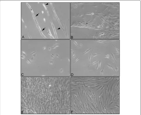
Morphological analysis by phase contrast microscopy After 4 days of co-culture, the MG-63s and the HFs showed a specific and characteristic organization based on cord-like formations (Figure 1A). After 7 days of co-culture, the cord-like formations were larger and the HFs were almost completely covered by MG-63 cells (Figure 1B).
On the contrary, control samples of the MG-63s and the HFs, grown separately, showed their characteristic cell morphology. The MG-63s had a polygonal morph-ology after 24 h of culture (Figure 1C) while the HFs showed a fibroblastic morphology with an elongated cell body after 24 h of culture (Figure 1D). At the end of 7 days of culture, both HFs and MG-63s had reached confluence (Figure 1E and F).
qRT-PCR of YKL-40 and VEGF in co-cultured HFs and MG-63 cells
To examine changes in protein expression during the coexistence of HFs and MG-63 cells, the mRNA expres-sion of YKL-40 and VEGF was evaluated using a quanti-tative real time polymerase chain reaction (qRT-PCR) in cells grown in co-culture, and the results were compared to the same cells grown separately as controls.
The expression of YKL-40 in HF cells seeded separately and used as controls was almost absent. Indeed, YKL-40 expression was the highest at 24 h; it subsequently de-creased and remained constant up to 96 h (Figure 2). In MG-63 cells, the expression of YKL-40 was very high at 24 h in controls seeded separately and then decreased until 96 h. In the MG-63 cells grown in co-culture, a similar trend to the HFs grown in co-culture was observed, but with a slightly lower intensity of expression (Figure 2).
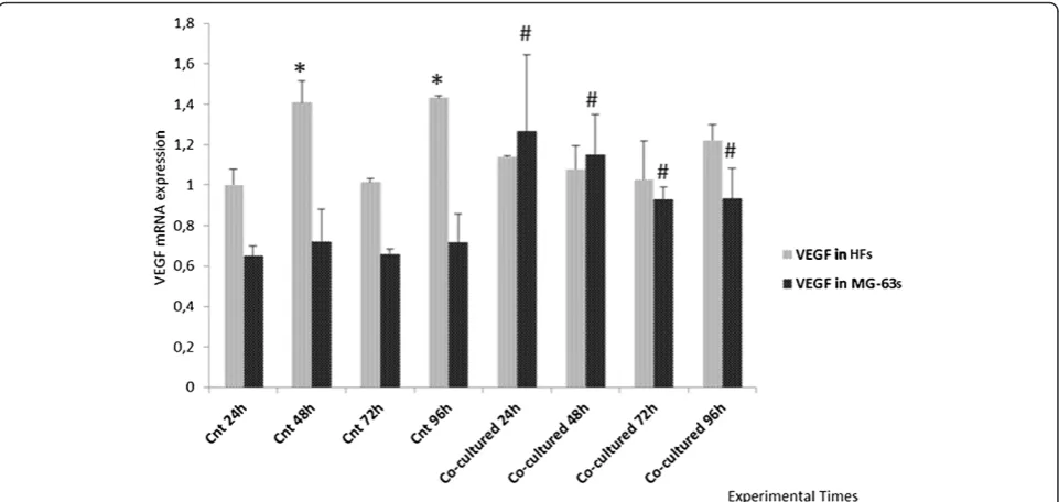
The VEGF showed constant expression in the HFs both grown in co-culture and separately while, in MG-63 cells, it was higher in co-culture with respect to the control cells seeded separately (Figure 3).
Western Blotting and densitometric analysis
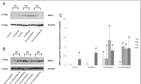
Control HFs showed a low level of MMP1 expression after 24 h of culture while it increased after 96 h. On the contrary, MG-63 control cells demonstrated a higher ex-pression of MMP1 protein after 24 h and 96 h of culture as compared to HFs (Figure 4A).
In the co-cultured cells, the intensity of the MMP1 bands increased considerably over time, reaching the highest intensity in HFs at 72 h and in MG-63s at 48 h (Figures 4A and B), as demonstrated by densitometric analysis (Figure 4C).
Discussion
For many years, laboratories have sought to under-stand the mechanisms by which cells respond to their
Salvatoreet al. Cancer Cell International2014,14:40 Page 2 of 8
microenvironment, and how these signals are integrated to specify programs of gene expression and, ultimately, tissue phenotype. Understanding these processes and their alteration will engender a greater appreciation of the mechanisms involved in tumor progression.
The scientific literature has shown how remodeling the ECM and producing aberrations can stimulate the development of cancer [21].
The aim of the present study was to develop an in vitro methodology for investigating the interactions between non-malignant cells and tumor cells, and to verify how the microenvironment influences cells in the co-culture system. During malignant transformation, healthy and tumor cells are increasingly described as networks of their physical
and signaling contacts [22]; with this network analysis approach, we chose to co-culture a healthy and non-malignant population of human fibroblast cells with a human osteosarcoma cell line, MG-63s, to study the morphological and molecular changes originated by this direct interaction.
The morphological analysis of cells independently co-cultured in monolayers demonstrated a well-defined and different cell morphology of MG-63s and HFs in contrast to monolayers of co-cultured HFs and MG-63s which showed cord-like structures after 4 days of culture, thick-ening after 7 days. We suggested that, in cord-like struc-tures, the fibroblasts worked as a climbing frame on which the MG-63 cells adhered and proliferated. The absence of
this type of organization in MG-63 cells and HFs grown separately suggests a morphological rearrangement due to modifications in the microenvironment of the co-culture system. Fauquette and colleagues [23] demonstrated that the cord-like morphogenesis was supported by changes in the cell surface receptor after contact between tumor cells and healthy cells, which was eventually associated with an elevated expression of adhesive molecules, leading to cell contact reorganization and cell-matrix changes. These data
correlated well with our results and with tumor invasive-ness ability.
To verify whether the microenvironment has an influ-ence on cells grown in co-culture, the expression of the YKL-40, VEGF and MMP1 proteins involved in the pro-gression of pathological conditions was analyzed. Some years ago, Johansen et al. [24] identified a protein called YKL-40, secreted in vitro in several types of solid tumors and in large amounts by MG-63 cells. On the contrary,
Figure 2Real Time PCR for YKL-40.Expression of HFs and MG-63 cells independently grown and co-cultured. *represents a significant difference from the MG-63 control cells, P < 0.05.
Figure 3Real Time PCR for VEGF.Expression of HFs and MG-63 cells independently grown and co-cultured. *represents a significant difference from the HF control sample at 24 h, P < 0.05. # represents a significant difference from the MG-63 control cells at 24 h, P < 0.05.
Salvatoreet al. Cancer Cell International2014,14:40 Page 4 of 8
there are no data regarding in vitro YLK-40 expression in HFs [16]. For this reason, we have chosen the YKL-40 protein as the main marker for verifying the influence of tumor cells grown in contact with healthy cells.
As reported in the literature [25], YKL-40 was identified as a tumor angiogenesis factor, inducing coordination of membrane-bound receptor syndecan 1 and integrinαvβ3, and activating an intracellular signaling cascade includ-ing FAK (focal adhesion kinase), Erk 1, and Erk 2 [26]. Furthermore, YKL-40 not only increased the expression of Flk-1/KDR (phosphatidylinositol 3-kinase/akt signal), VEGF receptor 2 which mediates VEGF angiogenesis [27], but also activated the tyrosine phosphorylated form of Flk-1/KDR, possibly leading to a synergistic effect on an-giogenic signaling activation. YKL-40 purified from the MG-63 osteosarcoma cell line has growth factor activity for fibroblast cell lines [15]. The YKL-40 secreted by can-cer cells has a role in the mutation of the fibroblasts sur-rounding the tumor, including the activation of fibroblast morphologic transformation, secretion of MMPs and neo-vascularization. Therefore, YLK-40 promotes the prolif-eration, differentiation and invasion of cancer cells and the destruction of stroma [28-30]. The overexpression of MMP1 has been shown in tumor tissues and has been suggested to be associated with tumor invasion and
metastasis [18]. The production of MMP1 is stimulated in fibroblasts by growth factors and cytokines; it is postulated that YKL-40 acts synergistically as a growth factor with VEGF, stimulating MMP1 expression [13,19].
Starting from these previous data, we investigated the expression of YKL-40, the VEGF and MMP1 using Real Time PCR and Western Blotting, first in cells grown separately and then in the co-culture system, in order to compare the protein expression trend in both cellular groups. The final goal was to verify whether HFs under-went changes after direct contact with tumoral cells.
Real Time results demonstrated that YKL-40 was expressed only in MG-63 control cells, in accordance with the literature [16], and was not expressed in HF control cells, during the entire experimental time. In addition, our data gave an unexpected result; in the co-culture system, we found the expression of YKL-40 even in HFs, although fibroblasts do not normally express this protein in vitro. The HFs co-cultured with MG-63s ef-fectively showed an elevated expression of YKL-40 after 24 h, and then remained almost constant during the experiment.
The expression of the VEGF is quite constant in the HFs used as controls and in co-cultures, but increases in MG-63 cells when co-cultured. We suggest that the
tumor cell-conditioned medium exhibited an impact on YKL-40 expression in HFs, and on VEGF expression in MG-63 cells, perhaps due to the growth factor activity of YKL-40 secreted in the tumor cell-conditioned medium and to its direct role in stimulating VEGF expression and ECM remodeling.
Western Blot analyses showed weak MMP1 expression in control HFs and a more elevated expression in con-trol MG-63 cells. In both samples, the level of MMP1 gradually increased during the time of culture. Co-cultures of HFs and MG-63 cells showed high levels of MMP1 after only 24 h and they greatly increased up to 96 h, suggesting a strong influence of the tumor micro-enviroment on the upregulation of the matrix metallo-proteinases responsible for the disruption of the ECM and tumor invasiveness [2]. In consideration of our re-sults, we could better postulate the role of YKL-40 in the tumor microenvironment.
Ngrnyuang et al. have recently reported [14] that the effects of YKL-40 are cell type-dependent, probably due to the different biochemical composition between cell lines. Human fibroblasts and MG-63s represent two bio-logically different cellular types involved in parallel roles during tumor progression. Tumor cells, releasing pro-inflammatory factors, are able to induce the activity of NF-kB (Nuclear Factor kappa B) in fibroblast cells which, in turn, induce the release of growth factors and cytokines in the adjacent extracellular matrix, enhancing an inflam-matory microenvironment and promoting changes in the ECM. Our hypothesis is that YKL-40 may have synergistic effects; on HFs, it can act similarly to insulin-like growth factor-1, leading to an inflammatory condition and pro-moting an up-regulation in MMP-1 expression, in order to remodel the extracellular matrix. In addition to the in-flammatory conditions, the expression of YKL-40 in MG-63s, correlated with those of the VEGF, suggests that YKL-40 acts as an angiogenic factor in tumor cells, as has been reported in the literature.
Following these preliminary data, it could be of great interest to examine the effect of the siRNA knockdown of YKL-40 in MG-63 cells on the changes reported in protein expression in the co-cultures. These experiments are currently in progress.
Conclusions
In conclusion, this study showed that cancer evolution can involve changes in the microenvironment as a result of oncogenic stress within healthy cells. The phenotypic reorganization between fibroblasts and MG-63 cells, extra-cellular matrix remodeling, changes in YLK-40 protein ex-pression and increased VEGF exex-pression are indications of cancer progression.
Our experimental approach suggested a realistic in vitro model of the tumor microenvironment, useful for better
investigating the complex mechanisms which transform a healthy tissue environment to a malignant state.
Methods
Primary culture of HFs
Human dental ligament (HDL) tissues were collected from the mid-third of roots of teeth extracted for ortho-dontic reasons, following informed consent of the pa-tients. After several washes in PBS, the HDL tissues were cut into small pieces and placed into culture dishes with 1 mL of Dulbecco’s Modified Essential Medium (DMEM) (Invitrogen, Carlsbad, CA, USA) supplemented with 10% (v/v) fetal bovine serum (FBS), penicillin (100 mg/mL) and streptomycin (10 mg/mL). The culture medium was chan-ged twice a week. When the HFs were subconfluent (70–80%) they were scraped off using 0.05% trypsin/ EDTA (Gibco, Grand Island, NE), washed and placed into T75 flasks. The cells obtained were cultured at 37°C in a humidified atmosphere of 5% CO2. Cells from passages 3 to 10 were utilized for the following experiments.
MG-63 cell culture
The MG-63 cell line was purchased from ATCC®
CRL-1427™ (USA) and cultivated in DMEM (Invitrogen,
Carlsbad, CA, USA) containing 10% FBS, supplemented with 10% (v/v) FBS, penicillin (100 mg/mL) and strepto-mycin (10 mg/mL) according to the recommendation of the supplier. The cells were cultured in T25 flasks (Nunc, USA) in a humidified incubator at 37°C in a 5% CO2 hu-midified atmosphere. For passaging, the cells were de-tached with trypsin/EDTA and subsequently replated.
Co-cultures of HFs and MG-63 cells
The HFs and the MG-63 cells were seeded at the same density (15 × 103 cells/mL each) in T75 flasks and cul-tured in DMEM F-12 supplemented with 10% (v/v) FBS, penicillin (100 mg/mL) and streptomycin (10 mg/mL) for 24 h, 48 h, 72 h and 96 h.
As controls, monocultures of MG-63 cells and HFs were seeded separately in T75 flasks and were cultured under the same conditions as the co-cultures. The cell medium was replaced twice a week.
Monostrate cultures and morphological analysis using phase contrast microscopy
Ten ×103cells/ml were seeded on glass slides in 6 multi-wells and co-cultured for 4 and 7 days. At the end of each experimental time point, the samples were observed using a phase contrast microscope Motic AE21. The images were recorded using Visicam 3.0 and analyzed by VisiCam Image Analyzer software, version 6.1.3.3.
The images of Figure 1 were representative for three independent experiments.
Salvatoreet al. Cancer Cell International2014,14:40 Page 6 of 8
Cell separation
At every experimental time point, the cells seeded in co-cultures were detached, collected and counted for separ-ation with MS Columns and Anti-Fibroblast Microbeads System (MACS, Miltenyi Biotec) according to the manu-facturer’s instructions. Briefly, the cells were centrifuged, and the pellets were suspended in MACS buffer solu-tion. The HFs were then magnetically labeled with hu-man Anti-Fibroblast MicroBeads while the unlabeled MG-63s were run through the column after 3 washes with MACS buffer. By so doing, the MG-63 fraction was de-pleted of HFs. After removing the column from the mag-netic field, the magmag-netically retained HFs were eluted as the positively-selected cell fraction. The separation effi-ciency guaranteed by the MACS Miltenyi System was more than 90%.
RNA extraction
Total RNA from control and co-cultured pellets was extracted using Nucleospin RNA II (Macherey-Nagel) and quantified using a NanoDrop® ND-1000 UV–vis Spectrophotometer (Thermo Scientific, Wilmington, DE, USA). Oneμg of total RNA was reverse transcribed using a high capacity cDNA Reverse Transcription kit (Applied Biosystem, Life Technologies, Monza Italy) according to the manufacturer’s instructions.
Real time PCR
The expression of mRNA was analyzed by quantitative Real Time PCR using 7500 Real Time PCR (Applied Biosystem, Life Technologies, Monza, Italy). All reactions were carried out in a 25μL reaction volume in triplicate. For the analysis, the following TaqMan assays (Applied Biosystems, Life Technologies, Monza, Italy) were used: Hs00609691_m1 for YKL-40 and Hs00900055_m1 for VGEF. All samples were normalized to glyceraldehyde 3-phosphate dehydrogenase (GAPDH, Hs99999905_m1) expression.
The data were representative of three independent ex-periments and showed the average of triplicates ± SD.
Western blot analysis and densitometric analysis
At each experimental time point, the cell pellets were lysed for 30 minutes using a RIPA extraction buffer (Invitrogen, Life Technologies, Monza Italy) supplemented with a pro-tease inhibitor cocktail (Sigma Aldrich, St Louis, Missouri, USA), 1 mM PMSF and 0.15%β-mercaptoethanol (Fluka, Sigma Aldrick, St Louis, Missouri, USA). The samples were centrifuged at 14,000 rpm for 10 minutes at 4°C, and the total protein amounts were assayed using Bradford re-agent (Sigma Aldrich, St. Louis, Missouri, USA).
Twentyμg of total protein were resolved on NuPAGE® SDS-PAGE pre-cast gels (4-12%) (Invitrogen, Life Tech-nologies, Monza, Italy), and the protein was transferred
to a nitrocellulose membrane (GE Healthcare Europe GmbH, Milan, Italy), blocked with no fat dry milk (Sigma Aldrich, St. Louis, Missouri, USA) for 30 min at room temperature (RT), and immunolabeled with anti-MMP1 1:500 in TBS pH7.5 and anti-β tubulin 1:10000 in TBS pH7.5 overnight at 4°C. The bands were visual-ized using an ECL Advanced TM Western blotting de-tection kit (GE Healthcare Europe GmbH, Milan, Italy) and the images were recorded with a Kodak digital image station (Eastman Kodak, Rochester, NY, USA).
Band densitometry was measured using Image J soft-ware (National Institutes of Health) and the intensities of the specific protein bands were corrected for equalβ -tubulin loading; they were expressed as relative to the intensity of the control sample. The relative quantitation of the western blots was expressed relative to the β -tubulin present on each blot.
The images of Figure 4 were representative of three in-dependent experiments. The densitometry data were representative of three independent experiments and showed the average of triplicates ± SD.
Statistical analysis
The statistical analysis was carried out using GRAPH PAD PRISM 5.0 software (San Diego, CA) by applying ANOVA and the Dunnet’s multiple comparison test. The differences were considered significant at p < 0.05.
Abbreviations
ECM:Extracellular matrix; FAK: Focal adhesion kinase; HDL: Human dental ligament; HFs: Human fibroblasts; MMP1: Matrix metalloprotease 1; MMPs: Matrix metalloproteinases; VEGF: Vascular Endothelial Growth Factor; YKL-40: Human cartilage glycoprotein–39 (N-term. Tyrosine (Y), Lysine (K) and Leucine (L) molecular mass 40 kDa (40).
Competing interests
The authors declare no financial interest or sources of research funding which could affect integrity of the scientific work presented.
Authors’contributions
VS, GT and MF participated in the design of the study and in the analysis of the data. VS and SB carried out the experimental work. VS and GT carried out the statistical analysis. The acquisition of data was carried out by SF, SD and MCM. They also helped to draft the manuscript. MF has given final approval of the version to be published. All the authors have read and approved the final manuscript.
Acknowledgements
This study was supported by the Italian Ministry of Research and Technology (MURST) with an FIRB grant [RBAP10MLK7_005], PRIN 2009 grant, and a Fondazione del Monte di Bologna and Ravenna 2012 grant.
Received: 21 November 2013 Accepted: 1 May 2014 Published: 13 May 2014
References
1. Bissell MJ, Radisky D:Putting tumours in context.Nat Rev Cancer2001,
1:46–54.
2. Mbeunkui F, Johann DJ:Cancer and the tumor microenvironment: a review of an essential relationship.Cancer Chemother Pharmacol
2009,63:571–582.
5. Hanahan D, Weinberg RA:The hallmarks of cancer.Cell2000,100:57–70. 6. Liotta LA, Kohn EC:The microenvironment of the tumour-host interface.
Nature2001,411:375–379.
7. Johansen JS, Schultz NA, Jensen BV:Plasma YKL-40: a potential new cancer biomarker?Future Oncol2009,5:1065–1082.
8. Lee CG, Da Silva CA, Dela Cruz CS, Ahangari F, Ma B, Kang MJ, He CH, Takyar S, Elias JA:Role of chitin and chitinase/chitinase-like proteins in inflammation, tissue remodeling, and injury.Annu Rev Physiol2011,
73:479–501.
9. Rehli M, Krause SW, Andreesen R:Molecular characterization of the gene for human cartilage gp-39 (CHI3L1), a member of the chitinase protein family and marker for late stages of macrophage differentiation. Genomics1997,43:221–225.
10. Hakala BE, White C, Recklies AD:Human cartilage gp-39, a major secretory product of articular chondrocytes and synovial cells, is a mammalian member of a chitinase protein family.J Biol Chem1993,
268:25803–25810.
11. Nishikawa KC, Millis AJ:gp38k (CHI3L1) is a novel adhesion and migration factor for vascular cells.Exp Cell Res2003,287:79–87.
12. Francescone RA, Scully S, Faibish M, Taylor SL, Oh D, Moral L, Yan W, Bentley B, Shao RJ:Role of YKL-40 in the angiogenesis, radioresistance, and progression of glioblastoma.Biol Chem2011,286:15332–15343. 13. Shao R:YKL-40 acts as an angiogenic factor to promote tumor
angiogenesis.Front Physiol2013, 10.3389/fphys.2013.00122.
14. Ngernyuang N, Francescone RA, Jearanaikoon P, Daduang J, Supoken A, Yan W, Shao R, Limpaiboon T:Chitinase 3 like 1 is associated with tumor angiogenesis in cervical cancer.Int J Biochem Cell Biol2014, 10.1016/j. biocel.2014.03.021.
15. Recklies AD, White C, Ling H:The chitinase 3-like protein human cartilage glycoprotein 39 (HC-gp39) stimulates proliferation of human connective-tissue cells and activates both extracellular signal-regulated kinase- and protein kinase B-mediated signalling pathways.Biochem J2002,365:119–126.
16. Johansen JS, Jensen BV, Roslind A, Nielsen D, Price PA:Serum YKL-40, a new prognostic biomarker in cancer patients?Cancer Epidemiol Biomarkers2006,15:194–202.
17. Mazor R, Alsaigh T, Shaked H, Altshuler AE, Pocock ES, Kistler EB, Karin M, Schmid-Schönbein GW:Matrix metalloproteinase-1-mediated up-regulation of vascular endothelial growth factor-2 in endothelial cells. J Biol Chem2013,288:598–607.
18. Sauter W, Rosenberger A, Beckmann L, Kropp S, Mittelstrass K, Timofeeva M, Wölke G, Steinwachs A, Scheiner D, Meese E, Sybrecht G, Kronenberg F, Dienemann H, LUCY-Consortium, Chang-Claude J, Illig T, Wichmann HE, Bickeböller H, Risch A:Matrix metalloproteinase 1 (MMP1) is associated with early-onset lung cancer.Cancer Epidemiol Biomarkers Prev2008,
17:1127–1135.
19. Wang H, Keiser JA:Vascular endothelial growth factor upregulates the expression of matrix metalloproteinases in vascular smooth muscle cells: role of flt-1.Circ Res1998,83:832–840.
20. Ferrara N, Gerber HP, LeCouter J:The biology of VEGF and its receptors. Nat Med2003,9:669–676.
21. Catalano V, Turdo A, Di Franco S, Dieli F, Todaro M, Stassi G:Tumor and its microenvironment: a synergistic interplay.Semin Cancer Biol
2013, 10.1016/j.semcancer.2013.08.007.
22. Csermelya P, Korcsmárosb T:Cancer-related networks: a help to understand, predict and change malignant transformation.Semin Cancer Biol2013,
23:209–212.
23. Fauquette W, Bourhis XDL, Delannoy-Courdent A, Boilly B, Desbiens X:
Characterization of morphogenetic and invasive abilities of human mammary epithelial cells: correlation with variations of urokinase-type plasminogen activator a&&y and type-l plasminogen activator inhibitor level.Biol Cell1997,89:453–465.
24. Johansen JS, Williamson MK, Rice JS, Price PA:Identification of proteins secreted by human osteoblastic cells in culture.J Bone Miner Res1992,
7:501–512.
25. Faibish M, Francescone RA, Bentley B, Yan W, Shao R:A YKL-40 neutralizing antibody blocks tumor angiogenesis and progression: a potential therapeutic agent in cancers.Mol Cancer Ther2011,10:742–751. 26. Shao R, Hamel K, Petersen L, Cao QJ, Arenas RB, Bigelow C, Bentley B, Yan
W:YKL-40, a secreted glycoprotein, promotes tumor angiogenesis. Oncogene2009,28:4456–4468.
27. Yan W, Bentley B, Shao R:Distinct angiogenic mediators are required for basic fibroblast growth factor- and vascular endothelial growth factor-induced angiogenesis: the role of cytoplasmic tyrosine kinase c-Abl in tumor angiogenesis.Mol Biol Cell2008,9:2278–2288. 28. Basset P, Bellocq JP, Wolf C, Stoll I, Hutin P, Limacher JM, Podhajcer OL,
Chenard MP, Rio MC, Chambon P:A novel metalloproteinase gene specifically expressed in stromal cells of breast carcinomas. Nature1990,348:699–704.
29. Grégoire M, Lieubeau B:The role of fibroblasts in tumor behavior. Cancer Metastasis Rev1995,14:339–350.
30. Rønnov-Jessen L, Petersen OW, Bissell MJ:Cellular changes involved in conversion of normal to malignant breast: importance of the stromal reaction.Physiol Rev1996,76:69–125.
doi:10.1186/1475-2867-14-40
Cite this article as:Salvatoreet al.:Simulating tumor microenvironment: changes in protein expression in an in vitro co-culture system.Cancer Cell International201414:40.
Submit your next manuscript to BioMed Central and take full advantage of:
• Convenient online submission
• Thorough peer review
• No space constraints or color figure charges
• Immediate publication on acceptance
• Inclusion in PubMed, CAS, Scopus and Google Scholar
• Research which is freely available for redistribution
Submit your manuscript at www.biomedcentral.com/submit
Salvatoreet al. Cancer Cell International2014,14:40 Page 8 of 8